Following the people and events that make up the research community at Duke

Students exploring the Innovation Co-Lab

Category: Cancer Page 2 of 6

The evolution of a tumor

The results of evolution are often awe-inspiring — from the long neck of the giraffe to the majestic colors of a peacock — but evolution does not always create structures of function and beauty.

In the case of cancer, the growth of a population of malignant cells from a single cell reflects a process of evolution too, but with much more harrowing results.

Johannes Reiter uses mathematical models to understand the evolution of cancer

Researchers like Johannes Reiter, PhD, of Stanford University’s Translational Cancer Evolution Laboratory, are examining the path of cancer from a single sell to many metastatic tumors. By using this perspective and simple mathematical models, Reiter interrogates the current practices in cancer treatment. He spoke at Duke’s mathematical biology seminar on Jan. 17.

 The evolutionary process of cancer begins with a single cell. At each division, a cell acquires a few mutations to its genetic code, most of which are inconsequential. However, if the mutations occur in certain genes called driver genes, the cell lineage can follow a different path of rapid growth. If these mutations can survive, cells continue to divide at a rate faster than normal, and the result is a tumor.

As cells divide, they acquire mutations that can drive abnormal growth and form tumors. Tumors and their metastases can consist of diverse cell populations, complicating treatment plans out patient outcomes. Image courtesy of Reiter Lab

With each additional division, the cell continues to acquire mutations. The result is that a single tumor can consist of a variety of unique cell populations; this diversity is called intratumoral heterogeneity (ITH). As tumors metastasize, or spread to other locations throughout the body, the possibility for diversity grows.

Intratumoral heterogeneity can exist within primary tumors, within metastases, or between metastases. Vogelstein et al., Science, 2013

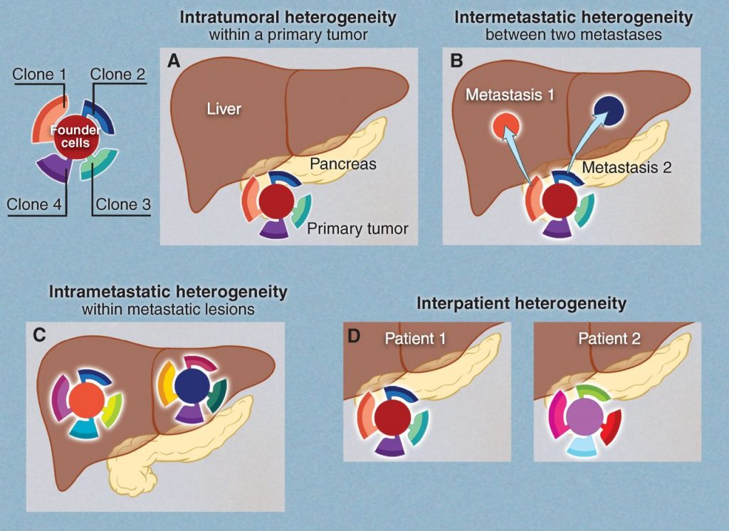
Reiter describes three flavors of ITH. Intra-primary heterogeneity describes the diversity of cell types within the initial tumor. Intrametastatic heterogeneity describes the diversity of cell types within a single metastasis. Finally, inter-metastatic heterogeneity describes diversity between metastases from the same primary tumor.

For Reiter, inter-metastatic heterogeneity presents a particularly compelling problem. If treatment plans are made based on biopsy of the primary tumor but the metastases differ from each other and from the primary tumor, the efficacy of treatment will be greatly limited.

With this in mind, Reiter developed a mathematical model to predict whether a cell sample collected by biopsy of just the primary tumor would provide adequate information for treatment.

Using genetic sequence data from patients who had at least two untreated metastases and a primary tumor, Reiter found that metastases and primary tumors overwhelmingly share a single driver gene. Reiter said this confirmed that a biopsy of the primary tumor should be sufficient to plan targeted therapies, because the risk of missing driver genes that are functional in the metastases proved to be negligible.

 In his next endeavors as a new member of the Canary Center for Cancer Early Detection, Reiter plans to use his knack for mathematical modeling to tackle problems of identifying cancer while still in its most treatable stage.  

Post by undergraduate blogger Sarah Haurin

Post by Sarah Haurin

Overdiagnosis and the Future of Cancer Medicine

For many years, the standard strategy for fighting against cancer has been to find it early with screening when the person is still healthy, then hit it with a merciless treatment regimen to make it go away.

But not all tumors will become life-threatening cancers. Many, in fact, would have caused no issues for the rest of the patients’ lives had they not been found by screening. These cases belong to the category of overdiagnosis, one of the chief complaints against population-level screening programs.

Scientists are reconsidering the way to treat tumors because the traditional hit-it-hard approach has often caused the cancer to seemingly go away, only to have a few cells survive and the entire tumor roar back later with resistance to previously effective medicine.

Dr. Marc Ryser, the professor who gave this meaty talk

In his May 23 talk to Duke Population Health, “Cancer Overdiagnosis: A Discourse on Population Health, Biologic Mechanism and Statistics,” Marc Ryser, an assistant professor at Duke’s Departments of Population Health Sciences and Mathematics, walked us through how parallel developments across different disciplines have been reshaping our cancer battle plan. He said the effort to understand the true prevalence of overdiagnosis is a point of focus in this shift.

Past to Future: the changing cancer battle plan
Credit: Marc Ryser, edit: Brian Du

Ryser started with the longstanding biological theory behind how tumors develop. Under the theory of clonal sweeps, a relatively linear progression of successive key mutations sweeps through the tumor, giving it increasing versatility until it is clinically diagnosed by a doctor as cancer.

Clonal sweeps model, each shade is a new clone that introduces a mutation credit: Sievers et al. 2016

With this as the underpinning model, the battle plan of screen early, treat hard (point A) makes sense because it would be better to break the chain of progression early rather than later when the disease is more developed and much more aggressive. So employing screening extensively across the population for the various types of cancer is the sure choice, right?

But the data at the population level for many different categories of cancers doesn’t support this view (point B). Excluding the cases of cervical cancer and colorectal cancer, which have benefited greatly from screening interventions, the incidence of advanced cases of breast cancer and other cancers have stayed at similar levels or actually continued to increase during the years of screening interventions. This has raised the question of when screening is truly the best option.

Scientists are thinking now in terms of a “benefit-harm balance” when mass-screening public health interventions are carried out. Overdiagnosis would pile up on the harms side, because it introduces unnecessary procedures that are associated with adverse effects.

Thinking this way would be a major adjustment, and it has brought with it major confusion.

Paralleling this recent development on the population level, new biological understanding of how tumors develop has also introduced confusion. Scientists have discovered that tumors are more heterogeneous than the clonal sweeps model would make it appear. Within one tumor, there may be many different subpopulations of cancer cells, of varying characteristics and dangerousness, competing and coexisting.

Additional research has since suggested a more complex, evolutionary and ecological based model known as the Big Bang-mutual evolution model. Instead of the “stepwise progression from normal to increasingly malignant cells with the acquisition of successive driver mutations, some cancers appear to evolve more like a Big Bang, where the malignant ability is already concentrated in the founder cell,” Ryser said.

As the first cell starts to replicate, its descendants evolve in parallel into different subpopulations expressing different characteristics. While more research has been published in favor of this model, some scientists remain skeptical.

Ryser’s research contributes to this ongoing discussion. In comparing the patterns by which mutations are present or absent in cancerous and benign tumors, he obtained results favoring the Big Bang-mutual evolution model. Rather than seeing a neat region of mutation within the tumor, which would align with the clonal sweeps model, he saw mutations dispersed throughout the tumor, like the spreading of newborn stars in the wake of the Big Bang.

How to think about mutations within a tumor
credit: NASA

The more-complicated Big Bang-mutual evolution model justifies an increasingly nuanced approach to cancer treatment that has been developing in the past few years. Known as precision medicine (point C), its goal is to provide the best treatment available to a person based on their unique set of characteristics: genetics, lifestyle, and environment. As cancer medicine evolves with this new paradigm, when to screen will remain a key question, as will the benefit-harm balance.

There’s another problem, though: Overdiagnosis is incredibly hard to quantify. In fact, it’s by nature not possible to directly measure it. That’s where another area of Ryser’s research seeks to find the answers. He is working to accurately model overdiagnosis to estimate its extent and impact.

Going forward, his research goal is to try to understand how to bring together different scales to best understand overdiagnosis. Considering it in the context of the multiscale developments he mentioned in his talk may be the key to better understand it.

Post by Brian Du

Don’t Drink the Tap

Have you ever questioned the quality of the water you drink every day? Or worried that cooking with tap water might be dangerous? For most of us, the answer to these questions is probably no. However, students from a Bass Connections team at Duke say we may want to think otherwise.

Image result for image of water

From bottle refilling stations to the tap, drinking water is so habitual and commonplace that we often take it for granted. Only in moments of crisis do we start worrying about what’s in the water we drink daily. The reality is that safe drinking water isn’t accessible for a lot of people.

Image result for pink hog farm water
Pig waste discoloring lagoon water

Images like this hog farm motivated the Bass Connections project team DECIPHER to take a closer look at the quality of water in North Carolina. On April 16 they presented their concerning findings from three case studies looking at lead contamination, coal ash impoundments, and aging infrastructure at the Motorco Music Hall.

Motorco in Durham. The talk was inside, though.

Nadratun Chowdhury, a Ph.D. student in Civil and Environmental Engineering, investigated lead contamination in water. Lead is an abundant and corrosion-resistant material, making it appealing for use in things like paint, batteries, faucets and pipes. While we’ve successfully removed lead from paint and gasoline, a lot of old water pipes in use today are still fashioned from lead. That’s not good – lead is very toxic and can leach into the water.

Just how toxic is it? Anything over a blood-lead level concentration of fifty parts per billion – fifty drops of water in a giant Olympic swimming pool – is considered dangerous. According to Duke graduate student Aaron Reuben, this much lead in one’s blood is correlated with downward social mobility, serious health concerns, diminished capacity to regulate thoughts and emotions, and hyperactivity. Lower income and minority areas are more at risk due to the higher likelihood of owning contaminated older homes.

Rupanjali Karthik, a Master of Laws student, conducted research on the intersection of water and aging infrastructure in Orange County. Breaks in water pipes are common and can result in serious consequences, like the loss of 9 million gallons of drinkable water. Sometimes it takes 8 or 9 months just to find the location of a broken pipe. In 2018, the UNC-Chapel Hill water main break caused a huge shortage on campus and at the medical center.

Excess fluoridation is also an issue caused by aging infrastructure. In February 2017, a combination of human and machine error caused an excessive fluoride concentration coming out of an Orange County Water Treatment Plant. People were advised not to use their water even to shower. A UNC basketball game had to move locations, and stores were completely swept of bottled water.

Another issue is that arsenic, a known carcinogen, is often used as the fluoridation agent. We definitely don’t want that in our drinking water. Fluoridation isn’t even that necessary these days when we have toothpaste and mouthwash that supports our dental health.

Tommy Lin, an undergraduate studying Chemistry and Computer Science, topped off the group’s presentation with findings surrounding coal ash in Belmont, NC. Coal ash, the residue after coal is burned in power plants, can pollute rivers and seep into ground water, affecting domestic wells of neighboring communities. This creates a cocktail of highly concentrated heavy metals and carcinogens. Drinking it can cause damage to your nervous system, cancer, and birth defects, among other things. Not so great.

The group’s presentation.

Forty-five plastic water bottles. That’s how much water it takes Laura, a Belmont resident, to cook her middle-sized family Thanksgiving. She knows that number because it’s been her family’s tradition the past three years. The Allen Plant Steam Station is a big culprit of polluting water with coal ash. Tons of homes nearby the station, like Laura’s, are told not to use the tap water. You can find these homes excessively stockpiled with cases on cases of plastic water bottles.

These issues aren’t that apparent to people unless they have been directly impacted. Lead, aging infrastructure, and coal ash all pose real threats but are also very invisible problems. Kathleen Burns, a Ph.D. student in English, notes that only in moments of crisis will people start to care, but by then it may be too late.

So, what can people do? Not much, according to the Bass Connections team. They noted that providing clean water is very much a structural issue which will require some complex steps to be solved. So, for now, you may want to go buy a Brita.

Will Sheehan
Post by Will Sheehan

Opportunities at the Intersection of Technology and Healthcare

What’d you do this Halloween?

I attended a talk on the intersection of technology and healthcare by Dr. Erich Huang, who is an assistant professor of Biostatistics & Bioinformatics and Assistant Dean for Biomedical Informatics. He’s also the new co-director of Duke Forge, a health data science research group.

This was not a conventional Halloween activity by any means, but I felt lucky to be exposed to this impactful research surrounded by views of the Duke forest in fall in Penn Pavilion at IBM-Duke Day.

Erich Huang

Erich Huang, M.D., PhD. is the co-director of Duke Forge, our new health data effort.

Dr. Huang began his talk with a statistic: only six out of 53 landmark cancer biology research papers are reproducible. This fact was shocking (and maybe a little bit scary?), considering  that these papers serve as the foundation for saving cancer patients’ lives. Dr. Huang said that it’s time to raise standards for cancer research.

What is his proposed solution? Using data provenance, which is essentially a historical record of data and its origins, when dealing with important biomedical data.

He mentioned Duke Data Service (DukeDS), which is an information technology service that features data provenance for scientific workflows. With DukeDS, researchers are able to share data with approved team members across campus or across the world.

Next, Dr. Huang demonstrated the power of data science in healthcare by describing an example patient. Mr. Smith is 63 years old with a history of heart attacks and diabetes. He has been having trouble sleeping and his feet have been red and puffy. Mr. Smith meets the criteria for heart failure and appropriate interventions, such as a heart pump and blood thinners.

A problem that many patients at risk of heart failure face is forgetting to take their blood thinners. Using Pillsy, a company that makes smart pill bottles with automatic tracking, we could record Mr. Smith’s medication taking and record this information on the blockchain, or by storing blocks of information that are linked together so that each block points to an older version of that information. This type of technology might allow for the recalculation of dosage so that Mr. Smith could take the appropriate amount after a missed dose of a blood thinner.

These uses of data science, and specifically blockchain and data provenance, show great opportunity at the intersection of technology and healthcare. Having access to secure and traceable data can lead to research being more reproducible and therefore reliable.

At the end of his presentation, Dr. Huang suggested as much collaboration in research between IBM and Duke as possible, especially in his field. Seeing that the Research Triangle Park location of IBM is the largest IBM development site in the world and is conveniently located to one of the best research universities in the nation, his suggestion makes complete sense.

By Nina Cervantes        

Creative Solutions to Brain Tumor Treatment

Survival rates for brain tumors have not improved since the 1960s; NIH Image Gallery.

Invasive brain tumors are among the hardest cancers to treat, and thus have some of the worst prognoses.

Dean of the Pratt School of Engineering, Ravi Bellamkonda, poses for his portrait inside and outside CIEMAS.

Displaying the survival rates for various brain tumors to the Genomic and Precision Medicine Forum on Thursday, Oct. 26, Duke professor Ravi Bellamkonda noted, “These numbers have not changed in any appreciable way since the 1960s.”

Bellakonda is the dean of the Pratt School of Engineering and a professor of biomedical engineering, but he is first a researcher. His biomedical engineering lab is working toward solutions to this problem of brain tumor treatment.

Unlike many other organs, which can sacrifice some tissue and remain functional, the brain does not perform the same way after removing the tumor. So a tumor without clearly defined boundaries is unsafe to remove without great risk to other parts of the patient’s brain, and in turn the patient’s quality of life.

Bellakonda hypothesized that brain tumors have characteristics that could be manipulated to treat these cancers. One key observation of brain tumors’ behavior is the tendency to form along white matter tracts. Put simply, tumors often spread by taking advantage of the brain’s existing structural pathways.

Bellakonda set out to build a device that would provide brain tumors a different path to follow, with the hope of drawing the tumor out of the brain where the cells could be killed.

The results were promising. Tests on rats and dogs with brain tumors showed that the device successfully guided out and killed tumor cells. Closer examination revealed that the cells killed were not cells that had multiplied as the tumor grew into the conduit, but were actually cells from the primary tumor.

The Bellamkonda lab’s device successfully guided and killed brain tumors in rats.

In addition to acting as a treatment device, Bellakonda’s device could be co-opted for other uses. Monitoring the process of deep brain tumors proves a difficult task for neurooncologists, and by bringing cells from deep within the tumor to the surface, this device could make biopsies significantly easier.

Although the device presents promising results, Bellakonda challenged his lab to take what they have learned from the device to develop a less invasive technique.

Another researcher in the Bellakonda lab, Tarun Saxena, engaged in research to utilize the body’s natural protection mechanisms to contain brain tumors. Creating scar tissue around tumors can trick the brain into treating the tumor as a wound, leading to immunological responses that effectively contain and suppress the tumor’s growth.

Visiting researcher Johnathan Lyon proposed utilizing electrical fields to lead a tumor to move away from certain brain regions. Moving tumors away from structures like the pons, which is vital for regulation of vital functions like breathing, could make formerly untreatable tumors resectable. Lyon’s 3D cultures using this technique displayed promising results.

Another Bellakonda lab researcher, Nalini Mehta, has been researching utilizing a surprising mechanism to deliver drugs to treat tumors throughout the brain: salmonella. Salmonella genetically engineered to not invade cells but to easily pass through the extracellular matrix of the brain have proven to be effective at delivering treatment throughout the brain.

While all of these therapies are not quite ready to be used to treat the masses, Bellakonda and his colleagues’ work presents reasonable hope of progress in the way brain tumors are treated.

By Sarah Haurin

Designing Drugs Aimed at a Different Part of Life’s Code

Individual RNA molecules fluoresce inside a breast cancer cell.

Individual RNA molecules fluoresce inside a breast cancer cell. Credit: Sunjong Kwon, Oregon Health & Science University, via Flickr.

Most drugs work by tinkering with the behavior of proteins. Like meddlesome coworkers, these molecules are designed to latch onto their target proteins and keep them from doing what they need to do.

If a protein is responsible for speeding up a reaction, the drug helps slow the reaction down. If a protein serves as a gatekeeper to a cell, regulating what gets in and what stays out, a drug changes how many molecules it lets through.

But proteins aren’t the only doers and shakers in our bodies. Scientists are finding that strings of RNA — known primarily for their role in shuttling genetic information from nucleus-bound DNA to the cell’s protein-manufacturing machinery — can also play a major role in regulating disease.

A portrait of Amanda Hargrove

Amanda Hargrove is an assistant professor of chemistry at Duke University.

“There has been what some people are calling an RNA revolution,” said Amanda Hargrove, assistant professor of chemistry at Duke. “In some diseases, non-coding RNAs, or RNAs that don’t turn into protein, seem to be the best predictors of disease, and even to be driving the disease.”

Hargrove and her team at Duke are working to design new types of drugs that target RNA rather than proteins. RNA-targeted drug molecules have the potential help treat diseases like prostate cancer and HIV, but finding them is no easy task. Most drugs have been designed to interfere with proteins, and just don’t have the same effects on RNA.

Part of the problem is that proteins and RNA have many fundamental differences, Hargrove said. While proteins are made of strings of twenty amino acids that can twist into myriad different shapes, RNA is made of strings of only four bases — adenine, guanine, cytosine and uracil.

“People have been screening drugs for different kinds of RNA for quite a while, and historically have not had a lot of success,” Hargrove said. “This begged the question, since RNA has such chemically different properties than proteins, is there something different about the small molecules that we need in order to target RNA?”

To find out, graduate student Brittany Morgan and research associate Jordan Forte combed the scientific literature to identify 104 small molecules that are known interact with specific types of RNA. They then analyzed 20 different properties of these molecules, and compared their properties to those of collections of drug molecules known to interact with proteins.

The team found significant differences in shape, atomic composition, and charge between the RNA-active molecules and the protein-active molecules. They plan to use the results to compile a collection of molecules, called a library, that are chosen to better “speak the language” of the RNA-active molecules. They hope this collection of molecules will be more likely to interact with RNA in therapeutically beneficial ways.

“We found that there are differences between the RNA-targeted molecules and the protein-targeted drugs, and some of them are pretty striking,” Hargrove said. “What that means is that we could start to enrich our screening libraries with these types of molecules, and make these types of molecules, to have better luck at targeting RNA.”

Discovery of Key Physicochemical, Structural, and Spatial Properties of RNA-Targeted Bioactive Ligands.” Brittany S. Morgan, Jordan E. Forte, Rebecca N. Culver, Yuqi Zhang and Amanda Hargrove. Angewandte Chemie, Sept. 18, 2017. DOI: 10.1002/anie.201707641

Kara J. Manke, PhDPost by Kara Manke

Rare Cancers and Precision Medicine in Southeast Asia

Data collected through genomics research is revolutionizing the way we treat cancer. But a large population of cancer patients are being denied the benefits of this research.

Patrick Tan MD, PhD is a professor of cancer and stem cell biology at Duke-NUS Medical School in Singapore.

In 2016, less than one percent of all the existing genomic data came from the 60% of the world population living outside of the US, Europe, and Japan. Furthermore, 70% of patients who die from cancer this year will come from Asia, Africa and Central and South America.

Patrick Tan, M.D., Ph.D., and the Duke-National University of Singapore (Duke-NUS) Medical School are key players in an effort to rectify this discrepancy, specifically as it exists in Southeast Asia.

In his talk, sponsored by the Duke Center for Applied Genomics and Precision Medicine, Tan focused specifically on his work in northeast Thailand with cholangiocarcinoma (CCA), or bile duct cancer.

Liver fluke

Liver flukes like this are parasites of fish that migrate to human hosts who eat the fish raw, leading to a form of bile duct cancer.

While CCA is rare in most of the world, it appears at 100 times the global rate in the region of Thailand where Tan and his colleagues work. Additionally, CCA in this region is of a separate and distinct nature.

CCA in this region is linked with a parasitic infection of the bile ducts called a liver fluke.  Residents of this area in Thailand have a diet consisting largely of raw fish, which can be infected by the liver fluke and transmitted to the person who eats the fish.

Because of the poverty in this area, encouraging people to avoid eating raw fish has proven ineffective. Furthermore, healthcare is not readily available, so by the time most patients are diagnosed, the disease has progressed into its later and deadly stage.

The life cycle of liver flukes. (Graphic U.S. Centers for Disease Control)

Tan’s genomic research has discovered certain factors at the gene level that make liver-fluke positive CCA different from other CCA. Thus genomic data specific to this population is vital to improve the outcomes of patients with CCA.

Duke-NUS Precision Medicine (PRISM) has partnered up with the National Heart Research Institute Singapore (NHRIS) in SPECTRA, a program designed to create a database of genomic data from the healthy Asian population. SPECTRA is sequencing the genomes of 5,000 healthy Asians in order to create a baseline to which they can compare the genomes of unhealthy individuals.

These and other programs are part of a larger effort to make precision medicine, or healthcare tailored to an individual based on factors like family history and genomic markers, accessible throughout southeast Asia.

By Sarah Haurin


A Summer Well-Spent In and Around Toxic Waste Sites

Edison, NJ is just 40 miles from Manhattan and 70 miles from Philadelphia. It’s also home to the US EPA’s Emergency Response Team (ERT), where I spent the summer as an intern.

Stella Wang and an EPA contractor used lifts to test oil being pumped out of these huge tanks. It was found to be contaminated with mercury, benzene and lead.

At the start of my internship, I had little idea of how ERT functioned. Unlike the 10 regional offices of the Environmental Protection Agency, ERT is a “headquarters” or Washington, DC-based group, which means it responds to incidents all over the country such as oil spills, train derailments, and natural disasters.

For example, my mentor, an air specialist who generally works from his cubicle in Edison, aided in the immediate aftermath of Hurricane Katrina by employing equipment to analyze air for hazardous pollutants. Other ERT team members have conducted sediment sampling to expedite the hazardous waste removal process, given consultation advice to other EPA members for long-term remedial site work, and led the innovation of new technology.

I was able to shadow and help my mentor and fellow ERT members with their Superfund site removal work. I created accurate maps showing injection well locations, learned how to use air monitoring instruments, and helped perform chemical lab experiments that will be employed for future site analysis.

Perhaps my favorite part of the internship was traveling to a myriad of active sites. At these sites, I not only got to see how ERT members worked with EPA’s on-scene coordinators, but also observed the physical removal and remediation processes. I was fortunate to visit a particular site multiple times — I witnessed the removal of contaminated oil from an abandoned lot as the summer progressed.

Stella Wang (left) and an EPA air specialist calibrating a air monitoring instrument before a public event.

At another site, I saw the beginning of an injection process intended to prevent the contamination of underground drinking water by hexavalent chromium. By pumping sodium lactate into underground wells, the hexavalent is converted into the insoluble and benign chromium-3 ion. If the injection process works, the community will no longer be threatened by this particular hazardous material.

ERT also acts in anticipation of possible contamination to protect the public. At largely attended events like the Democratic National Convention, a few ERT members will arrive with monitoring equipment. They pride themselves in their real-time data collection for a reason: throughout the event, they can detect whether a contaminant has been released and immediately instigate an emergency response to protect attendees.

Thanks to various ERT members, I felt accepted and welcome. They were open and patient with my never-ending questions about their career paths and other things. They’ve graciously taken me out to lunch so that they could get to know me better, ensuring my inclusion in their small community.

Of course, the experiences I had this summer, while brief, have taught me a tremendous amount and I have a clearer sense of how this division of the US federal government functions. But, it would be inaccurate and unjust to omit the impact that its people made on me.

Stella Wang, Duke 2019Guest post by Stella Wang, Class of 2019

If the Cancer Doesn't Kill You, the Drug Prices Might

The medical community is growing alarmed about a creeping malady that can diminish the quality of life for patients in treatment and even shorten their lives.

It’s found everywhere in the United States, but not to the same degree in other developed countries. They’re calling it “Financial Toxicity.”

Yousuf Zafar is an oncologist and health policy researcher.

A cancer diagnosis more than doubles an American’s chance of declaring bankruptcy, Duke medical oncologist  Yousuf Zafar, MD, MHS,  told an audience of nursing faculty and students at a May 10 luncheon lecture sponsored by the Duke Center for Community and Population Health Improvement. And that bankruptcy, in turn, has been shown to decrease survival rates.

In addition to treating cancer patients, Zafar studies access to care and the cost of care at the Duke Cancer Institute, the Sanford School of Public Policy, and the Margolis Center for Health Policy.

Zafar told personal stories of two patients who waved off treatments because of the financial hardship they feared.

Gleevec (Imatinib) is an oral chemotherapy made by Novartis.

One of them had a job with health insurance, but no prescription drug coverage, which put him on the hook for $4,000 in medications to treat his rectal cancer for just a few weeks. Had either the patient or Dr. Zafar brought the topic up, the costs might have been avoided, but they never talked about money, he said.

The other patient passed up another round of treatment for his pancreatic cancer, for fear of the bills his family would be saddled with when he died.

Chemotherapy for cancer would typically cost $100/month in the 1970s, Zafar said. But today that figure can be “ten, or tens, of thousands per month.” (Inflation would make that 1970 dollar about $6, not $600.)

“Pricing in the European Union and the rest of the world is a completely different picture,” he said.  In the US, pricing “simply reflects what the market will bear.”

Another source of the steep climb is the advent of biologic drugs, which are expensive to develop, use and store, but offer more targeted therapy for individual patients. One of the most successful of these is Gleevec (Imatinib) an oral chemotherapy that became 158 percent more expensive from 2007 to 2014, Zafar said.

If you do a Google search for Gleevec, the first thing you find is a Novartis page with the headline “Understand Your Out-Of-Pocket Costs For Gleevec” that includes a link to financial assistance resources.

In the face of outrageous costs and questionable benefits, a treatment team in many cases can help patients find other means of support or alternative treatments to achieve the same end with less financial damage. But they have to have the conversation, Zafar said. He’d like to see Duke’s Cancer Center become the first in the country to be totally transparent about costs, but he acknowledged that it may be a difficult quest.

To help enable those conversations, Zafar developed a mobile app called Pathlight to help patients make more informed decisions and plan better for the financial burden of treatment. For some of the technology used in the project, Zafar has partnered with a software company called Vivor, which has found innovative ways to help patients navigate to financial assistance programs. That part of the project is supported by the NIH’s National Cancer Institute.

Even for people not in treatment, drugs have become more costly. Healthcare premiums rose 182 percent from 1999 to 2013, with workers paying an increasing share of the cost of their own employee health plans.

Is this any way to run a health system?

“I don’t have all the answers – I don’t think anybody does,” Zafar said. “But I think we need to move toward a single-payer system.”

Post by Karl Leif Bates


Cells Need Their Personal Space

One of the body’s first lines of defense against harmful pathogens is the skin. The constant maintenance of this epithelial cell layer which serves as a barrier to infection  is essential to fighting off disease.

Jody Rosenblatt, an Associate Professor in the Department of Oncological Sciences at the University of Utah School of Medicine, has made it her lab’s mission to study the function of epithelia as a barrier, how this barrier is maintained, and what happens when it goes awry.

Jody Rosenblatt, PhD is an investigator for the Huntsman Cancer Institute at the University of Utah School of Medicine and a Howard Hughes Medical Institute Faculty Scholar

Rosenblatt recently spoke at Duke’s Developmental & Stem Cell Biology Colloquium where she presented some extraordinary findings about how epithelia can squeeze out  both healthy and dying cells  to preserve the protective barrier.

Some c cells commit suicide via programed cell death and are forced out of the cell layer because they are no longer functional. But in the case of forcing out living cells, “cell extrusion is more like a homicide” said Rosenblatt. The fact that perfectly functional living cells are pushed out of a cell layer perplexed her group until they discovered it was happening as a response to cell overcrowding.

Rosenblatt explained that like people, cells tend to like their personal space, so when this is compromised, live cells are actively pushed out of the cell layer, restoring balanced cell numbers.

Rosenblatt’s lab took this discovery a step farther and pinpointed the pathway that likely induces the extrusion of live cells.

Piezo1, a stretch-activated calcium ion channel present in epithelial cells, senses crowding and activates sphingosine-1-phosphate (S1P), the driver of epithelial cell extrusion. When Piezo1 channels are inhibited and don’t sense stretching, cells cannot extrude.

Using zebrafish, Rosenblatt showed that when extrusion was blocked by compromising the S1P2 pathway, epidermal cells form masses that are resistant to chemotherapy drugs and signals for programmed cell death.

Rosenblatt explains the importance of regulating cell extrusion in the epithelium to maintain the tissue’s function as a protective barrier for our organs. Misregulation of this function can result in diseases such as metastatic cancers.

This finding lead them to examine samples of human pancreatic, lung, colon, and breast tumors. They found that in all of these cancers, S1P2 is significantly reduced. But if they restored S1P2 activity in cell lines of these cancers, the extrusion pathway was rescued and tumor size and metastases were greatly decreased!

Rosenblatt and her colleagues have shown that the importance of cell extrusion cannot be overstated. If extrusion is compromised, cells can begin to pile up and move beneath the cell layer, which can lead to invasion of the tissues beneath the epithelium and metastasis to other sites in the body.

Now that we are uncovering more of the pathways involved in tumor formation and metastasis, we can develop new drugs that may be the key to fighting these devastating diseases.

Guest Post by Amanda Cox, PhD candidate in biology


Page 2 of 6

Powered by WordPress & Theme by Anders Norén