Following the people and events that make up the research community at Duke

Students exploring the Innovation Co-Lab

Author: Guest Post Page 1 of 10

Could Restoring Forests Reduce Disease Risk? A Case Study of Hantavirus in Madagascar

Sticky post
Forests and farmland meet in the SAVA region of northern Madagascar. New research suggests that wildlife-human interactions in such areas could spread disease. Credit: James Herrera, Duke Lemur Center

COVID-19 continues to plague us, Mpox is an emerging global threat, and the avian flu is decimating industrial poultry as well as endangered wildlife. What do all these epidemics have in common? They originated in wild animals and spread to domestic animals and people.

This pattern of spread is a trademark of many diseases, termed zoonoses or zoonotic diseases. Our new research shows that in rural settings of Madagascar where forested landscapes were converted to agriculture and settlements, the potential transmission of a deadly virus, Hantavirus, is likely facilitated by invasive rodents, especially the black rat. Also responsible for cyclically occurring plague events in Madagascar, the black rats could be transmitting multiple diseases to people in rural communities, based on our studies.

The work was published April 7 in the journal Ecology and Evolution.

People can get Hantavirus from the droppings or urine of rodents like rats and mice. Credit: Wikimedia Commons

Hantavirus is mainly spread from rodents to people via exposure to their urine and feces in the environment, and being bitten. It can cause severe and deadly disease of the lungs and kidneys, resulting in fever, fatigue, aches and pains, followed later by coughing, shortness of breath, and fluid in the lungs, causing death in almost 40% of people who experience later-stage symptoms. In rural settings like in Madagascar, there are no tests available to diagnose Hantavirus, and the generalized symptoms are often confused for influenza or other diseases. With no specific treatment, either, Hantavirus is an important, though neglected, zoonotic pathogen.

This research, funded by the U.S. National Institute of Health and National Science Foundation, as well as Duke University, connects scientists from around the world with diverse specialties, including field biology, infectious disease epidemiology, social sciences, veterinary health, and more.  Over the last eight years, our international and interdisciplinary team studied zoonotic pathogens in wildlife, domestic animals, and people. We compare how pathogens vary among different animals and in different landscapes.

Herrera and Malagasy student Tamby Ranaivoson check local mammals for pathogens.

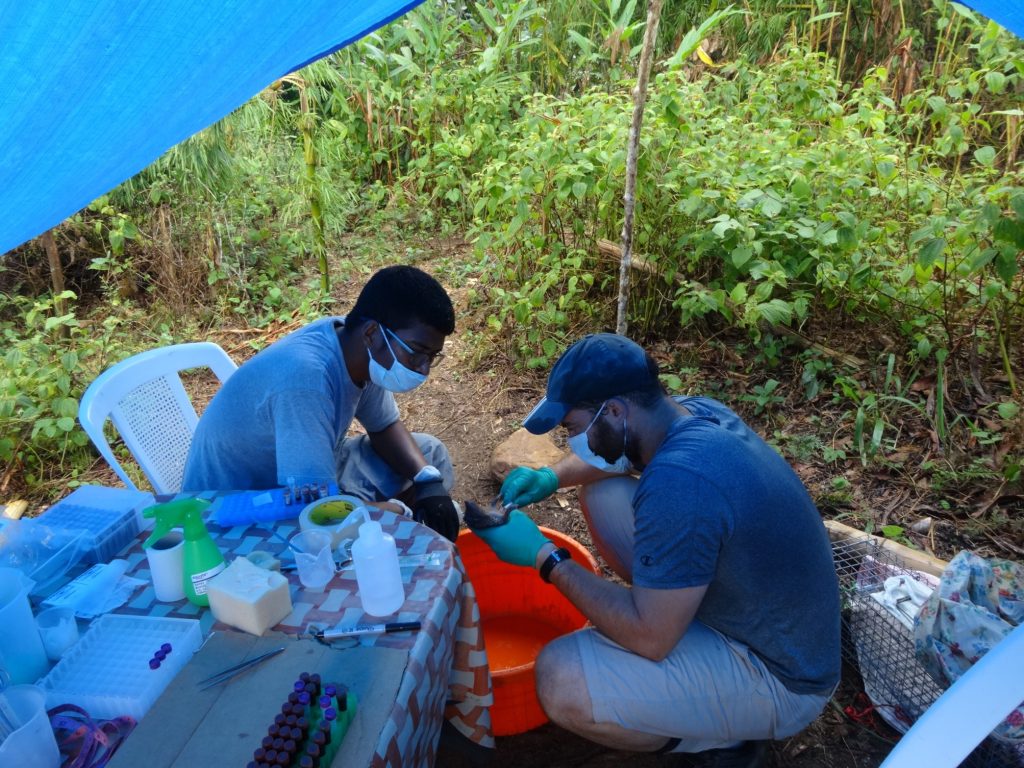
There are more than 29 species of small mammals and another 12 species of bats in these wildlife communities, including native rodents and animals that look like hedgehogs and shrews but are a unique group from Madagascar, the tenrecs. There are also ubiquitous introduced mammals, including black rats, the house mouse, and the shrew, which have spread around the world wherever almost everywhere people go. We studied natural, pristine rainforests and compared to different features of the agroecosystem including regenerating forests, agroforests, and rice fields. We captured rodents and shrews in people’s households, as well, to compare how small mammals and zoonotic pathogens change over this gradient of human land use.

Our results show that black rats were the only species in our system that were infected with Hantavirus, with 10% of sampled individuals infected. Rat abundance and infection were higher in agricultural settings, including rice fields and agroforests, where rats were larger. While some rats in people’s homes were infected, no infected individuals were found in the more mature forests. Hantavirus infection was lower in the homes than in the agricultural fields, but exposure to infected rats is likely higher in homes because of the close contact in enclosed settings. The results highlight how infectious disease risk varies across the landscape because of complex impacts of human land use on natural ecosystems.

The Hantavirus results closely mirror those our team have shown for other disease-causing emerging pathogens, including Astroviruses and Leptospira. Rats and the house mouse were the most commonly infected species, and in the case of Astrovirus, only a single individual of a native species was infected. While Astrovirus infection was more common in the regenerating scrubby environments, Leptospira infection was most common in seasonally flooded rice fields. These varying landscapes of disease risk have important implications for the emergence of zoonotic diseases as well as applications to policy for public health.

Preserving natural forest and facilitating the regeneration of transformed forests may decrease disease risk because infected individuals were rarely captured in natural forests. This may be because there are natural predators to keep rodent populations in check, though further research is needed. Calls to eradicate black rat populations have seldom been successful, but through nature-based solutions like restoration to encourage natural predators, it may be possible to decrease abundance of nuisance rodents. Awareness-raising campaigns to teach about the signs and symptoms of common rodent-borne diseases for rural communities will also be rolled out, and encouraging local health care workers to check for these symptoms in the community members they serve.

We share our results with the Ministry of Public Health and Ministry of Environment and Sustainable Development, and will be organizing more think-tank meetings with relevant actors to co-design intervention strategies that can address these potentially emerging threats to human well-being.

By James Herrera, Ph.D., Duke Lemur Center SAVA Conservation Initiative

Making Sure Drugs Work Where They’re Needed in the Brain

Sticky post

Treating Parkinson’s and other neurological conditions has been challenging due to a lack of tools capable of navigating the complexity of neural circuits. New precision tools like DART.2 help make those therapeutic aspirations a reality, one tethered drug at a time.

The brain is one of our most complex organs, full of neurons that are constantly communicating with one another at places called synapses. Synapses both release molecules called ligands and express cell surface receptors that the ligands bind to, prompting the cells to undergo various processes within. The kinds of ligands and receptors that are important to disease are often released by and found on a variety of cell types. It is crucial, therefore that when we use a drug that targets a certain receptor, we also make sure that they only interact with receptors on the desired cell type.

To achieve this, in 2018 a team led by Duke professor Mike Tadross introduced “Drugs Acutely Restricted to Tethering” or “DART,” a drug delivery system that allows researchers to administer drugs to specific neuronal cell types. In June of this year, the Tadross lab unveiled DART.2. Pairing the cell-type specificity provided by DART.2 with the cellular receptor-specificity already provided by a given drug is essential to treating diseases like Parkinson’s without severe off-target effects, something researchers have been unable to do until tools like DART entered the scene.

Think of it like cutting the water supply to an apartment that has had a pipe burst. You don’t want to cut the water to the entire building, just the flooding apartment, because the other tenants still need water. This newest version of DART increases the ability of researchers to flip the right switches.

With increased cell type specificity optimized for drugs targeting two different receptor types, enabling broader dosing techniques and opening the door for discoveries of unknown roles of well-known receptors, researchers have made DART.2 into an “even more subtle, refined, yet transformative drug delivery system, a marked improvement from more rudimentary options that operate more like sledgehammers,” said Brenda Shields, one of the lead scientists on the DART.2 project.

The HaloTag protein (HTP) helps recruit drugs tethered to the HaloTag ligand (RXDART) to the desired cell types. Courtesy of Erin Fykes.

This is, in part, owing to the use of natural, or endogenous, receptor machinery in its design. Organisms are infected with a virus that prompts only certain cell types to express HaloTag, a protein that sits on the surface of the cells of choice. A HaloTag ligand, or small molecule that binds specifically to the HaloTag surface protein, is tethered to a drug of choice. This allows the tethered drug to be selectively recruited to the cells that have the HaloTag protein, bringing the drug into closer proximity to its intended cellular receptor, discouraging it from binding to unintended receptors, and reducing the amount of drug needed for efficacy.

DART.2 is not changing receptors that are already present, nor is it affecting the signaling cascades activated by engagement with the receptors. Cell-specificity of drug delivery was improved in DART.2 by decreasing the time and drug concentration needed to achieve intended effects – it is 100 times more precise than the previous system, with desired effects achieved in just 15 minutes.

Using the previous version of DART, researchers tried delivering a tethered version of the drug gabazine to GABA receptors (neural receptors associated with inhibitory neurotransmission) on a select group of neurons – gabazine blocks GABA from binding to GABA receptors and subsequently increases neural activity. Unfortunately, DART did not achieve high enough cell-specificity and gabazine bound to enough off-target receptors to trigger epileptic responses in mouse models. DART.2, however, is capable of delivering gabazine without these effects. The original version of DART was only optimized to work with drugs targeting excitatory (AMPA receptors) neurotransmission. The ability of the current version to work with inhibitory (GABA) and excitatory (AMPA) neurotransmission makes this system useful for “bi-directional” modifications, greatly increasing its utility.

Interestingly, while testing the effects of the drug gabazine on GABA receptors in ventral tegmental area dopamine neurons, they found that GABA receptors on these cells actually suppress locomotion, opposite to findings in other studies that more broadly focused on GABA receptors in multiple cell types. This highlights the need for tools like DART.2 that allow us to understand diverse receptor/ligand dynamics on a cell-by-cell basis to gain more nuanced approaches to understanding and treating disease.

To visualize dispersion and binding of tethered drugs to HaloTag proteins versus off-target receptors, Tadross’s team developed a way of seeing where the tethered drugs accumulated by introducing a small percentage of HaloTag ligands bound to a fluorescent reporter rather than a drug into the pool of tethered drugs. This visualization further confirmed a significant increase in cell-type specificity and a decrease in off-target effects of DART.2.

With previous levels of cell specificity, local delivery of the tethered drugs via cannula insertion at the brain region of interest was necessary to ensure drugs made it to the right targets. With increased specificity and a new visualization method for seeing where DART.2 drugs bind, researchers were able to assess whether brain wide dosing would be possible, decreasing deleterious effects of pumping high concentrations of drug into a certain area. Excitingly, they found that broad administration of tethered drugs across large areas of the brain did not significantly increase binding at receptors on cells not expressing the HaloTag protein. Drug delivery in the brain is notoriously difficult, so having the flexibility to administer a drug from an easier delivery point without reducing binding at target sites translates to a greater chance of therapeutic success.  

For those unfamiliar with the process of drug and therapeutic development, the improvements presented in DART.2 represent a realistic look into the measure of scientific progress. Originally used to treat Parkinson’s mouse models, our conception of DART.2’s therapeutic relevance to other conditions is continually expanding. Shields shared that adaptions for conditions such as anxiety and depression may not be far off, just to name a few. DART.2 also makes it possible to use drugs like opioids in new ways. While helpful for pain management, the addictive potential of opioids sometimes renders them more harmful than helpful. Utilizing DART.2, opioids could be administered more specifically to cells that benefit from the drug, potentially reducing interactions with cell types involved in addiction development. Additionally, researchers are beginning to couple DART.2 with other tools on the market that can enhance its therapeutic promise and applicability (and vice versa). Each improvement of DART brings us closer to the reality of treating conditions we once deemed hopeless.

Post by Erin Fykes, Ph.D. student in cell and molecular biology

Determining Who’s White: How Vague Racial Categories Mask Health Vulnerabilities

Sticky post

Good healthcare decisions depend on good data – whether you’re making federal health policy or treating a single patient.

But the data is often incomplete – particularly when it comes to defining a group that still makes up the majority of the U.S. population — a ‘non-Hispanic White’ person. That’s the primary reference category used in health data.

“Nobody questions who’s white, but they should,” said Jen’nan Read, a Duke sociologist and lead author of new research recently published in the journal Demography. “The white category contains diverse ethnic subgroups, but because we lump them all together, we miss important health vulnerabilities for millions of Americans.”

Read and co-author Fatima Fairfax, a Duke doctoral student in sociology, analyzed data from the 2000 to 2018 waves of the National Health Interview Survey to compare the health of white adults born in the U.S., Europe, the Middle East, and the Former Soviet Union.

Duke sociology professor Jen’nan Read and PhD student Fatima Fairfax

Separating groups collapsed into the white category, they found that foreign-born Whites have a smaller health advantage over U.S.-born whites than is commonly assumed, and immigrants from the Former Soviet Union are particularly disadvantaged. Those immigrants report worse health, including higher rates of high blood pressure, compared to U.S.-born whites as well as people from Europe and the Middle East.

These findings illustrate how global events, such as the wars in the Ukraine and Syria, have contributed to changes in the composition of white immigrants over time.

Understanding these changes – and the distinct experiences of white immigrant subgroups – is vital to understanding long-term patterns in health disparities within the broad white category, the authors argue.

“If we truly care about reducing health disparities in this country, we need to know where the disparities are. And they get hidden when people are lumped into broad categories,” Read said. “Ukrainian immigrants, for example, we see in the news what they’re leaving. Death, destruction, their kids may have gone years now without education. This has lifelong impacts on their wellbeing. The physical consequences from stress are enormous–we know stress increases all sorts of physical health problems. High blood pressure, cholesterol, the list goes on.”

And the science is clear. The more accurate the information healthcare providers have on their patients, the better the outcomes.

“We’re missing health patterns here,” Read said. “Our country is extremely diverse, and not talking about diversity doesn’t change that fact. Health inequality costs us a lot–it costs the healthcare system and society as a whole.” 

“Health is arguably the most important indicator of how a society is doing, and paying more attention to diversity within broad categories will allow us to do better.” 

Post by Eric Ferreri, Duke Marketing & Communications

How the body’s own defense system plays a role in Alzheimer’s disease

Sticky post

Carol Colton, a distinguished professor in neurology and pathology and a member of the Duke Institute for Brain Sciences, is renowned for her groundbreaking research on the immune response’s role in the onset and progression of brain diseases, particularly Alzheimer’s disease (AD). She is a firm believer in using animals such as mice for scientific research, saying that progress in understanding and treating diseases like Alizheimer’s would not be possible without them. With a shorter life cycle than humans, mice can be studied throughout their whole life and across multiple generations. They are also biologically similar to humans and susceptible to many of the same health problems, such as Alzheimer’s. Her work has reshaped our understanding of the brain’s immune system, challenging the long-held notion that the brain is “immune privileged.” 

Carol Colton, PhD, professor of neurology and pathology at Duke

Central to Colton’s research is the role of “microglia,” the brain’s resident macrophages. Once thought to be passive observers in brain immunity, microglia are now recognized as active defenders, crucial in maintaining brain health. Colton’s early studies revealed that these cells not only eliminate harmful substances but also adapt to chronic conditions like Alzheimer’s. In this disease, microglia’s prolonged immune activity disrupts the brain’s metabolic balance, necessitating adaptations in neurons, astrocytes, and microglia themselves. She likens this adaptation to the brain coexisting with a parasite – functional but at a metabolic cost.  

Her research underscores how microglia can initially protect against Alzheimer’s by combating amyloid plaques and phospho-tau proteins but eventually contribute to the disease’s progression as metabolic disruptions intensify.

Colton’s approach integrates physiology and pathology, exploring how changes in normal physiological processes influence disease pathology. Her lab employs a variety of advanced techniques, from cellular microscopy to gene and protein analysis, to map the intricate relationships between brain metabolism and disease. This multidisciplinary approach enables a deeper understanding of how the brain’s unique environment shapes disease progression.

A cornerstone of Colton’s recent work is her discovery of “Radical S-Adenosyl domain 1 (RSAD1),” a mitochondrial protein found at the bottom of the ocean critical to understanding Alzheimer’s. RSAD1 is overexpressed in Alzheimer’s neurons, altering methionine metabolism and mitochondrial function. These disruptions contribute to the disease’s characteristic metabolic imbalance. By developing RSAD1-negative and RSAD1-overexpressing mouse models, her lab provides crucial tools to study the protein’s impact on neuronal and mitochondrial metabolism in the presence of amyloid plaques and phospho-tau.

RSAD1 also appears to be linked to methionine depletion in the brain, which may further exacerbate Alzheimer’s pathology. These findings pave the way for novel therapeutic targets aimed at restoring metabolic equilibrium in the brain.

Colton’s scientific journey is deeply influenced by her family’s academic legacy, particularly her mother, who earned a chemistry degree during an era when women faced significant barriers in science. Inspired by her mother’s determination, Colton is a passionate advocate for women scientists, often emphasizing the importance of diversity and mentorship in STEM fields.

Colton’s work highlights the slow, insidious nature of Alzheimer’s disease, driven by metabolic and immune system changes over decades. By asking fundamental questions, such as whether Alzheimer’s results from the loss of key metabolites or whether microglia contribute to this depletion, her research aims to uncover the mechanisms that underlie the disease and identify strategies for intervention.

In the fight against Alzheimer’s, Colton’s discoveries, particularly those surrounding RSAD1 and microglial activity, are setting the stage for innovative treatments. Her dedication to unraveling the complexities of brain metabolism and immune response solidifies her place as a leader in neurology and pathology, with an enduring impact on the field of Alzheimer’s research.

Post by Lydia Le, NCSSM class of 2026

Meet a Duke Expert on Pain: the Sixth Sense

Sticky post

Duke associate professor of anesthesiology Andrea Nackley is a kind, passionate scientist, although her most notable quality is determination. 

As a first generation college student, a mother of two teenagers, and a triathlon athlete, she is nothing but dedicated. She challenges herself not only in a professional environment, but strives for personal and physical growth in her free time as well. 

Andrea Nackley, PhD, Duke School of Medicine

I had the pleasure of interviewing Nackley in her office and labs, where we discussed her life as a scientist, mom, and leader. When asked how she manages her many responsibilities, she responded with a single word: “acceptance.” Nackley accepts her busy schedule and strives to prioritize daily to make the most of every moment. 

As a young adult, she initially pursued the pre-med psychology path, with support from her hard-working family. She remembers a pivotal moment in her journey, in a biopsychology class where she studied brain circuits and the brain-behavior connection. She found this class absolutely riveting, and knew that this is where her passion lied. 

She describes pain, her research’s current focus, as a sixth sense of sorts, not quite like touch but something different and intriguing. Her approach to studying chronic pain is collaborative and aims to make her findings applicable to medical pursuits regarding pain management. She has even worked closely with a clinical trial centered in Duke, an experience that directly exemplifies this bench-to-bedside approach. 

A scene from the Translational Pain Research Laboratory, which Nackley leads

After earning her PhD at the University of Georgia, she moved to UNC Chapel Hill to complete a postdoctoral fellowship. In 2016 she moved to Duke, where she now leads an open-floor Translational Pain Research Laboratory and promotes an extraordinarily collaborative lab environment.

She has received grants for her work in vulvodynia, vestibulodynia, and peripheral ADRB3. When asked what her favorite aspect of working at Duke is, she endearingly responded with, “all the people here are just so… nice.” 

Nackley is close-knit with the individuals in her lab, a group ranging from high school students to postdocs, but especially with her lab manager, Marguerita Klein. 

Outside of work, she enjoys open-water swimming, training for an Olympic-length triathlon, baking, and cooking. She said baking allows her creative side to emerge, an often uncultivated aspect of any scientist’s left-dominant brain. 

Meeting Nackley and touring the innovative lab she cultivates was a wonderful experience, and I’m sure the future output from her work and leadership will be invaluable.

Post by Abigail Keaton, NCSSM Class of 2026

Invincible Insect Pests Don’t Faze This Researcher

“My passion for what I do saved my life.”

Meet Ke Dong, a biology professor at Duke University. She’s a lover of nature, a great cook, and a Lupus survivor. About 20-25 years ago, she developed Lupus during her research years at Michigan State University. Her time with this autoimmune disease was not kind. “The Lupus brought depression,” she said. 

Fortunately, she was surrounded by amazing peers and her passion: research. Dong’s research focuses on ion channels and their reaction to various toxins and stimuli. These ion channels are incredibly important to the physiology of insects because of their impact on neuronal activity. 

Duke biology professor Ke Dong.

However, her passion didn’t develop from thin air. Dong grew up on a college campus in southeastern China. With both parents leading careers as professors — her father in history and her mother in biochemistry — she had the amazing opportunity to develop her passions early in childhood. 

Growing up, she “had never been afraid of insects” as her mother’s work focused on the development of an increased production rate of silk in silkworms. However, it was the incidents in the area around her that sparked her passion. People in the area were often poisoned from the consumption of insecticides from the rice they were growing. This piqued her interest in toxicology as she was curious about how these insecticides were toxic to the townspeople. 

Combining her fearlessness in the face of insects and her interest in toxicology, Dong has found the best of both worlds.

Dong also loves to dabble in the culinary worlds of a diverse range of cultures. As she travels from country to country, she brings with her the memorable flavors of each dish she tastes. Once arriving back home, she immediately purchases cookbooks from those countries to add to her rolodex of culinary skills. As she reads each recipe on her nightstand, she dreams of ways to introduce various flavors and techniques into her dishes. A creative cook, she has no time for following measurements. Her kitchen is her sandbox and allows her to dance with each flavor in her pot, adding less sugar but a little more salt. 

Dong has been through ups and downs in her life, but there’s nothing that’s going to stop her from her passion: research. 

Post by Eubey Kang, NCSSM Class of 2025

From Propulsion to Provost: A Conversation With Alec Gallimore

Sticky post

Science fiction may seem an unlikely source for research inspiration. But for Duke University Provost Alec Gallimore, it has been just that: inspiration for a career’s worth of electric propulsion research.

Alec Gallimore in his plasmadynamics and electric propulsion laboratory at the University of Michigan, where he was a faculty member and dean for more than 30 years before coming to Duke. (Credit: University of Michigan)

Gallimore said it was stories from science fiction authors like Arthur C. Clarke and Isaac Asimov that piqued his interests for fusion and other advanced propulsion technologies at a young age. It is an interest that led him to pursue studies in aerospace engineering with a focus in plasma physics at Princeton University and Rensselaer Polytechnic Institute.

He channeled those interests into the Plasmadynamics and Electric Propulsion Laboratory (PEPL) he founded as a professor of aerospace engineering at the University of Michigan. Focused on the development and testing of more efficient and powerful electric thrusters for spacecraft, the lab has long been at the forefront of electric propulsion research. 

High thrust, high efficiency electric propulsion systems are poised to transform space exploration. They are viable replacements for the inefficient, yet flight-proven chemical thrusters typically used on spacecraft. This is because the electric thrusters can operate over longer periods of time, providing sustained thrust that allows spacecraft to travel the solar system in record time. Electric propulsion systems are slated for use on countless future spacecraft, from the Gateway lunar space station to Mars orbiters.

Gallimore said he is proudest of the X3 Nested Channel Hall Thruster developed at PEPL. Weighing just a tenth the size of an SUV at 230 kg, the X3 is one of the largest, most powerful electric thrusters the lab has developed. It consists of three nested chambers in which ionized gases are accelerated by electric fields, generating thrust highly efficiently. Most Hall-effect thrusters – the category of electric thruster to which the X3 belongs – contain only one chamber. The X3’s three separate chambers help it generate substantially more thrust. That means it can be used to propel heavier spacecraft destined for more distant locations in the solar system. 

Low-power test run of the X3 Nested Channel Hall Thruster (Credit: PEPL)

Gallimore sees this as just the beginning for electric propulsion. Miniaturized electric thrusters will also, according to him, become mainstays on smaller satellites, providing them with the propulsion capabilities they have long lacked. More important will be future research on novel propellant types for electric thrusters, specifically water. “Water is the answer,” Gallimore said. 

“Water is all over the place in the solar system, and so you are able to develop an infrastructure where you can tank up as you need to with water as your propellant,” he explained. “It opens everything up in the solar system so that, by the second half of the century, you can have an amazing infrastructure throughout the inner part of the solar system with water as a propellant.”

Leading research advancements such as these comprised much of Gallimore’s work at PEPL, experience that has informed his work at Duke, where he became provost in July 2023. “Genius is 10% inspiration, 90% perspiration,” he said. Having a team of people fully committed to their research and a common mission was vital to him.

So was having a diversity of opinions. PEPL hosted researchers from varying disciplines such as applied physics and aerospace engineering, as well as diverse life experiences and identities. That promoted a culture of “mutual respect” in “intangible ways” that drove innovation and staved off “group think,” he said. 

That philosophy of thoughtful discussion and collaboration is one Gallimore has taken to Duke, informing the Office of the Provost’s efforts to advance academic excellence and improve campus community.

Whether as an electric propulsion researcher developing the thrusters that will take humans to Mars or as Duke University Provost, working to invigorate the school community, Gallimore has pushed boldly forward. In a future perhaps defined by advanced human space exploration and a more just world, we will no doubt have some small thanks to pay to Gallimore.

Post by Adrian Tejada, NCSSM class of 2025. 

What can we learn from watching a fish’s ear take shape? You might be surprised

Sticky post

Dr. Akankshi Munjal is a developmental biology researcher at Duke University, who studies the development and mutations of inner ear tissue in zebrafish, and how that may be caused by genome disorders. 

Akankshi Munjal, assistant professor of cell biology

From a young age, Munjal has been fascinated by watching things being built and developed. Her grandfather was a civil engineer, and she was inspired by the many blueprints littering his home. Growing up, she wanted to be an architect. 

Though she found inspiration elsewhere, and did not pursue architecture, in a way, her career mirrors this, “I guess I am not an architect, but I still watch embryos being built, so that kept with me – how you shape things.” 

The inspiration of Munjal’s current career came to her in high school. Growing up, she lived in a large city in India, and did not have much exposure to science fields and research. “If you don’t see it around you, it’s not something you see as an option.” 

However, she was able to find inspiration from a few of her instructors, “There were some teachers who were very inspiring in exposing that there is research out there, that you can be at the bench, ask questions, and address them using experiments.” 

She was also involved with a project dealing with bacteria that could process heavy metals in the Yamuna river near Delhi, India, and this helped introduce the idea of research as a potential career path. 

Though most of Munjal’s work has moved toward lab management, the research is what she really loves, “I could spend days in the microscope room, watching development happen.”

The interesting thing about zebrafish, is that their eggs are transparent, and develop outside of the parent organism. This provides an incredible way to observe the development of tissues under a microscope. Zebrafish also share 70% of the DNA of humans, which makes them a great model organism to observe human disorders and how they affect tissue. The ability to witness this development is Munjal’s favorite part of the job,“It’s why I love what I do, we are able to watch these things happen, in the lab.”

When asked what she wished she would’ve learned earlier on, she mentioned the classic comparison of teaching a man to fish, as opposed to giving him a fish. She applies this saying to the process of learning. In her earlier education, there was an emphasis on collecting and memorizing information and facts, rather than learning how to gather knowledge. An emphasis on academic intelligence, as opposed to emotional intelligence. 

Looking back, this presentation and memorization of facts was less helpful, “Some of them are not facts, some of them are interpretations, so if there was more information on collecting knowledge, that would be more helpful.”

Munjal loves to watch things being developed. This not only applies to her research in developmental biology, and her former passion for architecture, but also to her love of collecting knowledge. 

Guest post by Rhynn Alligood, NCSSM class of 2025

Advancing Immunotherapy for Glioblastoma

Sticky post
Vidyalakshmi Chandramohan, associate professor in neurosurgery and pathology, and member of the Duke Cancer Institute. Credit: Duke Department of Neurosurgery

Duke professor Vidyalakshmi Chandramohan is a pioneering neuro-oncologist whose work is redefining the future of glioblastoma (GBM) treatment. As a researcher in the Department of Neurosurgery at Duke, she is driven by a profound commitment to improving patient outcomes and providing new hope for those battling one of the most aggressive forms of brain cancer.

Her journey into science began with an interest in immunology and oncology, which led her to earn a Ph.D. and conduct postdoctoral research focused on the complex relationship between cancer and immune responses. Her fascination with GBM stemmed from the urgent need to develop innovative treatments for a disease with limited therapeutic options. Today, her groundbreaking research offers new avenues for fighting GBM and improving patient survival.

PET scan showing glioblastoma brain cancer. Credit: Wikimedia Commons.

Chandramohan’s work is centered on immunotherapy, particularly the development of D2C7-IT, a dual-specific immunotoxin currently in Phase I clinical trials for recurrent GBM patients. This precision medicine approach targets tumors with remarkable specificity, minimizing damage to healthy tissue. Her ongoing research aims to enhance the efficacy of D2C7-IT and expand its potential as a viable alternative to traditional treatments.

For Chandramohan, translating her research into tangible solutions is essential. “Developing a therapy is one thing, but making sure it works in the real world is another,” she says. She is exploring ways to combine D2C7-IT with other therapies to improve treatment outcomes and minimize side effects, pushing the boundaries of what is possible in GBM care.

A critical aspect of her research involves identifying biomarkers that predict patient responses to treatment, enabling personalized therapies. “Personalized medicine is the future,” she believes. “Tailoring treatment to each patient’s unique response will improve survival rates and outcomes.”

Collaboration is at the heart of Chandramohan’s work. She fosters an interdisciplinary environment where scientists, clinicians, and engineers work together toward a shared goal. “No one person can do it all,” she emphasizes. “It takes a community of experts to make breakthroughs happen.”

Despite the challenges of translating research into clinical practice, Chandramohan remains unwavering in her determination. “When our work leads to better treatment options, it reminds us why we do this every day,” she says. Her dedication to improving patient care fuels her optimism for the future of GBM treatment.

Looking ahead, Chandramohan is hopeful that the integration of immunotherapy, precision medicine, and innovative technologies will revolutionize the field of neuro-oncology. “We’re just scratching the surface,” she says, confident in the potential to improve outcomes for GBM patients.

Through her relentless pursuit of excellence, Chandramohan is not only advancing the science of glioblastoma treatment but also inspiring the next generation of researchers to push the boundaries of what is possible in the fight against cancer.

Blog post by Adarsh Magesh, NCSSM Class of 2025


Advancing Care and Research in Traumatic Brain Injury

Sticky post

Meet a trailblazer in the realm of neurocritical care and emergency medicine: Dr. Katharine Rose Colton, MD. Balancing roles as a clinician, researcher, and educator, Colton serves as an Assistant Professor of Neurology and Neurosurgery at Duke University. Her dedication to understanding and treating traumatic brain injury (TBI) exemplifies her commitment to improving the lives of patients facing severe neurological challenges.

TBI is a significant public health issue, often resulting from falls, motor vehicle accidents, or sports injuries. It can range from mild concussions to severe brain trauma, leaving patients in comas or with long-term disabilities. While treatments for TBI have evolved, many gaps remain in understanding how to optimize recovery and outcomes. Colton’s work bridges this divide, combining cutting-edge research with compassionate patient care.

Colton’s journey into medicine wasn’t linear. A Canadian native, she initially pursued an eclectic range of interests, including ethnobotany and anthropology, during her undergraduate studies. She pivoted to medicine, taking the MCAT on a whim and earning her M.D. from Duke University School of Medicine.

Her first exposure to TBI occurred during a research year at the University of Maryland’s Shock Trauma Center. A project initially focused on trauma surgery shifted to neurocritical care, igniting her passion for studying brain injuries. “I loved it,” she recalls. “It was a completely different way of looking at medicine.”

Colton’s clinical path led her to a residency in Emergency Medicine at Northwestern University and a fellowship in Neurocritical Care. While she enjoyed the fast-paced decision-making of emergency medicine, she found herself drawn to the intricate details of critical care. “I struggled with letting patients go and handing them off to others,” she says. “I wanted to stay involved and see the whole story unfold.”

Now focused primarily on neurocritical care, Colton dedicates a third of her time to research, primarily on clinical trials targeting severe TBI. She has worked on large-scale, multi-site studies investigating drug therapies and monitoring systems to optimize treatment for comatose patients.

Her approach to research is pragmatic: “I’m a clinician first. I want to know how the things we do today will benefit the patient tomorrow.” For instance, her current trials explore the potential of older, cost-effective drugs previously overlooked by pharmaceutical companies to improve outcomes in TBI patients. These trials adopt adaptive designs, allowing for real-time adjustments based on early results to maximize impact.

Colton is also a strong advocate for personalizing TBI treatment. “TBI is an incredibly heterogeneous condition,” she explains. “We can’t treat a 20-year-old in a car accident the same as a 70-year-old who fell. They have completely different recovery pathways.” Her work aims to identify biomarkers and refine classifications of TBI to develop more targeted interventions.

One of the most memorable cases from Colton’s career underscores her dedication to patient care. A young woman struck by a car in Chicago arrived at the ICU in a deep coma, with little hope of recovery. Months later, to Colton’s astonishment, the patient returned to work and resumed her life. “You just don’t know,” she reflects. “That case taught me the importance of patience and persistence in medicine.”

Colton’s role extends beyond the ICU, often involving interactions with patients’ families during some of their most vulnerable moments. “Families often show incredible grace, even in tragedy,” she says. “It’s humbling to see their resilience and willingness to contribute to research, even when it might not help their loved one directly.”

Despite the challenges of long, emotionally taxing weeks in the ICU, Colton finds fulfillment in both the technical and human aspects of her work. “There’s something beautiful about the physiology — adjusting treatments and seeing how the body responds,” she explains. Yet, she never loses sight of the bigger picture: the patient. “Numbers on a screen don’t matter if we’re not improving their lives.”

Outside of work, Colton enjoys paddleboarding, camping, and spending time with her two young children. Her background in ethnobotany and love for snowboarding reflect her multifaceted personality, blending curiosity, determination, and a deep appreciation for life.

Dr. Katharine Colton is shaping the future of TBI care through her dedication to research, her patients, and the families she serves. Her journey is a testament to the impact of resilience, curiosity, and compassion in medicine.

Written by Amy Lei, NCSSM class of 2025

Page 1 of 10

Powered by WordPress & Theme by Anders Norén