If there was a contest for biggest female bullies of the animal world, lemurs would be near the top of the list. In these distant primate cousins, it’s the ladies who call the shots, relying on physical aggression to get their way and keep males in line.
Not all lemur societies are built about female rule, however. In one branch of the lemur family tree, some species have evolved, within the last million years, to have a more harmonious relationship between the sexes.
Now, new findings suggest that this amiable shift in lemurs was at least partly driven by changes in the action of the “love hormone” oxytocin inside their brains. The research could shed light on how hormones influence behavior in humans and other animals.
In a study published in the journal Biology Letters, Duke University researchers studied seven closely related lemur species in the genus Eulemur, noting which ones had domineering females and which were more egalitarian.
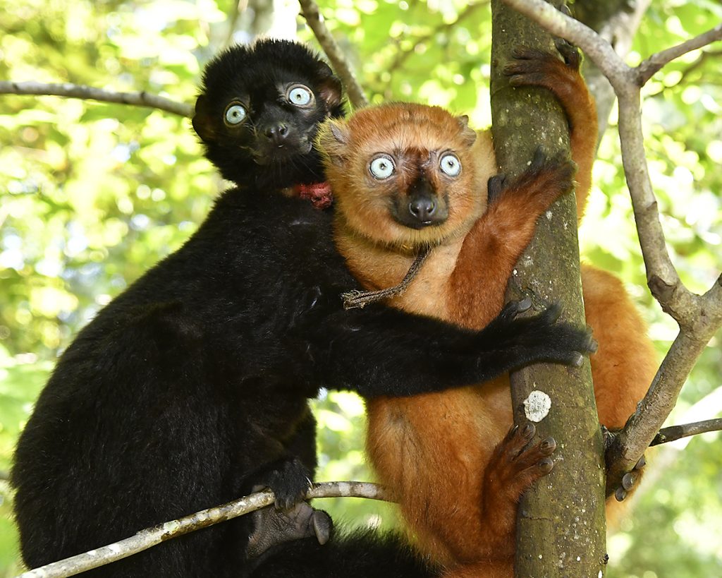
Take blue-eyed black lemurs, for example. Females get first dibs on food and prime resting spots; smacking, biting and chasing the males to get their way.
Their behavior isn’t the fierce protectiveness of a mother defending her babies, said senior author Christine Drea, a professor of evolutionary anthropology at Duke. Aggression in these females can be entirely unprovoked, simply to remind others who’s in charge.
“Males let females have priority access to whatever they want,” Drea said.
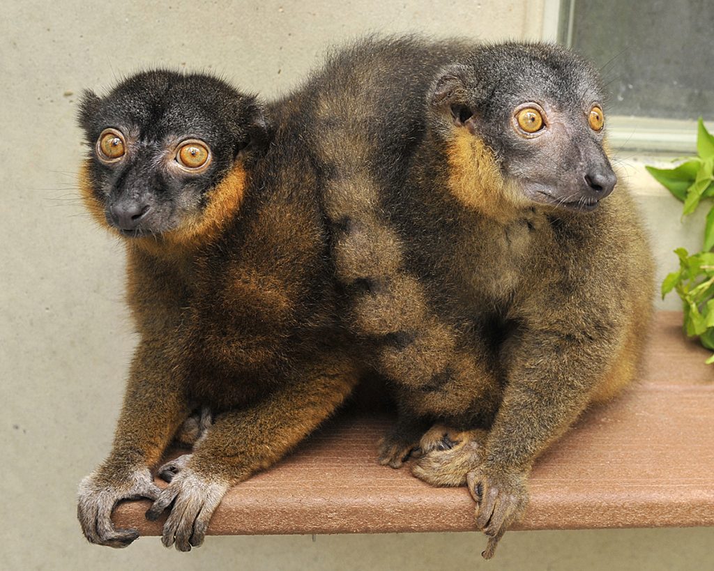
Others species, like the collared lemurs, are more peaceful and egalitarian, with males and females sharing equal status. “It’s more of an even playing field,” said first author Allie Schrock, who earned her Ph.D. in the Drea lab.
The lemurs in the study died of natural causes some time ago, but their tissues live on, thanks to a bank of tissues from these endangered primates kept frozen at the Duke Lemur Center. Using an imaging technique called autoradiography, the researchers mapped brain binding sites for oxytocin, a hormone involved in social behaviors like trust and bonding.
The results revealed a striking pattern.
The researchers found that the more recently evolved egalitarian species had more oxytocin receptors than the others, essentially giving them more targets for oxytocin to act on.
The key difference was in the amygdala, a region of the brain typically associated with emotions such as fear, anxiety and anger.
The pattern held up for both sexes, suggesting that egalitarian species achieved gender parity by becoming less aggressive towards others overall, rather than males ramping up their aggression to match their female counterparts, Drea said.

The potential implications go beyond lemurs, the researchers said. Problems with oxytocin signaling in the brain have been linked to aggression, personality disorders and autism in humans, rodents and other animals.
Next, the researchers plan to examine links between hormone receptors and additional aspects of social behavior in lemurs, such as whether they are solitary or social.
“There’s a lot more that we can learn from lemurs about how the brain regulates behavior,” Schrock said.
CITATION: “Neuropeptide Receptor Distributions in Male and Female Eulemur Vary Between Female-Dominant and Egalitarian Species,” Allie E. Schrock, Mia R. Grossman, Nicholas M. Grebe, Annika Sharma, Sara M. Freeman, Michelle C. Palumbo, Karen L. Bales, Heather B. Patisaul and Christine M. Drea. Biology Letters, March 19, 2025. DOI:10.1098/rsbl.2024.0647