The Web of Science ranking of the world’s most highly-cited scientists was released this morning, telling us who makes up the top 1 percent of the world’s scientists. These are the authors of influential papers that other scientists point to when making their arguments.
EDITOR’S NOTE! — Web of Science shared last year’s data! We apologize. List below is now corrected, changes to copy in bold. We’re so sorry.
Twenty-three of the citation laureates are Duke scholars or had a Duke affiliation when the landmark works were created over the last decade.
A couple of these Duke people disappeared from this year’s list, but we’re still proud of them.
Dan Scolnic of Physics returns as our lone entry in Space Science, which just makes Duke sound cooler all around, don’t you think?
This is a big deal for the named faculty and an impressive line on their CVs. But the selection process weeds out “hyper-authorship, excessive self-citation and anomalous citation patterns,” so don’t even think about gaming it.
Fifty-nine nations are represented by the 6,636 individual researchers on this year’s list. About half of the citation champions are in specific fields and half in ‘cross-field’ — where interdisciplinary Duke typically dominates. The U.S. is still the most-cited nation with 36 percent of the world’s share, but shrinking slightly. Mainland China continues to rise, claiming second place with 20 percent of the cohort, up 2.5 percent from just last year. Then, in order, the UK, Germany and Australia round out the top five.
In fact, five Duke NUS faculty made this year’s list: Antonio Bertoletti, Derek Hausenloy and Jenny Guek-Hong Low for cross-field; Carolyn S. P. Lam for clinical medicine, and the world famous “Bat Man,” Lin-Fa Wang, for microbiology.
It sounds fantastical, but it’s a reality for the scientists who work at the world’s largest particle collider:
In an underground tunnel some 350 feet beneath the France–Switzerland border, a huge device called the Large Hadron Collider sends beams of protons smashing into each other at nearly the speed of light, creating tiny eruptions that mimic the conditions that existed immediately after the Big Bang.
Scientists like Duke physicist Ashutosh Kotwal think the subatomic debris of these collisions could contain hints of the universe’s “missing matter.” And with some help from artificial intelligence, Kotwal hopes to catch these fleeting clues on camera.
A view inside the ATLAS detector at the Large Hadron Collider. Akin to a giant digital camera, physicists hope to use the detector in the quest to find dark matter, the mysterious stuff that fills the universe but no one has ever seen it. Credit: CERN.
Ordinary matter — the stuff of people and planets — is only part of what’s out there. Kotwal and others are hunting for dark matter, an invisible matter that’s five times more abundant than the stuff we can see but whose nature remains a mystery.
Scientists know it exists from its gravitational influence on stars and galaxies, but other than that we don’t know much about it.
The Large Hadron Collider could change that. There, researchers are looking for dark matter and other mysteries using detectors that act like giant 3D digital cameras, taking continuous snapshots of the spray of particles produced by each proton-proton collision.
Only ordinary particles trigger a detector’s sensors. If researchers can make dark matter at the LHC, scientists think one way it could be noticeable is as a sort of disappearing act: heavy charged particles that travel a certain distance — 10 inches or so — from the point of collision and then decay invisibly into dark matter particles without leaving a trace.
If you retraced the paths of these particles, they would leave a telltale “disappearing track” that vanishes partway through the detector’s inner layers.
When beams collide at the Large Hadron Collider, they split into thousands of smaller particles that fly out in all directions before vanishing. Scientists think some of those particles could make up dark matter, and Duke physicist Ashutosh Kotwal is using AI and image recognition to help in the hunt. Credit: Pcharito.
But to spot these elusive tracks they’ll need to act fast, Kotwal says.
That’s because the LHC’s detectors take some 40 million snapshots of flying particles every second.
That’s too much raw data to hang on to everything and most of it isn’t very interesting. Kotwal is looking for a needle in a haystack.
“Most of these images don’t have the special signatures we’re looking for,” Kotwal said. “Maybe one in a million is one that we want to save.”
Researchers have just a few millionths of a second to determine if a particular collision is of interest and store it for later analysis.
“To do that in real time, and for months on end, would require an image recognition technique that can run at least 100 times faster than anything particle physicists have ever been able to do,” Kotwal said.
Kotwal thinks he may have a solution. He has been developing something called a “track trigger,” a fast algorithm that is able to spot and flag these fleeting tracks before the next collision occurs, and from among a cloud of tens of thousands of other data points measured at the same time.
Ashutosh Kotwal is the Fritz London Distinguished Professor of Physics at Duke University.
His design works by divvying up the task of analyzing each image among a large number of AI engines running simultaneously, built directly onto a silicon chip. The method processes an image in less than 250 nanoseconds, automatically weeding out the uninteresting ones.
Kotwal first described the approach in a sequence of two papers published in 2020 and 2021. In a more recent paper published this May in Scientific Reports, he and a team of undergraduate student co-authors show that his algorithm can run on a silicon chip.
Kotwal and his students plan to build a prototype of their device by next summer, though it will be another three or four years before the full device — which will consist of about 2000 chips — can be installed at detectors at the LHC.
As the performance of the accelerator continues to crank up, it will produce even more particles. And Kotwal’s device could help make sure that, if dark matter is hiding among them, scientists won’t miss it.
“Our job is to ensure that if dark matter production is happening, then our technology is up to snuff to catch it in the act,” Kotwal said.
It’s that most wonderful time of the year: The official list of Clarivate’s Most Highly Cited Scientists came out this morning. Scientists all over the world came racing down the stairs in their PJs to see if Clarivate had left a treat under the tree for them.
L-R: Odgers, Scolnic, Dong, Hernandez, Harrington, Smith, Ostrom and Lopes.
Good news – there are 30 Duke names on the list!
Being highly cited is a point of pride for researchers. To make the cut, a paper has to be ranked in the top 1 percent for its field for the last decade. Clarivate’s “Institute for Scientific Information” crunches all the numbers.
Mostly, the names on this year’s list of Duke authors are the usual titans. Oddly, some returning names have changed categories since last year — but that’s okay, they’re still important.
And there are three fresh faces: Cardiologist Renato Delascio Lopes, MD Ph.D., who studies atrial fibrillation; David R. Smith Ph.D. of physics and electrical engineering, who’s a leading light in the field of metamaterials; and Dan Scolnic Ph.D. of physics, who’s measuring the expansion of the universe and trying to figure out the dark energy that apparently drives it.
Five of the Duke names on the list this year are co-authors in the Terrie Moffit and Avshalom Caspi lab, a hugely influential group of psychologists and social scientists. Honnalee Harrington, Renate Houts, Caspi, Moffitt, and UC Irvine professor and Duke adjunct Candice Odgers are studying human development from cradle to grave using two cohorts of life-long study participants in New Zealand and England.
Two other longitudinal scientists, Jane Costello and William Copeland of the Great Smoky Mountains Study, are also on the list.
There are 6,938 highly cited scientists this year, from 69 countries and regions. Several appear in more than one division. The United States still dominates with 38 percent of the honorees, but Chinese scientists are on the rise at 16 percent.
The most highly cited Duke authors are:
Biology and Biochemistry
Charles A. Gersbach
Clinical Medicine
Christopher Bull Granger
Adrian F. Hernandez
Renato D. Lopes
Cross-Field
Stefano Curtarolo
Xinnian Dong
HonaLee Harrington
Renate Houts
Tony Jun Huang
Ru-Rong Ji
Robert Lefkowitz
Jason Locasale
David B. Mitzi
Christopher B. Newgard
Michael J. Pencina
Bryce B. Reeve
Pratiksha I. Thakore
Mark R. Wiesner
Microbiology
Barton F. Haynes
Neuroscience and Behavior
Quinn T. Ostrom
Pharmacology and Toxicology
Evan D. Kharasch
Physics
David R. Smith
Plant and Animal Science
Sheng Yang He
Psychiatry and Psychology
Avshalom Caspi
E. Jane Costello
Terrie E. Moffitt
Space Science
Dan Scolnic
Duke Affiliated:
Cross Field
Po-Chun Hsu – University of Chicago, Adjunct Assistant Professor in Mechanical Engineering and Materials Science at Pratt School of Engineering
Candice Odgers, UC Irvine, Adjunct at Duke
Environment and Ecology
Robert B. Jackson, Stanford University, Adjunct Professor of Earth and Ocean Science at Nicholas School of the Environment
William E. Copeland, University of Vermont, adjunct in psychiatry and behavioral sciences, School of Medicine.
The finding of natural quasicrystals is a tale of “crazy stubborn people or stubbornly crazy people,” said physicist and Princeton professor, Paul J. Steinhardt, who spoke at Duke University on October 10 regarding his role in their discovery.
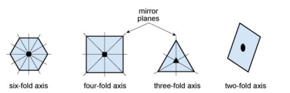
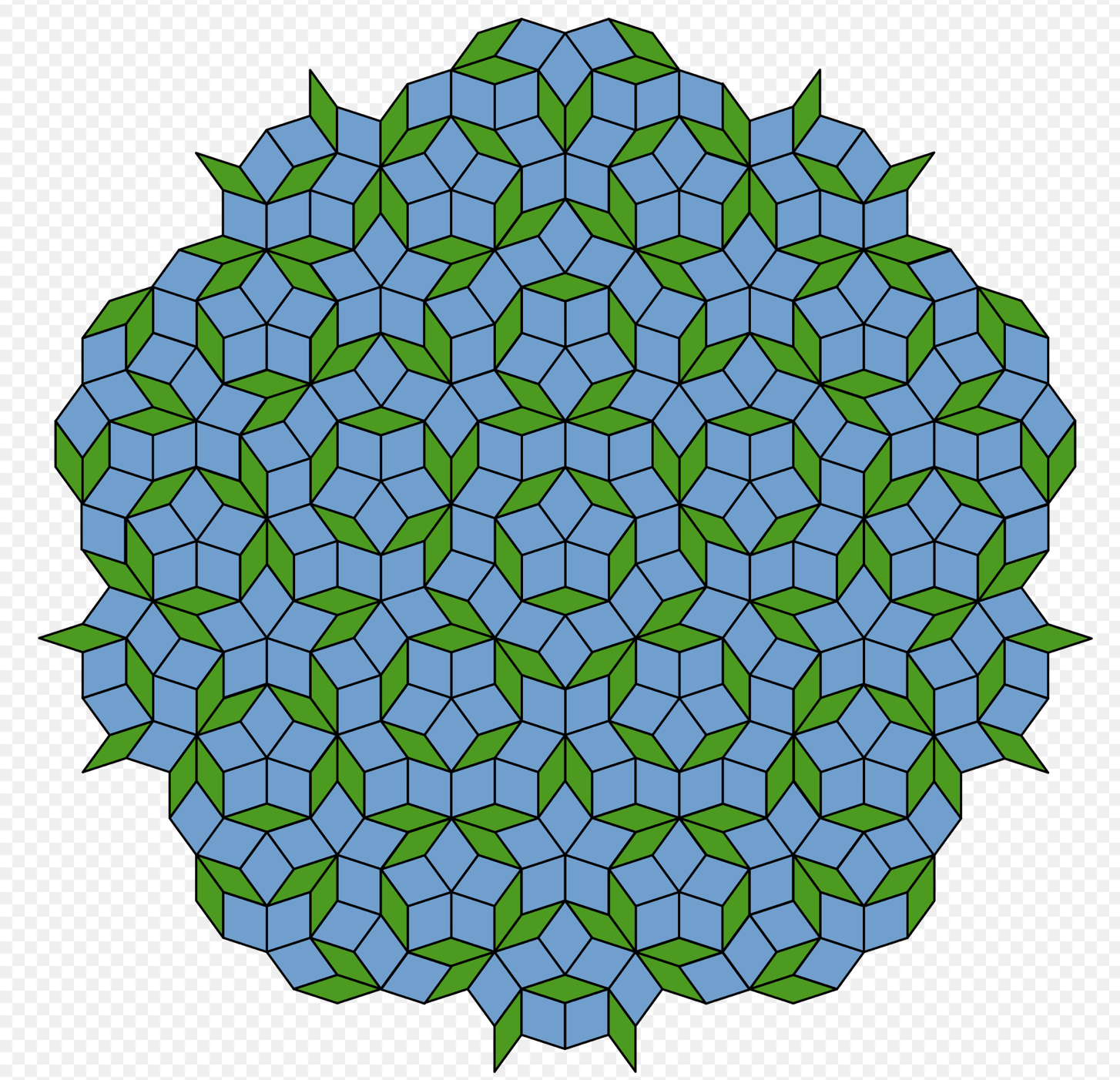
Quasicrystals were once thought to be impossible, as crystals were the only stable form of matter. Crystals allow for periodic patterns of atoms while quasicrystals allow for an ordered, yet non-periodic pattern that results in rotational symmetry. Crystals only allow for two-, three-, four-, and six-fold symmetry and create the geographical shapes of squares/rectangles, triangles, hexagons, and rhombuses (Figure 1). However, quasicrystals allow for ten-fold symmetry with unlimited layers of quasicrystal patterns and various shapes. The penrose tiles (Figure 2) is an example of one-dimensional quasicrystal pattern, while the kitchen tiles of your home is an example of a traditional crystal pattern.
After the discovery of man-made quasicrystals from a fellow scientist, Steinhardt wanted to find quasicrystals in nature as opposed to laboratories. He began this by contacting museums with global mineral samples in case they contained undiscovered quasicrystals. This did not yield any results.
Luca Bindi, who then worked for the Museum of Natural History at the University of Florence in Italy, discovered that Steinhardt was searching for natural quasicrystal and wanted to join his endeavors. Bindi found the first interesting sample at the museum he worked in through the rare mineral, khatyrkite, from the Koryak Mountains of Chukotka, Russia. They analyzed the tip of this sample, the width was that of a strand of hair, and discovered the most perfect ten-fold, rotationally symmetric pattern of a quasicrystal from minerals in nature. Even more interesting was that the chemical compound of this quasicrystal, Al63Cu24Fe13, was the exact composition of quasicrystals created in a Japanese laboratory, now found in a rock.
Steinhardt then took these findings to Lincoln Hollister, a renowned geologist, for his expert opinion. Hollister proceeded to tell Steinhardt that this discovery is impossible as its chemical composition of metallic aluminum cannot be created in nature. Steinhardt wondered if this sample came from a meteorite, which was an “ignorant, stupid suggestion, but Lincoln didn’t know that,” Steinhardt said. Lincoln refers Steinhardt to Glenn Macpherson, an expert meteorologist, who further elaborated that metallic aluminum from meteorites is, once again, impossible.
Two renowned experts in their fields describing the impossibility of Steinhardt and Bindi’s hypotheses was not enough for them to quit. Their next step was to trace Bindi’s khatyrkite to obtain more samples. Firstly, they attempted to find Nico Koekkoek, a Dutch mineral collector who had sold innumerable mineral samples to various museums. Dead end. Then they wrote to museums globally regarding their khatyrkite samples and discovered four potential samples. All fakes. Yet another dead end. Next was to analyze the legitimate sample in St. Petersburg because any sample of a newly discovered mineral must be given to a museum. The uncooperative discoverer, Leonid Razin, had immigrated to Israel and refused to let anyone touch the sample. They had hit a dead end again.
Bindi relayed this story to his sister and her friend over dinner. The friend’s neighbor shared the same common last name as the Dutch mineral collector, so the friend decided to ask his neighbor if it was an unlikely connection. Miraculously, the neighbor was the widow of the Dutch mineral collector and, after much persuading, handed over her late-husband’s secret diary. The diary reveals a mineral smuggler named Tim from Romania whom he received the khatyrkite. They were unable to locate Tim until Koekkoek’s widow relented yet another secret diary, which revealed that Tim had received these minerals from ‘L. Razin.’ The same Leonid Razin who refused them to view the sample! Eventually, Steinhardt discovered that Leonid Razid had sent a man named Valery Kryachko on an expedition for platinum. While he did not find platinum, he gave his samples to Leonid Razin, which astoundingly contained the natural quasicrystals that Steinhardt had searched for decades. Kryachko was completely unaware of its journey and even provided the remaining sample, which Steinhardt and his team used for testing.
Steinhardt’s original “ignorant, stupid suggestion” proved remarkably accurate, as they discovered that a meteorite hit Chukotka and resulted in natural metallic aluminum.
Steinhardt and his dream team needed more samples of khatyrkite to conduct further research. Therefore, seven Russians, five Americans, one Italian, and a cat named Buck set forth the scientific Mission Impossible for natural quasicrystals. They came back with several million grains and after a few weeks, found a sample of clay layer that had not been touched in 10,000 years. This was the first quasicrystal to be declared a natural mineral. They ultimately discovered a total of nine quasicrystal samples, each from a different part of the meteorite.
Steinhardt and his team’s analysis of quasicrystals is still not over and his book, “The Second Kind of Impossible,” delves further into the outlandish details of the over 30 years of research. This extraordinary journey of passion and ambition allows for the thrilling hope for the future of scientific discovery.
One of downtown Durham’s most memorable landmarks, the Chesterfield building looks like it was aesthetically designed to maintain the country’s morale during World War II. On the former cigarette factory’s roof rests a brilliant red sign that’s visible from miles away:
But don’t mistake the building’s quaint exterior for antiquity: the Chesterfield Building is home to one of the nation’s most powerful quantum computers. Managed by the Duke Quantum Center, the computer is part of Duke’s effort to bolster the Scalable Quantum Computing Laboratory (SQLab).
On February 2nd, the lab’s director – Christopher Monroe – joined engineering professor Michael Reiter and English professor Charlotte Sussman in a Research Week panel to discuss the growing presence of computation at Duke and in research institutions across the country. (View the panel.)
Chris Monroe
Monroe opened by detailing the significance of quantum computing in the modern world. He explained that quantum mechanics are governed by two golden rules: first, that quantum objects are waves and can be in superposition, and second, that the first rule only applies when said objects are not being measured.
The direct impact of quantum mechanics is that electrons can be in two orbits at the same time, which revolutionizes computing. Quantum computers factor numbers exponentially faster than classical computers, converge to more desirable solutions in optimization problems and have been shown to bolster research in fields like biomolecular modeling.
Still, Monroe insists that the future reach of quantum computing is beyond anyone’s current understanding. Says Monroe, “quantum computing is an entirely new way of dealing with information, so we don’t know all the application areas it will touch.” What we do know, he says, is that quantum computers are poised to take over where conventional computers and Moore’s Law leave off.
While Monroe discussed computing innovations, Michael Reiter – James B. Duke Professor of Computer Science and Electrical and Computer Engineering – demonstrated the importance of keeping computing systems safe. By pointing to the 2010 Stuxnet virus, a series of cyberattacks against Iranian nuclear centrifuges, and the 2017 Equifax Data Breach, which stole the records of 148 million people, Dr. Reiter provided evidence to show that modern data systems are vulnerable and attractive targets for cyber warfare.
Michael Reiter
To show the interdisciplinary responsibilities associated with the nation’s cybersecurity needs, Reiter posed two questions to the audience. First, what market interventions are appropriate to achieve more accountability for negligence in cybersecurity defenses? Second, what are the rules of war as it relates to cyber warfare and terrorism?
After Reiter’s presentation, Charlotte Sussman transitioned the conversation from the digital world to the maritime world. A professor of English at Duke, Sussman has always been interested in ways to both memorialize and understand the middle passage, the route slave trading ships took across the Atlantic from Africa to the Americas. Through the University’s Bass Connections and Data+ research programs, she and a group of students were able to approach this problem through the unlikely lens of data science.
Sussman explained that her Data+ team used large databases to find which areas of the Atlantic Ocean had the highest mortality rates during the slave trade, while the Bass Connections team looked at a single journey to understand one young migrant’s path to the bottom of the sea.
Professor Sussman (second from right), and the Bass Connections/Data+ Team.
Monroe, Reiter, and Sussman all showed that the applications of computing are growing without bound. Both the responsibility to improve computing infrastructures and the ability to leverage computing resources are rapidly expanding to new fields, from medicine and optimization to cybersecurity and history.
With so many exciting paths for growth, one point is clear about the future of computing: it will outperform anyone’s wildest expectations. Be prepared to find computing in academia, business, government, and other settings that require advanced information.
Many of these areas, like the Chesterfield Building, will probably see the impact of computing before you know it.
Russia sucessfully tested a direct-ascent anti-satellite missile on Monday, creating a debris field of more than 1,500 pieces of trackable orbital debris — space junk — whizzing around the planet. The crew aboard the International Space Station was ordered into their spacesuits to help them survive if one of the shards hit their home.
The Russian test, which has been strongly condemned by US officials, has created extreme hazards for satellites. US Space Command Commander General James Dickinson stated that “Russia has demonstrated a deliberate disregard for the security, safety, stability, and long-term sustainability of the space domain for all nations.”
Benjamin Schmitt PhD, a postdoctoral research fellow at the Harvard-Smithsonian Center for Astrophysics, facilitated the group conversation, which featured Hugh Lewis PhD, Professor of Astronautics and Head of the Astronautics Research Group at the University of Southampton. Schmitt stated that for the last two weeks, people around the world have paused to look up at the climate with the proceedings of COP26, but they “should also tilt their heads back a bit further” and consider the problem of space junk.
The challenge of space debris requires technical and diplomatic solutions, which are often complex. This has been effectively demonstrated by the Russian launch and resultant global reactions to the “irresponsibility” of the maneuver.
Schmitt and Lewis were joined by Brit Lundgren PhD, Laura Newburgh PhD, and W. Robert Pearson JD. Lundgren is an Associate Professor of Physics and Astronomy at the University of North Carolina at Asheville, Newburgh is an Assistant Professor of Physics at Yale University, and Pearson is a retired U.S. Ambassador and current Duke University Center for International and Global Studies Fellow.
Space experts engaged in Friday’s conversation
“The space debris problem is a wicked problem,” Lewis said. And the problem is this: According to the European Space Agency, there are over 36,500 objects larger than 10cm, 1,000,000 objects over 1cm, and more than one-third of a billion objects over 1mm in size in orbit around the Earth. These numbers, though bewilderingly large, are posed to expand.
As all this junk collides with itself, there are more and more fragments and particles in space. Lewis said that unlike climate change, there is not a “tipping point.” There will not be a warning or any sudden event that pushes us into the exponential growth phase – it will just, sort of, happen.
These pieces of debris pose substantial risks to the space systems that our modern societies have come to rely on, like piloting and navigation, communication, and many forms of entertainment like television. “Without those services, all of us, the entire planet, would suffer,” Lewis said.
A visualization of the space debris currently rotating around Earth.
But this issue of space debris likely feels entirely disconnected and irrelevant for most of the world’s population. “For us down here on Earth, we are really not aware of this growing problem … and we are really not able to connect to it,” Lewis said. “Unless we make that human connection, it’s not something we would be able to address.”
The panelists all agreed that making the connections between space debris and the current functioning of our globe is a critical step to getting the public to engage with the space debris challenge.
There are also other important reasons to care about space debris. Lundgren pointed out that there has already been a global 10% increase in brightness relative to the natural, dark sky because of light-reflecting space debris. This is the kind of light pollution that you cannot escape, Lundgren stated, “You can’t just drive away like with city pollution.” For communities of people, like the Indigenous, this is also having severe impact on the cultural ways in which they use nighttime skies.
Newburgh’s scientific research uses a particular satellite frequency for data collection. This wavelength was just sold to a communication company, meaning that eventually, she will no longer be able to do her work. The frequencies used for satellites are limited, and thus an extremely valuable and expensive, monopolizable commodity. Scientists like Newburgh are gravely concerned about the protection of the future of their work and worried that we might “lose out on science.”
This was a very important tenet of the discussion: “[Space debris] is not just a technical problem we have to solve, but a social one as well.” While technical solutions are needed to constrain the exponential growth of space debris, the even bigger challenge seems to lie in answering questions like “Who gets to use the remaining capacity in lowest orbit and how do we decide?” that Lewis asked. “Lots of companies, governments, and so on want to use space,” Lewis said.
Starlink satellites are changing to night sky. The company’s satellites can be seen traveling through space.
Ambassador Pearson said that this issue could be resolved by starting with a shared interest in the space debris issue and working outwards to points of change that are important across nations. The result would not ultimately be the full wish of any singular entity. Pearson also emphasized the pertinence of action: “It’s one thing to talk about what ought to be done and to talk about what we will do.”
While Pearson says that he does not believe there is a way to avoid national competition in space, it is essential to develop rules to mitigate and govern international interactions in space. This is likely to be a long process and has been on the minds of experts for decades already. But as Pearson reminded the audience it took almost 40 years to “get the ball rolling on climate change” and 10 years for the first nuclear disarmament.
The conversation ultimately kept returning to the need to engage the public and the impact that unconstrained space debris would have on their lives. Pearson said it is important to let the public know that the access to health, technology, communications, and many facets of society people had come to expect in their lives, would be severely impacted by damage to our space infrastructure.
“Whenever you think about the environment down here that we all occupy, that we are all connected to, we have to also think about the environment in space,” Lewis said.
He ended the conversation with a quote from the science fiction movie, Terminator 2: There is no fate but what we make for ourselves. This fate is dependent on cooperation between scientists, diplomats, regulatory and technical experts, and the public around the world.
Peak achievement in the sciences isn’t measured by stopwatches or goals scored, it goes by citations – the number of times other scientists have referenced your findings in their own academic papers. A high number of citations is an indication that a particular work was influential in moving the field forward.
Nobel laureate Bob Lefkowitz made the list in two categories this year.
And the peak of this peak is the annual “Highly Cited Researchers” list produced each year by the folks at Clarivate, who run the Institute for Scientific Information. The names on this list are drawn from publications that rank in the top 1% by citations for field and publication year in the Web of Science™ citation index – the most-cited of the cited.
Duke has 38 names on the highly cited list this year — including Bob Lefkowitz twice because he’s just that good — and two colleagues at the Duke NUS Medical School in Singapore. In all, the 2021 list includes 6,602 researchers from more than 70 countries.
The ISI says that US scientists are a little less than 40 percent of the highly cited list this year – and dropping. Chinese researchers are gaining, having nearly doubled their presence on the roster in the last four years.
“The headline story is one of sizeable gains for Mainland China and a decline for the United States, particularly when you look at the trends over the last four years,” said a statement from David Pendlebury, Senior Citation Analyst at the Institute for Scientific Information. “(This reflects) a transformational rebalancing of scientific and scholarly contributions at the top level through the globalization of the research enterprise.”
Without further ado, let’s see who our champions are!
Engineers, medical students, ecologists, political scientists, ethicists, policymakers — come one, come all to the Duke Space Initiative (DSI), “the interdisciplinary home for all things space at Duke.”
At Duke Polis’ “Perspectives on Space: Introducing the Duke Space Initiative” on Sept. 9, DSI co-founder and undergraduate student Ritika Saligram introduced the initiative and moderated a discussion on the current landscape of space studies both at Duke and beyond.
William R. & Thomas L. Perkins Professor of Law Jonathan Wiener began by expressing his excitement in the amount of interest he’s observed in space at Duke.
One of these interested students was Spencer Kaplan. Kaplan, an undergraduate student studying public policy, couldn’t attend Wiener’s Science & Society Dinner Dialogue about policy and risk in the settlement of Mars. Unwilling to miss the learning opportunity, Kaplan set up a one-on-one conversation with Wiener. One thing led to another: the two created a readings course on space law — Wiener hired Kaplan as a research assistant and they worked together to compile materials for the syllabus — then thought, “Why stop there?”
Wiener and Kaplan, together with Chase Hamilton, Jory Weintraub, Tyler Felgenhauer, Dan Buckland, and Somia Youssef, created the Bass Connections project “Going to Mars: Science, Society, and Sustainability,” through which a highly interdisciplinary team of faculty and students discussed problems ranging from the science and technology of getting to Mars, to the social and political reality of living on another planet.
The team produced a website, research papers, policy memos and recommendations, and a policy report for stakeholders including NASA and some prestigious actors in the private sector. According to Saligram, through their work, the team realized the need for a concerted “space for space” at Duke, and the DSI was born. The Initiative seeks to serve more immediately as a resource center for higher education on space, and eventually as the home of a space studies certificate program for undergraduates at Duke.
Wiener sees space as an “opportunity to reflect on what we’ve learned from being on Earth” — to consider how we could avoid mistakes made here and “try to do better if we settle another planet.” He listed a few of the many problems that the Bass Connections examined.
The economics of space exploration have changed: once, national governments funded space exploration; now, private companies like SpaceX, Blue Origin, and Virgin Galactic seek to run the show. Space debris, satellite and launch junk that could impair future launches, is the tragedy of the commons at work — in space. How would we resolve international disputes on other planets and avoid conflict, especially when settlements have different missions? Can we develop technology to ward off asteroids? What if we unintentionally brought microorganisms from one planet to another? How will we make the rules for the settlement of other planets?
These questions are vast — thereby reflecting the vastness of space, commented Saligram — and weren’t answerable within the hour. However, cutting edge research and thinking around them can be found on the Bass Connections’ website.
Earth and Climate Sciences Senior Lecturer Alexander Glass added to Wiener’s list of problems: “terraforming” — or creating a human habitat — on Mars. According to Glass, oxygen “isn’t a huge issue”: MOXIE can buzz Co2 with electricity to produce it. A greater concern is radiation. Without Earth’s magnetosphere, shielding of some sort will be necessary; it takes sixteen feet of rock to produce the same protection. Humans on Mars might have to live underground.
Glass noted that although “we have the science to solve a lot of these problems, the science we’re lagging in is the human aspects of it: the psychological, of humanity living in conditions like isolation.” The engineering could be rock solid. But the mission “will fail because there will be a sociopath we couldn’t predict beforehand.”
Bass Connections project leader and PhD candidate in political science Somia Youssef discussed the need to examine deeply our laws, systems, and culture. Youssef emphasized that we humans have been on Earth for six million years. Like Wiener, she asked how we will “apply what we’ve learned to space” and what changes we should make. How, she mused, do prevailing ideas about humanity “transform in the confines, the harsh environment of space?” Youssef urged the balancing of unity with protection of the things that make us different, as well as consideration for voices that aren’t being represented.
Material Science Professor, Assistant Professor of Surgery, and NASA Human System Risk Manager Dr. Dan Buckland explained that automation has exciting potential in improving medical care in space. If robots can do the “most dangerous aspects” of mission medical care, humans won’t have to. Offloading onto “repeatable devices” will reduce the amount of accidents and medical capabilities needed in space.
Multiple panelists also discussed the “false dichotomy” between spending resources on space and back home on Earth. Youssef pointed out that many innovations which have benefited (or will benefit) earthly humanity have come from the excitement and passion that comes from investing in space. Saligram stated that space is an “extension of the same social and policy issues as the ones we face on Earth, just in a different context.” This means that solutions we find in our attempt to settle Mars and explore the universe can be “reverse engineered” to help Earth-dwelling humans everywhere.
Saligram opened up the panel for discussion, and one guest asked Buckland how he ended up working for NASA. Buckland said his advice was to “be in rooms you’re not really supposed to be in, and eventually people will start thinking you’re supposed to be there.”
Youssef echoed this view, expressing the need for diverse perspectives in space exploration. She’s most excited by all the people “who are interested in space, but don’t know if there’s enough space for them.”
If this sounds like you, check out the Duke Space Initiative. They’ve got space.
Collaborating with a colleague in Shanghai, we recently published an article that explains the mathematical concept of ‘in-betweening,’in images – calculating intermediate stages of changes in appearance from one image to the next.
Our equilibrium-driven deformation algorithm (EDDA) was used to demonstrate three difficult tasks of ‘in-betweening’ images: Facial aging, coronavirus spread in the lungs, and continental drift.
Part I. Understanding Pneumonia Invasion and Retreat in COVID-19
The pandemic has influenced the entire world and taken away nearly 3 million lives to date. If a person were unlucky enough to contract the virus and COVID-19, one way to diagnose them is to carry out CT scans of their lungs to visualize the damage caused by pneumonia.
However, it is impossible to monitor the patient all the time using CT scans. Thus, the invading process is usually invisible for doctors and researchers.
To solve this difficulty, we developed a mathematical algorithm which relies on only two CT scans to simulate the pneumonia invasion process caused by COVID-19.
We compared a series of CT scans of a Chinese patient taken at different times. This patient had severe pneumonia caused by COVID-19 but recovered after a successful treatment. Our simulation clearly revealed the pneumonia invasion process in the patient’s lungs and the fading away process after the treatment.
Our simulation results also identify several significant areas in which the patient’s lungs are more vulnerable to the virus and other areas in which the lungs have better response to the treatment. Those areas were perfectly consistent with the medical analysis based on this patient’s actual, real-time CT scan images. The consistency of our results indicates the value of the method.
The COVID-19 pneumonia invading (upper panel) and fading away (lower panel) process from the data-driven simulations. Red circles indicate four significant areas in which the patient’s lungs were more vulnerable to the pneumonia and blue circles indicate two significant areas in which the patient’s lungs had better response to the treatment. (Image credit: Gao et al., 2021)We also applied this algorithm to simulate human facial changes over time, in which the aging processes for different parts of a woman’s face were automatically created by the algorithm with high resolution. (Image credit: Gao et al., 2021. Video)
Part II. Solving the Puzzle of Continental Drift
It has always been mysterious how the continents we know evolved and formed from the ancient single supercontinent, Pangaea. But then German polar researcher Alfred Wegener proposed the continental drift hypothesis in the early 20th century. Although many geologists argued about his hypothesis initially, more sound evidence such as continental structures, fossils and the magnetic polarity of rocks has supported Wegener’s proposition.
Our data-driven algorithm has been applied to simulate the possible evolution process of continents from Pangaea period.
The underlying forces driving continental drift were determined by the equilibrium status of the continents on the current planet. In order to describe the edges that divide the land to create oceans, we proposed a delicate thresholding scheme.
The formation and deformation for different continents is clearly revealed in our simulation. For example, the ‘drift’ of the Antarctic continent from Africa can be seen happening. This exciting simulation presents a quick and obvious way for geologists to establish more possible lines of inquiry about how continents can drift from one status to another, just based on the initial and equilibrium continental status. Combined with other technological advances, this data-driven method may provide a path to solve Wegener’s puzzle of continental drift.
The theory of continental drift reconciled similar fossil plants and animals now found on widely separated continents. The southern part after Pangaea breaks (Gondwana) is shown here evidence of Wegener’s theory. (Image credit: United States Geological Survey)The continental drift process of the data-driven simulations. Black arrow indicates the formation of the Antarctic. (Image credit: Gao et al., 2021)
The study was supported by the Department of Mathematics and Physics, Duke University.
CITATION: “Inbetweening auto-animation via Fokker-Planck dynamics and thresholding,” Yuan Gao, Guangzhen Jin & Jian-Guo Liu. Inverse Problems and Imaging, February, 2021, DOI: 10.3934/ipi.2021016. Online: http://www.aimsciences.org/article/doi/10.3934/ipi.2021016
Yuan Gao
Yuan Gao is the William W. Elliot Assistant Research Professor in the department of mathematics, Trinity College of Arts & Sciences.
Jian-Guo Liu is a Professor in the departments of mathematics and physics, Trinity College of Arts & Sciences.
Most of Physics Professor Haiyan Gao’s students see their doctoral dissertations posted on her lab’s web site very soon after they have been awarded their Ph.Ds.
But Yang Zhang, Ph.D. 2018, had to wait two years, because his thesis work had a very good chance of being accepted by a major journal. And this week, it has been published in the journal Science.
What Zhang did was to create the world’s most precise value for a subatomic nuclear particle called a neutral pion. It’s a quark and an antiquark comprising a meson. The neutral pion (also known as p0) is the lightest of the mesons, but a player in the strong attractive force that holds the atom’s nucleus together.
Haiyan Gao (left) with newly-minted physics Ph.D. Yang Zhang in 2018. (Photo courtesy of Min Huang, Ph.D. ’16)
And that, in turn, makes it a part of the puzzle Gao and her students have been trying to solve for many years. The prevailing theory about the strong force is called quantum chromodynamics (QCD), and it’s been probed for years by high-energy physics. But Gao, Zhang and their collaborators are trying to study QCD under more normal energy states, a notoriously difficult problem.
Yang Zhang spent six years analyzing and writing up the data from a Primakoff (PrimEx-II) experiment in Hall B at Thomas Jefferson National Accelerator Facility (Jefferson Lab) in Newport News, VA. His work was done on equipment supported by both the National Science Foundation and the Department of Energy.
This is the quark structure of the positive pion – an up quark and an anti-down quark. The strong force is from gluons, represented as the wavy lines (Arpad Horvath via Wikimedia Commons)
In a Primakoff experiment, a photon beam is directed on a nuclear target, producing neutral pions. In both the PrimEx-I and PrimEx-II experiments at Jefferson Lab, the two photons from the decay of a neutral pionwere subsequently detected in an electromagnetic calorimeter. From that, Zhang extracted the pion’s ‘radiative decay width.’ That decay width is a handy thing to have, because it is directly related to the pion’s life expectancy, and QCD has a direct prediction for it.
Zhang’s hard-won answer: The neutral pion has a radiative decay width of 7.8 electron-volts, give or take. That makes it an important piece of the dauntingly huge puzzle about QCD. Gao and her colleagues will continue to ask the fundamental questions about nature, at the finest but perhaps most profound scale imaginable.
The PrimEx-I and PrimEx-II collaborations were led by Prof. Ashot Gasparian from North Carolina A&T State University. Gao and Zhang joined the collaboration in 2011.
“Precision Measurement of the Neutral Pion Lifetime,” appears in Science May 1. Dr. Yang Zhang is now a quantitative researcher at JPMorgan Chase & Co.