Following the people and events that make up the research community at Duke

Students exploring the Innovation Co-Lab

Author: Kara Manke Page 1 of 4

Teaching a Machine to Spot a Crystal

A collection of iridescent crystals grown in space

Not all protein crystals exhibit the colorful iridescence of these crystals grown in space. But no matter their looks, all are important to scientists. Credit: NASA Marshall Space Flight Center (NASA-MSFC).

Protein crystals don’t usually display the glitz and glam of gemstones. But no matter their looks, each and every one is precious to scientists.

Patrick Charbonneau, a professor of chemistry and physics at Duke, along with a worldwide group of scientists, teamed up with researchers at Google Brain to use state-of-the-art machine learning algorithms to spot these rare and valuable crystals. Their work could accelerate drug discovery by making it easier for researchers to map the structures of proteins.

“Every time you miss a protein crystal, because they are so rare, you risk missing on an important biomedical discovery,” Charbonneau said.

Knowing the structure of proteins is key to understanding their function and possibly designing drugs that work with their specific shapes. But the traditional approach to determining these structures, called X-ray crystallography, requires that proteins be crystallized.

Crystallizing proteins is hard — really hard. Unlike the simple atoms and molecules that make up common crystals like salt and sugar, these big, bulky molecules, which can contain tens of thousands of atoms each, struggle to arrange themselves into the ordered arrays that form the basis of crystals.

“What allows an object like a protein to self-assemble into something like a crystal is a bit like magic,” Charbonneau said.

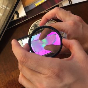
Even after decades of practice, scientists have to rely in part on trial and error to obtain protein crystals. After isolating a protein, they mix it with hundreds of different types of liquid solutions, hoping to find the right recipe that coaxes them to crystallize. They then look at droplets of each mixture under a microscope, hoping to spot the smallest speck of a growing crystal.

“You have to manually say, there is a crystal there, there is none there, there is one there, and usually it is none, none, none,” Charbonneau said. “Not only is it expensive to pay people to do this, but also people fail. They get tired and they get sloppy, and it detracts from their other work.”

Three microscope images of protein crystallization solutions

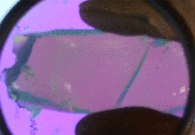
The machine learning software searches for points and edges (left) to identify crystals in images of droplets of solution. It can also identify when non-crystalline solids have formed (middle) and when no solids have formed (right).

Charbonneau thought perhaps deep learning software, which is now capable of recognizing individual faces in photographs even when they are blurry or caught from the side, should also be able to identify the points and edges that make up a crystal in solution.

Scientists from both academia and industry came together to collect half a million images of protein crystallization experiments into a database called MARCO. The data specify which of these protein cocktails led to crystallization, based on human evaluation.

The team then worked with a group led by Vincent Vanhoucke from Google Brain to apply the latest in artificial intelligence to help identify crystals in the images.

After “training” the deep learning software on a subset of the data, they unleashed it on the full database. The A.I. was able to accurately identify crystals about 95 percent of the time. Estimates show that humans spot crystals correctly only 85 percent of the time.

“And it does remarkably better than humans,” Charbonneau said. “We were a little surprised because most A.I. algorithms are made to recognize cats or dogs, not necessarily geometrical features like the edge of a crystal.”

Other teams of researchers have already asked to use the A.I. model and the MARCO dataset to train their own machine learning algorithms to recognize crystals in protein crystallization experiments, Charbonneau said. These advances should allow researchers to focus more time on biomedical discoveries instead of squinting at samples.

Charbonneau plans to use the data to understand how exactly proteins self-assemble into crystals, so that researchers rely less on chance to get this “magic” to happen.

“We are trying to use this data to see if we can get more insight into the physical chemistry of self-assembly of proteins,” Charbonneau said.

CITATION: “Classification of crystallization outcomes using deep convolutional neural networks,” Andrew E. Bruno, et al. PLOS ONE, June 20, 2018. DOI: 10.1371/journal.pone.0198883


Post by Kara Manke

Stretchable, Twistable Wires for Wearable Electronics

A new conductive “felt” carries electricity even when twisted, bent and stretched. Credit: Matthew Catenacci

The exercise-tracking power of a Fitbit may soon jump from your wrist and into your clothing.

Researchers are seeking to embed electronics such as fitness trackers and health monitors into our shirts, hats, and shoes. But no one wants stiff copper wires or silicon transistors deforming their clothing or poking into their skin.

Scientists in Benjamin Wiley’s lab at Duke have created new conductive “felt” that can be easily patterned onto fabrics to create flexible wires. The felt, composed of silver-coated copper nanowires and silicon rubber, carries electricity even when bent, stretched and twisted, over and over again.

“We wanted to create wiring that is stretchable on the body,” said Matthew Catenacci, a graduate student in Wiley’s group.

The conductive felt is made of stacks of interwoven silver-coated copper nanotubes filled with a stretchable silicone rubber (left). When stretched, felt made from more pliable rubber is more resilient to small tears and holes than felts made of stiffer rubber (middle). These tears can be seen in small cavities in the felt (right). Credit: Matthew Catenacci

To create a flexible wire, the team first sucks a solution of copper nanowires and water through a stencil, creating a stack of interwoven nanowires in the desired shape. The material is similar to the interwoven fibers that comprise fabric felt, but on a much smaller scale, said Wiley, an associate professor of chemistry at Duke.

“The way I think about the wires are like tiny sticks of uncooked spaghetti,” Wiley said. “The water passes through, and then you end up with this pile of sticks with a high porosity.”

The interwoven nanowires are heated to 300 F to melt the contacts together, and then silicone rubber is added to fill in the gaps between the wires.

To show the pliability of their new material, Catenacci patterned the nanowire felt into a variety of squiggly, snaking patterns. Stretching and twisting the wires up to 300 times did not degrade the conductivity.

The material maintains its conductivity when twisted and stretched. Credit: Matthew Catenacci

“On a larger scale you could take a whole shirt, put it over a vacuum filter, and with a stencil you could create whatever wire pattern you want,” Catenacci said. “After you add the silicone, so you will just have a patch of fabric that is able to stretch.”

Their felt is not the first conductive material that displays the agility of a gymnast. Flexible wires made of silver microflakes also exhibit this unique set of properties. But the new material has the best performance of any other material so far, and at a much lower cost.

“This material retains its conductivity after stretching better than any other material with this high of an initial conductivity. That is what separates it,” Wiley said.

Stretchable Conductive Composites from Cu-Ag Nanowire Felt,” Matthew J. Catenacci, Christopher Reyes, Mutya A. Cruz and Benjamin J. Wiley. ACS Nano, March 14, 2018. DOI: 10.1021/acsnano.8b00887

Post by Kara Manke

MRI Tags Stick to Molecules with Chemical “Velcro®”

An extremely close-up view of Velcro

In the new technique, MRI chemical tags attach to a target molecule and nothing else – kind of like how Velcro only sticks to itself. Credit: tanakawho, via Flickr.

Imagine attaching a beacon to a drug molecule and following its journey through our winding innards, tracking just where and how it interacts with the chemicals in our bodies to help treat illnesses.

Duke scientists may be closer to doing just that. They have developed a chemical tag that can be attached to molecules to make them light up under magnetic resonance imaging (MRI).

This tag or “lightbulb” changes its frequency when the molecule interacts with another molecule, potentially allowing researchers to both locate the molecule in the body and see how it is metabolized.

“MRI methods are very sensitive to small changes in the chemical structure, so you can actually use these tags to directly image chemical transformations,” said Thomas Theis, an assistant research professor in the chemistry department at Duke.

Chemical tags that light up under MRI are not new. In 2016, the Duke team of Warren S. Warren’s lab and Qiu Wang’s lab created molecular lightbulbs for MRI that burn brighter and longer than any previously discovered.

A photo of graduate students Junu Bae and Zijian Zhou in front of a bookshelf.

Junu Bae and Zijian Zhou, the co-first authors of the paper. Credit: Qiu Wang, Duke University.

In a study published March 9 in Science Advances, the researchers report a new method for attaching tags to molecules, allowing them to tag molecules indirectly to a broader scope of molecules than they could before.

“The tags are like lightbulbs covered in Velcro,” said Junu Bae, a graduate student in Qiu Wang’s lab at Duke. “We attach the other side of the Velcro to the target molecule, and once they find each other they stick.”

This reaction is what researchers call bioorthogonal, which means that the tag will only stick to the molecular target and won’t react with any other molecules.

And the reaction was designed with another important feature in mind — it generates a rare form of nitrogen gas that also lights up under MRI.

“One could dream up a lot of potential applications for the nitrogen gas, but one that we have been thinking about is lung imaging,” Theis said.

Currently the best way to image the lungs is with xenon gas, but this method has the downside of putting patients to sleep. “Nitrogen gas would be perfectly safe to inhale because it is what you inhale in the air anyways,” Theis said.

A stylized chemical diagram of the hyperpolarization process

In the new technique, a type of molecule called a tetrazine is hyperpolarized, making it “light up” under MRI (illustrated on the left). It is then tagged to a target molecule through a what is called a bioorthogonal reaction. The reaction also generates a rare form of nitrogen gas that can be spotted under MRI (illustrated on the right). Credit: Junu Bae and Seoyoung Cho, Duke University.

Other applications could include watching how air flows through porous materials or studying the nitrogen fixation process in plants.

One downside to the new tags is that they don’t shine as long or as brightly as other MRI molecular lightbulbs, said Zijian Zhou, a graduate student in  Warren’s lab at Duke.

The team is tinkering with the formula for polarizing, or lighting up, the molecule tags to increase their lifetime and brilliance, and to make them more compatible with chemical conditions in the human body.

“We are now developing new techniques and new procedures which may be helpful for driving the polarization levels even higher, so we can have even better signal for these applications,” Zhou said.

15N4-1,2,4,5-tetrazines as potential molecular tags: Integrating bioorthogonal chemistry with hyperpolarization and unearthing para-N2,” Junu Bae, Zijian Zhou, Thomas Theis, Warren S. Warren and Qiu Wang. Science Advances, March 9, 2018. DOI: 10.1126/sciadv.aar2978

Post by Kara Manke

How Earth’s Earliest Lifeforms Protected Their Genes

A colorful hot spring in Yellowstone National Park

Heat-loving thermophile bacteria may have been some of the earliest lifeforms on Earth. Researchers are studying their great great great grandchildren, like those living in Yellowstone’s Grand Prismatic Spring, to understand how these early bacteria repaired their DNA.

Think your life is hard? Imagine being a tiny bacterium trying to get a foothold on a young and desolate Earth. The earliest lifeforms on our planet endured searing heat, ultraviolet radiation and an atmosphere devoid of oxygen.

Benjamin Rousseau, a research technician in David Beratan’s lab at Duke, studies one of the molecular machines that helped these bacteria survive their harsh environment. This molecule, called photolyase, fixes DNA damaged by ultraviolet (UV) radiation — the same wavelengths of sunlight that give us sunburn and put us at greater risk of skin cancer.

“Anything under the sun — in both meanings of the phrase — has to have ways to repair itself, and photolyase proteins are one of them,” Rousseau said. “They are one of the most ancient repair proteins.”

Though these proteins have been around for billions of years, scientists are still not quite sure exactly how they work. In a new study, Rousseau and coworkers, working with Professor David Beratan and Assistant Research Professor Agostino Migliore, used computer simulations to study photolyase in thermophiles, the great great great great grandchildren of Earth’s original bacterial pioneers.

The study appeared in the Feb. 28 issue of the Journal of the American Chemical Society.

DNA is built of chains of bases — A, C, G and T — whose order encodes our genetic information. UV light can trigger two adjacent bases to react and latch onto one other, rendering these genetic instructions unreadable.

Photolyase uses a molecular antenna to capture light from the sun and convert it into an electron. It then hands the electron over to the DNA strand, sparking a reaction that splits the two bases apart and restores the genetic information.

A ribbon diagram of a photolyase protein

Photolyase proteins use a molecular antenna (green, blue and red structure on the right) to harvest light and convert it into an electron. The adenine-containing structure in the middle hands the electron to the DNA strand, splitting apart DNA bases. Credit: Benjamin Rousseau, courtesy of the Journal of the American Chemical Society.

Rousseau studied the role of a molecule called adenine in shuttling the electron  from the molecular antenna to the DNA strand. He looked at photolyase in both the heat-loving ancestors of ancient bacteria, called thermophiles, and more modern bacteria like E. Coli that thrive at moderate temperatures, called mesophiles.

He found that in thermophiles, adenine played a role in transferring the electron to the DNA. But in E. coli, the adenine was in a different position, providing mainly structural support.

The results “strongly suggest that mesophiles and thermophiles fundamentally differ in their use of adenine for this electron transfer repair mechanism,” Rousseau said.

He also found that when he cooled E. Coli down to 20 degrees Celsius — about 68 degrees Fahrenheit — the adenine shifted back in place, resuming its transport function.

“It’s like a temperature-controlled switch,” Rousseau said.

Though humans no longer use photolyase for DNA repair, the protein persists in life as diverse as bacteria, fungi and plants — and is even being studied as an ingredient in sunscreens to help repair UV-damaged skin.

Understanding exactly how photolyase works may also help researchers design proteins with a variety of new functions, Rousseau said.

“Photolyase does all of the work on its own — it harvests the light, it transfers the electron over a huge distance to the other site, and then it cleaves the DNA bases,” Rousseau said. “Proteins with that kind of plethora of functions tend to be an attractive target for protein engineering.”

Post by Kara Manke

How A Zebrafish’s Squiggly Cartilage Transforms into a Strong Spine

A column of green cartilage cells divides into an alternating pattern of green cartilage and red vertebra

Our spines begin as a flexible column called the notochord. Over time, cells on the notochord surface divide into alternating segments that go on to form cartilage and vertebrae.

In the womb, our strong spines start as nothing more than a rope of rubbery tissue. As our bodies develop, this flexible cord, called the notochord, morphs into a column of bone and cartilage sturdy enough to hold up our heavy upper bodies.

Graduate student Susan Wopat and her colleagues in Michel Bagnat’s lab at Duke are studying the notochords of the humble zebrafish to learn how this cartilage-like rope grows into a mature spine.

In a new paper, they detail the cellular messaging that directs this transformation.

It all comes down to Notch receptors on the notochord surface, they found. Notch receptors are a special type of protein that sits astride cell membranes. When two cells touch, these Notch receptors link up, forming channels that allow messages to rapidly travel between large groups of cells.

Notch receptors divide the outer notochord cells into two alternating groups – one group is told to grow into bone, while the other is told to grow into cartilage. Over time, bone starts to form on the surface of the notochord and works its way inward, eventually forming mature vertebrae.

X-ray images of four zebrafish spines

Meddling with cellular signaling on the notochord surface caused zebrafish spines to develop deformities. The first and third image show healthy spines, and the second and fourth image show deformed spines.

When the team tinkered with the Notch signaling on the surface cells, they found that the spinal vertebrae came out deformed – too big, too small, or the wrong shape.

“These results demonstrate that the notochord plays a critical role in guiding spine development,” Wopat said. “Further investigation into these findings may help us better understand the origin of spinal defects in humans.”

Spine patterning is guided by segmentation of the notochord sheath,” Susan Wopat, Jennifer Bagwell, Kaelyn D. Sumigray, Amy L. Dickson, Leonie F. Huitema, Kenneth D. Poss, Stefan Schulte-Merker, Michel Bagnat. Cell, February 20, 2018. DOI: 10.1016/j.celrep.2018.01.084

Post by Kara Manke

Duke Scholars Bridge Disciplines to Tackle Big Questions

A visualization showing faculty as dots that are connected by lines

This visualization, created by James Moody and the team at the Duke Network Analysis Center, links faculty from across schools and departments who serve together on Ph.D. committees. An interactive version is available here.

When the next big breakthrough in cancer treatment is announced, no one will ask whether the researchers are pharmacologists, oncologists or cellular biologists – and chances are, the team will represent all three.

In the second annual Scholars@Duke Visualization Challenge, Duke students explored how scholars across campus are drawing from multiple academic disciplines to tackle big research questions.

“I’m often amazed at how gifted Duke faculty are and how they can have expertise in multiple fields, sometimes even fields that don’t seem to overlap,” said Julia Trimmer, Director of Faculty Data Systems and Analysis at Duke.

In last year’s challenge, students dug into Scholars@Duke publication data to explore how Duke researchers collaborate across campus. This year, they were provided with additional data on Ph.D. dissertation committees and asked to focus on how graduate education and scholarship are reaching across departmental boundaries.

“The idea was to see if certain units or disciplines contributed faculty committee members across disciplines or if there’s a lot of discipline ‘overlap.’” Trimmer said.

The winning visualization, created by graduate student Matthew Epland, examines how Ph.D. committees span different fields. In this interactive plot, each marker represents an academic department. The closer together markers are, the more likely it is that a faculty member from one department will serve on the committee of a student in the other department.

Epland says he was intrigued to see the tight-knit community of neuroscience-focused departments that span different schools, including psychology and neuroscience, neurobiology, neurology and psychiatry and behavioral Sciences. Not surprisingly, many of the faculty in these departments are members of the Duke Institute for Brain Sciences (DIBS).

Duke schools appear as dots and are connected by lines of different thicknesses

Aghil Abed Zadeh and Varda F. Hagh analyzed publication data to visualize the extent to which faculty at different Duke schools collaborate with one another. The size of each dot represents the number of publications from each school, and thickness of each line represents the number of faculty collaborations between the connected schools.

Sociology Professor James Moody and the team at the Duke Network Analysis Center took a similar approach, creating a network of individual faculty members who are linked by shared students. Faculty who sit on committees in only one field are bunched together, highlighting researchers who bridge different disciplines. The size of each marker represents the extent to which each researcher sits “between” two fields.

The map shows a set of strong ties within the natural sciences and within the humanities, but few links between the two groups. Moody points out that philosophy is a surprising exception to this rule, lying closer to the natural sciences cluster than to the humanities cluster.

“At Duke, the strong emphasis on philosophy of science creates a natural link between philosophy and the natural sciences,” Moody said.

Duke graduate student Aghil Abed Zadeh teamed up with Varda F. Hagh, a student at Arizona State University, to create elegant maps linking schools and departments by shared authorship. The size of each marker represents the number of publications in that school or department, and the thickness of the connecting lines indicate the number of shared authorships.

“It is interesting to see how connected law school and public policy school are. They collaborate with many of the sciences as well, which is a surprising fact,” Zadeh said. “On the other hand, we see Divinity school, one the oldest at Duke, which is isolated and not connected to others at all.”

The teams presented their visualizations Jan. 20 at the Duke Research Computing Symposium.

Post by Kara Manke


Farewell, Electrons: Future Electronics May Ride on New Three-in-One Particle

“Trion” may sound like the name of one of the theoretical particles blamed for mucking up operations aboard the Starship Enterprise.

But believe it or not, trions are real — and they may soon play a key role in electronic devices. Duke researchers have for the first time pinned down some of the behaviors of these one-of-a-kind particles, a first step towards putting them to work in electronics.

A carbon nanotube, shaped like a rod, is wrapped in a helical coating of polymer

Three-in-one particles called trions — carrying charge, energy and spin — zoom through special polymer-wrapped carbon nanotubes at room temperature. Credit: Yusong Bai.

Trions are what scientists call “quasiparticles,” bundles of energy, electric charge and spin that zoom around inside semiconductors.

“Trions display unique properties that you won’t be able to find in conventional particles like electrons, holes (positive charges) and excitons (electron-hole pairs that are formed when light interacts with certain materials),” said Yusong Bai, a postdoctoral scholar in the chemistry department at Duke. “Because of their unique properties, trions could be used in new electronics such as photovoltaics, photodetectors, or in spintronics.”

Usually these properties – energy, charge and spin – are carried by separate particles. For example, excitons carry the light energy that powers solar cells, and electrons or holes carry the electric charge that drives electronic devices. But trions are essentially three-in-one particles, combining these elements together into a single entity – hence the “tri” in trion.

A diagram of how a trion is formed in carbon nanotubes.

A trion is born when a particle called a polaron (top) marries an exciton (middle). Credit: Yusong Bai.

“A trion is this hybrid that involves a charge marrying an exciton to become a uniquely distinct particle,” said Michael Therien, the William R. Kenan, Jr. Professor of Chemistry at Duke. “And the reason why people are excited about trions is because they are a new way to manipulate spin, charge, and the energy of absorbed light, all simultaneously.”

Until recently, scientists hadn’t given trions much attention because they could only be found in semiconductors at extremely low temperatures – around 2 Kelvin, or -271 Celcius. A few years ago, researchers observed trions in carbon nanotubes at room temperature, opening up the potential to use them in real electronic devices.

Bai used a laser probing technique to study how trions behave in carefully engineered and highly uniform carbon nanotubes. He examined basic properties including how they are formed, how fast they move and how long they live.

He was surprised to find that under certain conditions, these unusual particles were actually quite easy to create and control.

“We found these particles are very stable in materials like carbon nanotubes, which can be used in a new generation of electronics,” Bai said. “This study is the first step in understanding how we might take advantage of their unique properties.”

The team published their results Jan. 8 in the Proceedings of the National Academy of Sciences.

Dynamics of charged excitons in electronically and morphologically homogeneous single-walled carbon nanotubes,” Yusong Bai, Jean-Hubert Olivier, George Bullard, Chaoren Liu and Michael J. Therien. Proceedings of the National Academy of Sciences, Jan. 8, 2018 (online) DOI: 10.1073/pnas.1712971115

Post by Kara Manke

Glitter and Jell-O Reveal the Science of Oobleck

A black and white image showing a circular disk dropping into a container of oobleck

Mixing black glitter with oobleck allowed researchers to track the movement of individual cornstarch particles after a sudden impact. A computer program locked onto pieces of glitter and illustrated their motion. Credit: Melody Lim.

What do gelatin and glitter have to do with serious science? For some experiments, a lot! Duke alumna Melody Lim used jiggly Jell-O and a just a pinch of glitter to solve a scientific mystery about the curious goo many like to call oobleck.

To the uninitiated, oobleck is almost magic. The simple mixture of cornstarch and water feels solid if you squeeze it, but moments later runs through your fingers like water. You can dance across a bathtub full of oobleck, but stand still for too long and you will be sucked into a goopy mess. Not surprisingly, the stuff is a Youtube favorite.

Oobleck is an example of what scientists call a non-Newtonian fluid, a liquid whose viscosity – how easily it changes shape and flows – depends upon the force that is applied. But exactly how it is that this material switches from solid to liquid and back again has remained a mystery to scientists.

A piece of gelatin being squeezed viewed through a circular polarizer

This blogger mixed up a batch of jello to see the photoelastic effect for herself. When viewed with polarized light – from an iPhone screen and a circular polarizer – the jello changes color when squeezed.

“Water is simple to understand, and so is cornstarch,” said Lim, ’16, who is currently a graduate student at the University of Chicago. “However, a combination of the two produces this ‘liquid’ that ripples and flows, solidifies beneath your feet if you run on it, then turns back into a liquid if you stop running and stand still. I wanted to know why.”

The question beguiling scientists was whether sudden impact causes the cornstarch particles to “jam” into a solid like cement, or whether the suspension remains liquid but simply moves too slowly for its liquid-like properties to be apparent — similar to what happens if you skip a rock off the surface of a lake.

“There are these two opposing pictures,” said Robert Behringer, James B. Duke Professor of Physics at Duke. “Either you squish the material and turn it into cement temporarily, or you simply transmit the stress from the impactor straight to the boundary.”

Lim did two sets of experiments to find out which way oobleck works. In one experiment, she mixed black glitter into a transparent channel filled with oobleck, and then used a high-speed camera to watch how the material responded to the impact. The glitter let her track the motion of individual particles after the disc hit.

A piece of gelatin changes color when you squeeze it.

The photoelastic effect in gelatin.

Her video shows that the particles near the impact site jam and become solid, forming what the researchers call a “mass shock” wave that travels slowly through the suspension.

In a second set of experiments, Lim placed the oobleck in a container lined with gelatin, the main ingredient in Jell-O – besides sugar and food dye, of course. Gelatin is what is called a photoelastic material, which means that applying pressure bends light that travels through it, like a prism.

“Next time you eat Jell-O, get out your sunglasses and get somebody else’s sunglasses and look between them,” Behringer said. “Because if you give it a shake you should see all these stress patterns bouncing around.”

After the metal disc hit the oobleck, the gelatin let Lim see how fast the resulting pressure wave traveled through the material and reached the boundary.

A black and white image showing pressure waves traveling through a transparent material after impact

The researchers poured oobleck into a clear container lined with gelatin, a material that bends light when a pressure is applied to it. They saw that the force of a sudden impact is rapidly transmitted through the oobleck and to the boundary with the gelatin. Credit: Melody Lim.

They found that when the impact is sudden, the pressure wave traveled to the gelatin boundary faster than the “mass shock” wave. This means that the reason oobleck appears solid after a sudden impact is because the force of the collision is quickly transmitted to a solid boundary.

“If you are running across the water, that actually puts you into an impact velocity range where the pressure wave is significantly faster than the mass shock,” Behringer said. “Whereas if you try to walk across it, the impact speeds are slow, and the system actually doesn’t have the ability to transport the momentum quickly through the material and so you just sink in.”

“If you’d told me when I started that I would line a narrow container with Jell-o, add cornstarch, water, and black glitter, drop a piece of metal on it, then publish a paper on the results, I would have laughed at you,” Lim said.

CITATION: “Force and Mass Dynamics in Non-Newtonian Suspensions,” Melody X. Lim, Jonathan Barés, Hu Zheng and Robert P. Behringer. Physical Review Letters, Nov. 3, 2017. DOI: 10.1103/PhysRevLett.119.184501

Post by Kara Manke

Cheating Time to Watch Liquids do the Slow Dance

Colorful spheres simulating liquid molecules shift around inside a cube shape

The team’s new algorithm is able to simulate molecular configurations of supercooled liquids below the glass transition. The properties of these configurations are helping to solve a 70-year paradox about the entropy of glasses. Credit: Misaki Ozawa and Andrea Ninarello, Université de Montpellier.

If you could put on a pair of swimming goggles, shrink yourself down like a character from The Magic School Bus and take a deep dive inside a liquid, you would see a crowd of molecules all partying like it’s 1999.

All this frenetic wiggling makes it easy for molecules to rearrange themselves and for the liquid as a whole to change shape. But for supercooled liquids — liquids like honey that are cooled below their freezing point without crystallizing – the lower temperature slows down the dancing like Etta James’ “At Last.” Lower the temperature enough, and the slow-down can be so dramatic that it takes centuries or even millennia for the molecules to rearrange and the liquid to move.

Scientists can’t study processes that last longer than their careers. But Duke chemists and their Simons Foundation collaborators have found a way to cheat time, simulating the slow dance of deeply supercooled liquids. Along the way, they have found new physical properties of “aged” supercooled liquids and glasses.

A droplet rises above a surface of water

Credit: Ruben Alexander via Flickr.

To understand just how slow deeply supercooled liquids move, consider the world’s longest-running experiment, the University of Queensland’s Pitch Drop Experiment. A single drop of pitch forms every eight to thirteen years — and this pitch is moving faster than deeply supercooled liquids.

“Experimentally there is a limit to what you can observe, because even if you managed to do it over your entire career, that is still a maximum of 50 years,” said Patrick Charbonneau, an associate professor of chemistry and physics at Duke. “For many people that was considered a hard glass ceiling, beyond which you couldn’t study the behavior of supercooled liquids.”

Charbonneau, who is an expert on numerical simulations, said that using computers to simulate the behavior of supercooled liquids has even steeper time limitations. He estimates that, given the current rate of computer advancement, it would take 50 to 100 years before computers would be powerful enough for simulations to exceed experimental capabilities – and even then the simulations would take months.

To break this glass ceiling, the Charbonneau group collaborated with Ludovic Berthier and his team, who were developing an algorithm to bypass these time constraints. Rather than taking months or years to simulate how each molecule in a supercooled liquids jiggles around until the molecules rearrange, the algorithm picks individual molecules to swap places with each other, creating new molecular configurations.

This allows the team to explore new configurations that could take millennia to form naturally. These “deeply supercooled liquids and ultra-aged glasses” liquids are at a lower energy, and more stable, than any observed before.

“We were cheating time in the sense that we didn’t have to follow the dynamics of the system,” Charbonneau said. “We were able to simulate deeply supercooled liquids well beyond is possible in experiments, and it opened up a lot of possibilities.”

Two columns of blue and red spheres represent simulations of vapor-deposited glasses.

Glasses that are grown one layer at a time have a much different structure than bulk glasses. The team used their new algorithm to study how molecules in these glasses rearrange, and found that at low temperatures (right), only the molecules at the surface are mobile. The results may be used to design better types of glass for drug delivery or protective coatings. Credit: Elijah Flenner.

Last summer, the team used this technique to discover a new phase transition in low-temperature glasses. They recently published two additional studies, one of which sheds light on the “Kauzmann paradox,” a 70-year question about the entropy of supercooled liquids below the glass transition. The second explores the formation of vapor-deposited glasses, which have applications in drug delivery and protective coatings.

“Nature has only one way to equilibrate, by just following the molecular dynamics,” said Sho Yaida, a postdoctoral fellow in Charbonneau’s lab. “But the great thing about numerical simulations is you can tweak the algorithm to accelerate your experiment.”

Configurational entropy measurements in extremely supercooled liquids that break the glass ceiling.” Ludovic Berthier, Patrick Charbonneau, Daniele Coslovich, Andrea Ninarello, Misaki Ozawa and Sho Yaida. PNAS, Oct. 24, 2017. DOI: 10.1073/pnas.1706860114

The origin of ultrastability in vapor-deposited glasses.” Ludovic Berthier, Patrick Charbonneau, Elijah Flenner and Francesco Zamponi. PRL, Nov. 1, 2017. DOI: 10.1103/PhysRevLett.119.188002

Post by Kara Manke

Designing Drugs Aimed at a Different Part of Life’s Code

Individual RNA molecules fluoresce inside a breast cancer cell.

Individual RNA molecules fluoresce inside a breast cancer cell. Credit: Sunjong Kwon, Oregon Health & Science University, via Flickr.

Most drugs work by tinkering with the behavior of proteins. Like meddlesome coworkers, these molecules are designed to latch onto their target proteins and keep them from doing what they need to do.

If a protein is responsible for speeding up a reaction, the drug helps slow the reaction down. If a protein serves as a gatekeeper to a cell, regulating what gets in and what stays out, a drug changes how many molecules it lets through.

But proteins aren’t the only doers and shakers in our bodies. Scientists are finding that strings of RNA — known primarily for their role in shuttling genetic information from nucleus-bound DNA to the cell’s protein-manufacturing machinery — can also play a major role in regulating disease.

A portrait of Amanda Hargrove

Amanda Hargrove is an assistant professor of chemistry at Duke University.

“There has been what some people are calling an RNA revolution,” said Amanda Hargrove, assistant professor of chemistry at Duke. “In some diseases, non-coding RNAs, or RNAs that don’t turn into protein, seem to be the best predictors of disease, and even to be driving the disease.”

Hargrove and her team at Duke are working to design new types of drugs that target RNA rather than proteins. RNA-targeted drug molecules have the potential help treat diseases like prostate cancer and HIV, but finding them is no easy task. Most drugs have been designed to interfere with proteins, and just don’t have the same effects on RNA.

Part of the problem is that proteins and RNA have many fundamental differences, Hargrove said. While proteins are made of strings of twenty amino acids that can twist into myriad different shapes, RNA is made of strings of only four bases — adenine, guanine, cytosine and uracil.

“People have been screening drugs for different kinds of RNA for quite a while, and historically have not had a lot of success,” Hargrove said. “This begged the question, since RNA has such chemically different properties than proteins, is there something different about the small molecules that we need in order to target RNA?”

To find out, graduate student Brittany Morgan and research associate Jordan Forte combed the scientific literature to identify 104 small molecules that are known interact with specific types of RNA. They then analyzed 20 different properties of these molecules, and compared their properties to those of collections of drug molecules known to interact with proteins.

The team found significant differences in shape, atomic composition, and charge between the RNA-active molecules and the protein-active molecules. They plan to use the results to compile a collection of molecules, called a library, that are chosen to better “speak the language” of the RNA-active molecules. They hope this collection of molecules will be more likely to interact with RNA in therapeutically beneficial ways.

“We found that there are differences between the RNA-targeted molecules and the protein-targeted drugs, and some of them are pretty striking,” Hargrove said. “What that means is that we could start to enrich our screening libraries with these types of molecules, and make these types of molecules, to have better luck at targeting RNA.”

Discovery of Key Physicochemical, Structural, and Spatial Properties of RNA-Targeted Bioactive Ligands.” Brittany S. Morgan, Jordan E. Forte, Rebecca N. Culver, Yuqi Zhang and Amanda Hargrove. Angewandte Chemie, Sept. 18, 2017. DOI: 10.1002/anie.201707641

Kara J. Manke, PhDPost by Kara Manke

Page 1 of 4

Powered by WordPress & Theme by Anders Norén