Following the people and events that make up the research community at Duke

Students exploring the Innovation Co-Lab

Category: Behavior/Psychology Page 13 of 28

Understanding the Link Between ADHD and Binge Eating Could Point to New Treatments


Binge eating disorder is the most prevalent eating disorder in the United States. Infographic courtesy of Multi-Service Eating Disorders Association

With more than a third of the adult population of the United States meeting criteria for obesity, doctors are becoming increasingly interested in behaviors that contribute to these rates.

Allan Kaplan is interested in improving treatment of binge eating disorder.

Allan Kaplan, MD, of the University of Toronto, is interested in eating disorders, specifically binge eating disorder, which is observed in about 35 percent of people with obesity.

Binge eating disorder (BED) is a pattern of disordered eating characterized by consumption of a large number of calories in a relatively short period of time. In addition to these binges, patients report lack of control and feelings of self-disgust. Because of these patterns of excessive caloric intake, binge eating disorder and obesity go hand-in-hand, and treatment of the disorder could be instrumental in decreasing rates of obesity and improving overall health.

In addition to the health risks associated with obesity, binge eating disorder is associated with anxiety disorders, affective disorders, substance abuse and attention deficit hyperactivity disorder (ADHD) – in fact, about 30 percent of individuals with binge eating disorder also have a history of ADHD.

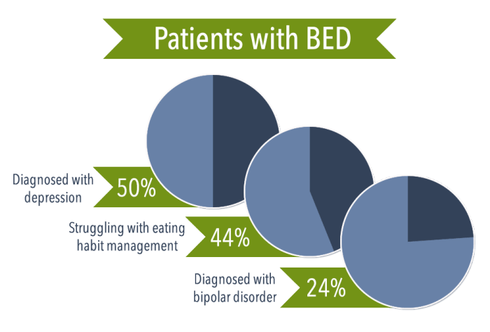
Binge eating disorder displays a high comorbidity with mood and affective disorders. Infographic courtesy of American Addiction Centers.

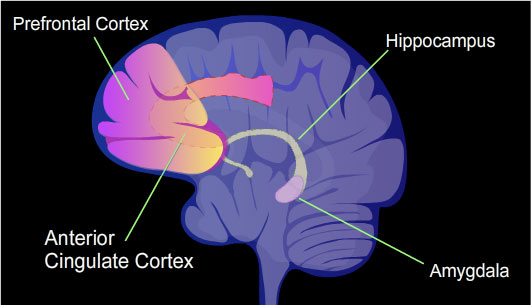
ADHD is characterized by inability to focus, hyperactivity, and impulsivity, and substance abuse involves cravings and patterns of losing control followed by regret. These patterns of mental and physiological sympoms resemble those seen in patients with binge eating disorder. Kaplan and other researchers are linking the neurological patterns observed in these disorders to better understand BED.

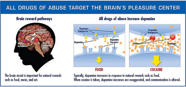
Researchers have found that the neurological pathways become active when a patient with binge eating disorder is provided with a food-related stimulus. Individuals with the eating disorder are more sensitive to food-related rewards than most people. Researchers have also identified a genetic basis — certain genes make individuals more susceptible to reward and thus more likely to engage in binges.

Because patients with ADHD exhibit similar neurological patterns, doctors are looking to drugs already approved by the FDA to treat ADHD as possible treatments for binge eating disorder. The first of these approved drugs, Vyvanse, has proven not much better than the traditional form of treatment, cognitive behavioral therapy, a form of talk therapy that aims to identify and correct dysfunctions in behavior and thought patterns that lead to disordered behaviors.

Another drug, however, proved promising in a study conducted by Kaplan and his colleagues. The ADHD drug methylphenidate, combined with CBT, led to significant clinical outcomes — pateints engaged in less binges and cravings and body mass index decreased. Kaplan argues that the most effective treatment would reduce binges, treat physiological symptoms like obesity, improve psychological disturbances like low self-esteem, and, of course, be safe. So far, the combination of psychostimulants like methylphenidate and CBT have met these criteria.

Kaplan emphasized a need to make information about binge eating disorder and its treatments more available. Most individuals currently being treated for BED do not obtain treatment knowing they have an eating disorder — they are usually diagnosed only after seeking help with obesity-related health issues or help in weight loss. Making clinicians more familiar with the disorder and its associated behaviors as well as encouraging patients to seek treatment could prove instrumental in combating the current healthcare issue of obesity.

By Sarah Haurin

Mice, motor learning, and making decisions

Advanced imaging techniques allow neuroscientists to better understand how the motor outputs we observe are created in the brain.

Early understandings of the brain viewed it as a black box that takes sensory input and generates a motor response, with the in-between functioning of the brain as a mystery.

Takaki Komiyama is curious about how the brain produces the stereotypical movements characteristic of motor learning.

Takaki Komiyama of the University of California, San Diego is curious about the relationship between sensory input, motor output, and what happens in between. “What fascinates me the most is the flexibility of this dynamic… this flexibility of the relationship between the environment and the brain is the key element of my research,” Komiyama said to an audience of Duke neuroscience researchers.

Komiyama and his lab have designed experiments to watch how the brain changes as mice learn. Specifically, they train mice to complete a lever-pushing task in response to an auditory stimulus and then use an advanced imaging technique to watch the activity of specific populations of neurons.

Komiyama based his experimental design on a hallmark of motor learning: An “expert” mouse will hear the auditory stimulus and produce a motor response that is exactly the same each time. Komiyama’s team was curious about how these reproducible movements are learned.

Focusing on the primary motor cortex, called M1 for short, Komiyama observed many different neuronal firing patterns as the mouse learned the motion of lever-pushing. As the mouse ventured into “expert” territory, usually after about two weeks of training, this variation was replaced by an activity pattern that is the same from trial to trial. In addition to being consistent, this final pattern starts earlier after the stimulus and takes less time to complete than earlier patterns. In other words, during learning, the brain tries out different pathways for the goal action and then converges on the most efficient way of producing the desired response.

Komiyama then turned his focus to M2, the secondary motor cortex, which he observed to be one of the last areas activated during early learning trials but one of the first activated during late trials. To test M2’s role in learning, Komiyama inactivated the region in trained mice and subjected them to the same stimulus-motor response trial.

The mice with inactivated M2’s missed more trials, took longer to initiate movement, and completed the lever pushing less efficiently. Essentially, the mice behaved as if they had never learned the movement, suggesting that M2 is crucial for coordinating learned motor behavior.

In addition to identifying crucial patterns of motor learning, Komiyama and his team are working to understand decision making. After designing a more complex lever-pushing task that required pushing a joystick in different directions depending on the visual stimulus, Komiyama observed the mice’s accuracy plateaued around 60%.

The mice’s internal biases prevented them from achieving better results in the visual stimuli task.

Komiyama hypothesized that this pattern of inaccuracy could be explained by the mice’s internal biases from previous trials’ outcomes. He designed a statistical model that incorporated the previous trials’ outcomes. With further testing, the model accurately predicted the mice’s wrong choices.

The posterior parietal cortex (PPC) is an area of the brain that has been found to be involved in decision making tasks. Komiyama observed neurons in the PPC that predicted which direction the mice would push the joystick. In addition to being active before the motor response during trials, these neurons were also active in the time between trials.

Seeing this as a neural correlate for internal biases, Komiyama hypothesized that inactivating this region would decrease the influence of bias on the mice’s choices. Sure enough, inactivating the PPC led to more accurate responses in the mice, thus confirming the PPC as a neural source of bias.

 By Sarah Haurin

How A Bat’s Brain Navigates

Most of what we know about how the hippocampus, a region of the brain associated with memory formation and spatial representations, comes from research done on rodents. Rat brains have taught us a lot, but researchers in Israel have found an interesting alternative model to understanding how the hippocampus helps mammals navigate: Bats.

The Egyptian fruit bat proved the perfect subject for studies of mammalian navigation.

Weizmann Institute neurophysiologist Nachum Ulanovsky, PhD, and his team have looked to bats to understand the nuances of navigation through space. While previous research has identified specific cells in the hippocampus, called place cells, that are active when an animal is located in a specific place, there is not much literature describing how animals actually navigate from point A to point B.

Nachum Ulanovsky

Ulanovsky believes that bats are an ingenious model to study mammalian navigation. While bats have the same types of hippocampal neurons found in rats, the patterns of bats’ neurons’ firings more closely match that of humans than rats do.

Ulanovsky sought to test how bats know where they are going. Using GPS tracking equipment, his team found that wild bats that lived in a cave would travel up to 20 kilometers to forage fruit from specific trees. Night after night, these bats followed similar routes past perfectly viable food sources to the same tree over and over again.

The understanding of hippocampal place cells firing at specific locations doesn’t explain the apparent guided travel of the bat night after night, and other explanations like olfactory input do not explain why the bats fly over good food sources to their preferred tree.

The researchers designed an experiment to test how bats encode the 3D information necessary for this navigation. By letting the bats fly around and recording brain activity, Ulanovsky and team found that their 3D models are actually spherical in shape. They also found another type of hippocampal cells that encode the orientation the bat is facing. These head direction cells operate in a coordinate system that allows for a continuity of awareness of its orientation as the animal moves through space.

http://www.cell.com/cms/attachment/2091916945/2076305003/gr1_lrg.jpg

Ulanovsky found bats relied on memory to navigate toward the goal.

To understand how the bats navigate toward a specific goal, the researchers devised another experiment. They constructed a goal with a landing place and a food incentive. The bat would learn where the goal was and find it. In order to test whether the bats’ ability to find the goal was memory-based, or utilized the hippocampus, the researchers then conducted trials where the goal was hidden from the bats’ view.

To test whether the bats’ relied on memory, the Ulvanosky team measured the goal direction angle, or the angle between the bat’s head orientation and the goal. After being familiarized with the location of the goal, the bats tended toward a goal-direction angle of zero, meaning they oriented themselves toward the goal even when the goal was out of sight.

Continued research identified cells that encode information about the distance the bat is from the goal, the final piece allowing bats to navigate to a goal successfully. These hippocampal cells selectively fire when the bat is within specific distances of the goal, allowing for an awareness of location over distance.

While Ulanovsky and his team have met incredible success in identifying new types of cells as well as new functions of known cells in the hippocampus, further research in a more natural setting is required.

“If we study only under these very controlled and sterile environments, we may miss the very thing we are trying to understand, which is behavior,” Ulanovsky concluded.

By Sarah Haurin

Hearing Loss and Depression Are Connected

Jessica West is a PhD candidate in sociology.

Jessica West, a PhD student in sociology at Duke, has found that hearing loss creates chronic stress but that high levels of social support – from family, friends and others – can help alleviate depression. Given that hearing loss is a growing social and physical health problem, her study suggests a need for increased vigilance regarding hearing loss among older adults, West said.

Her study was published in the November issue of Social Science & Medicine and is available here.

Here, West discusses her research.

Your research examines the correlation between hearing loss and depression. That seems a logical connection: why study it in the way you did?

Despite how common hearing loss is, it is actually quite understudied. A handful of studies have looked at the relationship between hearing loss and mental health over time, but the results from these studies are mixed: some find a relationship between hearing loss and more depressive symptoms, while others do not. On top of the mixed findings, most studies have been based overseas, and studies based in the U.S. have tended to use state-specific datasets, like the Alameda County Study, which drew from Oakland and Berkeley, CA.

I use the Health and Retirement Study, which is nationally representative of adults aged 50 and older in the U.S., and therefore more generalizable to the U.S. population.

I frame hearing loss as a physical health stressor that can impact mental health, and that social support can alter this relationship by preventing a person from experiencing stress or reducing the severity of a reaction to it. To the best of my knowledge, this is the first paper to link hearing loss to health outcomes in this way.

What might surprise people about your findings?

More than one-fifth of the people in my sample have fair to poor hearing (23.12% or 1,405 people in the first wave). Hearing loss is really common in the U.S.

Also, I found that social support is most beneficial in easing the burden of hearing loss among people with significant hearing loss. Overall, this suggests that hearing loss is a chronic stressor in people’s lives and that responses to this stressor will vary by the level of social resources that people have available to them.

What does ‘social support’ mean in real terms? What can the family and friends do for a person with hearing loss to help them?

For people with hearing loss, it’s important that they feel able to lean on, talk to, and rely on family, friends, spouses or partners, and children. And going a step further, people with hearing loss need to know that these important people in their lives truly understand the struggles they face. What this means is that people with hearing loss can benefit quite a lot from having a network of people that they feel comfortable discussing things with or reaching out to when needed.

Do people with hearing loss have adequate mental health resources or care available to them?

My research shows that social support is really important for people with hearing loss. One suggestion I make in my paper is that audiologic – or hearing — rehabilitation programs could include educational training for significant others, like spouses or friends, to emphasize the importance of supporting people with hearing impairment. Audiologists, primary care physicians, family, and friends are all key resources that could be targeted in such rehabilitation programs.

 What is your next project related to hearing loss?

 I am currently working on several projects related to hearing loss. In one, I am looking at the relationship between an individual’s hearing loss and his/her spouse’s mental health outcomes. Few population-based studies have examined the relationship between hearing loss and spousal mental health longitudinally, so I hope this study will shed light on the experience of spousal disability within marriages.

Another project I am working on looks at hearing loss from a life course perspective. In other words, I am looking at people who self-reported hearing loss before the age of 16 and seeing how their hearing loss influenced their marriages, academics and careers. A better understanding of how early life hearing loss influences later life outcomes has implications for earlier identification of hearing loss and/or the use of assistive technology to help people remain socially, academically, and economically engaged.

CITATION: West, Jessica S. 2017. “Hearing Impairment, Social Support, and Depressive Symptoms among U.S. Adults: A Test of the Stress Process Paradigm.” Social Science & Medicine 192(Supplement C):94-101.

 Read the paper 

Guest post by Eric Ferreri, News and Communications

Dopamine, Drugs, and Depression

The neurotransmitter dopamine plays a major role in mental illnesses like substance abuse disorders and depressive disorders, as well as a more general role in reward and motivational systems of the brain. But there are still certain aspects of dopamine activity in the brain that we don’t know much about.

Nii Antie Addy and his lab are interested in the role of dopamine in substance abuse and mood disorders.

Duke graduate Nii Antie Addy, PhD, and his lab at Yale School of Medicine have been focusing on dopamine activity in a specific part of the brain that has not been studied: the ventral tegmental area (VTA).

To understand the mechanisms underlying this association, Addy and his team looked at cue-induced drug-seeking behavior. Using classical conditioning, rats can be trained to pair certain cues with the reward of drug administration. When a rat receives an unexpected award, dopamine activity increases. After conditioning, dopamine is released in response to the cue more  than to the drug itself. Looking at the patterns of dopamine release in rats who are forced to undergo detoxification can thus provide insight into how these cues and neurotransmitter activity relate to relapse of substance abuse.

When rats are taught to self-administer cocaine, and each administration of the drug is paired with the cue, after a period of forced detoxification, the rodents continue to try to self-administer the drug, even when the drug is withheld and only the cue is presented. This finding again demonstrates the connection between the cue and drug-seeking behavior.

Studying the activity in the VTA gave additional insights into the regulation of this system. During the period of abstinence, when the rodents are forced to detox, researchers observed an increase in the activity of cholingergic neurons, or neurons in the brain system that respond to the neurotransmitter acetylcholine.

Using these observations, Addy and his team sought to identify which of the various receptors that respond to acetylcholine can be used to regulate the dopamine system behind drug-seeking behaviors. They discovered that a specific type of acetylcholine receptor, the muscarinic receptor, is involved in more general reward-seeking behaviors and thus may be a target for therapies.

Using Isradipine, a drug already approved by the FDA for treatment of high blood pressure, Addy designed an experiment to test the role of these muscarinic receptors. He co-opted the drug to act as a calcium antagonist in the VTA and thus increase dopamine activity in rodents during their forced detox and before returning them to access to cocaine. The outcome was promising: administration of Isradipine was associated with a decrease in the coke-seeking behavior of rodents then placed in the chamber with the cue.

The understanding of the role of cholinergic neurons in regulation of dopamine-related mental illnesses like substance-abuse also contributes insights into depressive and anxiety disorders. If the same pathway implicated in cue-induced drug-seeking were involved in depressive and anxious behaviors, then increasing cholinergic activity should increase pro-depressive behavior.

Addy’s experiment yielded exactly these results, opening up new areas to be further researched to improve the treatment of mood disorders.

Post by Sarah Haurin


Meet Africa’s Bird Master of Vocal Imitation

The red-capped robin-chat (Cossypha natalensis) can mimic the songs and calls of dozens of other bird species – even their duets, says Duke researcher Tom Struhsaker.

The red-capped robin-chat (Cossypha natalensis) can mimic the songs and calls of dozens of other bird species – even their duets, says Duke researcher Tom Struhsaker.

Singing a duet in a foreign language isn’t just for opera stars — red-capped robin-chats do it too. These orange-brown birds with grey wings can imitate the sounds of 40 other bird species, even other species’ high-speed duets.

The latter finding comes from Tom Struhsaker, adjunct professor of evolutionary anthropology at Duke. Struhsaker didn’t set out to study robin-chats. His interest in their vocal abilities developed while studying monkeys in Kibale Forest in Uganda, where he lived for nearly two decades from 1970 to 1988.

Their typical song “sounds like a long, rambling human-like whistle,” Struhsaker said. But during the 18 years he spent studying and living in Kibale, Struhsaker also heard these birds impersonate the tambourine-like courtship call of the crested guineafowl, the crow of a rooster, and the “puweepuweepuwee” of a crowned eagle, among others.

“The robin-chat’s ability to imitate is so good that many a bird watcher has looked skyward vainly searching for a crowned eagle performing its aerial display, when in fact the source of the eagle’s undulating whistle was a robin-chat in the nearby understory,” Struhsaker said.

He also noticed that if he whistled, eavesdropping robin-chats would approach and call back, and if he tweaked the pitch and sequence of notes in his whistle, the birds sometimes changed their reply.

This suggests red-capped robin-chats may be lifelong learners, unlike many other bird species that only learn songs during critical time windows, Struhsaker said.

But the robin-chat doesn’t stop at mimicking others’ solo performances. Notably, Struhsaker also heard them imitate the duet of the black-faced rufous warbler.

Black-faced rufous warblers sing a rapid-fire “seee-oooo-ee” duet with their mates. The two birds take turns such that the male sings the “seee,” the female chimes in with the “oooo” and the male fires back with the final “ee,” with no pauses between the three notes. The partners sing back and forth so seamlessly that they are often mistaken for a single bird.

“In order to do this, birds have an incredibly rapid reaction time, much greater than that of humans,” Struhsaker said.

On two occasions he heard single robin-chats sing both the male and female parts of the warbler duet by themselves. On another occasion he heard two robin-chats make music together as the warblers do, with one singing the male warbler’s part and the other singing the female part.

“This suggests these birds have an unusually high level of auditory perception and reaction time and cognitive ability,” Struhsaker said.

CITATION:  “Two Red-Capped Robin-Chats Cossypha Natalensis Imitate Antiphonal Duet of Black-Faced Rufous Warblers Bathmocercus rufus,” Thomas Struhsaker. Journal of East African Natural History, Dec. 2017. https://doi.org/10.2982/028.106.0201.


On a Mission to Increase Exercise

Dr. Zachary Zenko of the Center for Advanced Hindsight at Duke is on a mission to get people to exercise. He shared this mission and his research aimed at achieving this mission at Duke’s Exercise and the Brain Symposium on December 1st.

Dr. Zenko started out with a revealing statistic: Only 1 in 10 people meet the United States activity guidelines for exercise. While I wasn’t completely shocked at this fact as the U.S. is known for its high rates of obesity and easy access to fast-food, I was definitely eager to learn more about how Dr. Zenko planned to fight this daunting statistic.

Next, Dr. Zenko pointed out a common assumption in exercise psychology: if people know how good exercise is for them, they will exercise. However, this hasn’t proven to be true. Most people already know that they should be exercising, but don’t. And those that do often quickly drop out.

Then Dr. Zenko began to break down Dual-Process Theory in Behavioral Economics. Type 1 Processes are those that are fast and non-conscious, while Type 2 Processes are those that are controlled and conscious. While research on exercise usually focuses on Type 2 processes, Zenko believes that we must focus on both.

Ideally, exercise would involve the affect heuristic, which is a mental shortcut in which an emotional response drives an individual. This heuristic involves Type 1 processes. Dr. Zenko’s goal was to shift away from only considering Type 2 processes, and instead focus on using Type 1 processes to make exercise more appealing.

How did he propose doing this? By continually decreasing the difficulty of exercise. By changing the slope of the intensity of a workout and having a continually declining heart rate, exercisers could have a more pleasant experience. In addition, this positive experience could influence memory and make an individual more likely to exercise in the future.

Dr. Zenko put this hypothesis to the test by having unfit adults exercise while continually decreasing the intensity throughout the workout. While test subjects exercised, he measured the amount of pleasure experienced by asking “How do you feel right now?” at certain intervals. This new exercise method  has the most potential when starting at the highest intensity levels because it leaves more room to change the slope of the workout intensity throughout, leading to an overall more pleasurable workout.

Looking forward, this new method of exercise could possibly change the way we think about exercise. It may not only involve doing the right amount of exercise, but also doing the right kind of exercise that leaves us more likely to exercise in the future. Considering that traditional methods of promoting exercise, such as educating people about its benefits, have not been particularly successful thus far, Dr. Zenko’s method is very exciting.

Dr. Zenko wrapped up his talk by suggesting that people consider exercise prescriptions that are safe, effective, pleasant and enjoyable. As exercise has become a huge part of my weekly routine throughout college, I will definitely take this advice to heart. Maybe even look out for me lowering my intensity during a workout soon in a gym near you?

By Nina Cervantes

Exercise is Good for Your Head and Might Fight Alzheimer’s

Recent studies have confirmed that exercising is just about the best thing you can do for your brain health.

Dan Blazer, MD is a psychiatrist who studies aging.

On Dec. 1 during the DIBS event, Exercise and the Brain, Duke psychiatrist Dan Blazer reported findings about the relationship between physical activity and brain health. After lots of research, study groups at the National Academy of Medicine  concluded that their number one recommendation to those experiencing “cognitive aging” is exercise.

Processing speed, memory, and reasoning decline over time in every one of us. But thankfully, simple things like riding a bike or playing pick up basketball can help keep our minds fresh and at their best possible level.

One cool thing a committee conducting the research did to advertise their findings was create keychains saying “take your brain for a walk.” There’s a little image of a brain with legs walking. They wanted to get the word out that physical activity has another benefit than just staying in shape — it can also support your cognitive health.

However, the committees are having a hard time motivating people to exercise in the first place. Even after hearing their findings, it’s not like people everywhere are suddenly going to get off their couches and hit the gym. A world with healthier people — both physically and mentally — sounds nice, but getting there is much more than a matter of publishing these studies.

And, as always, too much of a good thing can make it harmful. While there does seem to appear a potential “biological gradient,” where greater physical activity correlated with better outcomes, you can’t just run a marathon every day of the week and then ~boom~ aging hardly affects your brain anymore. You don’t want to do that to yourself. Just get a healthy amount of exercise and you’ll be keeping your brain young and smart.

One of the best parts about why exercising is so great for you and your brain is because it helps you sleep (and we all know how important sleep is). If you ever have trouble going to bed or are having disrupted sleeps, physical activity could be your savior. It’s a much healthier option for your brain than taking stuff like melatonin, and you’ll get fit in the process.

Regarding exercising and Alzheimer’s, a disease where vital mental functions deteriorate, studies have unfortunately been insufficient to conclude anything. But if getting Alzheimer’s is your worst fear, I’m sure staying active can’t hurt as a preventative. More research on this topic is being conducted as we speak.

When is the best time to start exercising, in order to reap the maximum cognitive benefits, you ask? Well, the sooner the better. As Blazer said, “exercising helps in maintaining or improving cognitive function in later life,” so you better get on that. Go outside and get moving!

Will Sheehan      Post by Will Sheehan


How We Know Where We Are

The brain is a personalized GPS. It can keep track of where you are in time and space without your knowledge.

The hippocampus is a key structure in formation of memories and includes cells that represent a person’s environment.

Daniel Dombeck PhD, and his team of researchers at Northwestern University have been using a technique designed by Dombeck himself to figure out how exactly the brain knows where and when we are. He shared his methods and findings to a group of researchers in neurobiology at Duke on Tuesday.

Domeck and his lab at Northwestern are working at identifying exactly how the brain represents spatial environments.

The apparatus used for these experiments was adapted from a virtual reality system. They position a mouse on a ball-like treadmill that it manipulates to navigate through a virtual reality field or maze projected for the mouse to see. Using water as a reward, Dombeck’s team was able to train mice to traverse their virtual fields in a little over a week.

In order to record data about brain activity in their mice as they navigated virtual hallways, Dombeck and his team designed a specialized microscope that could record activity of single cells in the hippocampus, a deep brain structure previously found to be involved in spatial navigation.

The device allows researchers to observe single cells as a mouse navigates through a simulated hallway.

Previous research has identified hippocampal place cells, specialized cells in the hippocampus that encode information about an individual’s current environment. The representations of the environment that these place cells encode are called place fields.

Dombeck and his colleague Mark Sheffield of the University of Chicago were interested in how we encode new environments in the hippocampus.

Sheffield studied the specific neural mechanisms behind place field formation.

After training the mice to navigate in one virtual environment, Sheffield switched the virtual hallway, thus simulating a new environment for the mouse to navigate.

They found that the formation of these new place cells uses existing neural networks initially, and then requires learning to adapt and strengthen these representations.

After identifying the complex system representing this spatial information, Dombeck and colleagues wondered how the system of representing time compared.

Jim Heys, a colleague of Dombeck, designed a new virtual reality task for the lab mice.

In order to train the mice to rely on an internal representation of passing time, Heys engineered a door-stop task, where a mouse traversing the virtual hallway would encounter an invisible door. If the mouse waited 6 seconds at the door before trying to continue on the track, it would be rewarded with water. After about three months of training the mice, Heys was finally able to collect information about how they encoded the passing of time.

Heys indentified cells in the hippocampus that would become active only after a certain amount of time had passed – one cell would be active after 1 second, then another would become active after 2 seconds, etc. until the 6-second wait time was reached. Then, the mouse knew it was safe to continue down the hallway.

When comparing the cells active in each different task, Dombeck and Heys found that the cells that encode time information are different from the cells that encode spatial information. In other words, the cells that hold information about where we are in time are separate from the ones that tell us where we are in space.

Still these cells work together to create the built-in GPS we share with animals like mice.

By Sarah Haurin

Duke’s Researchers Are 1 Percent of the Top 1 Percent

This year’s listing of the world’s most-cited researchers is out from Clarivate Analytics, and Duke has 34 names on the list of 3,400 researchers from 21 fields of science and social science.

Having your publication cited in a paper written by other scientists is a sign that your work is significant and advances the field. The highly-cited list includes the top 1 percent of scientists cited by others in the years 2005 to 2015.

“Citations by other scientists are an acknowledgement that the work our faculty has published is significant to their fields,” said Vice Provost for Research Lawrence Carin. “In research, we often talk about ‘standing on the shoulders of giants,’ as a way to explain how one person’s work builds on another’s. For Duke to have so many of our people in the top 1 percent indicates that they are leading their fields and their work is indeed something upon which others can build.”

In addition to the Durham researchers, Duke-NUS, our medical school in Singapore,  claims another 13 highly cited scientists.

The highly-cited scientists on the Durham campus are:

Barton Haynes

CLINICAL MEDICINE
Robert Califf
Christopher Granger
Kristin Newby
Christopher O’Connor
Erik Magnus Ohman
Manesh Patel
Michael Pencina
Eric Peterson

ECONOMICS AND BUSINESS
Dan Ariely
John Graham
Campbell Harvey

Drew Shindell

ENVIRONMENT/ECOLOGY
John Terborgh
Mark Wiesner

GEOSCIENCES
Drew Shindell

IMMUNOLOGY
Barton Haynes

MATHEMATICS
James Berger

Georgia Tomaras

Georgia Tomaras

MICROBIOLOGY
Bryan Cullen
Barton Haynes
David Montefiori
Georgia Tomaras

PHARMACOLOGY & TOXICOLOGY
Robert Lefkowitz

PHYSICS
David R. Smith

PLANT AND ANIMAL SCIENCE
Philip Benfey

Terrie Moffitt

Terrie Moffitt

PSYCHIATRY & PSYCHOLOGY
Angold, Adrian
Caspi, Avshalom
Copeland, William E
Costello, E J
Dawson, Geraldine
Keefe, Richard SE
McEvoy, Joseph P
Moffitt, Terrie E

SOCIAL SCIENCES (GENERAL)
Deverick Anderson
Kelly Brownell
Michael Pencina

Page 13 of 28

Powered by WordPress & Theme by Anders Norén