Research Blog

Following the people and events that make up the research community at Duke

Students exploring the Innovation Co-Lab

David Carlson: Engineering and Machine Learning for Better Medicine

How can we even begin to understand the human brain?  Can we predict the way people will respond to stress by looking at their brains?  Is it possible, even, to predict depression based on observations of the brain?

These answers will have to come from sets of data, too big for human minds to work with on our own. We need mechanical minds for this task.

Machine learning algorithms can analyze this data much faster than a human could, finding patterns in the data that could take a team of researchers far longer to discover. It’s just like how we can travel so much faster by car or by plane than we could ever walk without the help of technology.

David Carlson Duke

David Carlson in his Duke office.

I had the opportunity to speak to David Carlson, an assistant professor of Civil and Environmental Engineering with a dual appointment at the Department of Biostatistics and Bioinformatics at Duke University.  Through machine learning algorithms, Carlson is connecting researchers across campus, from doctors to statisticians to engineers, creating a truly interdisciplinary research environment around these tools.

Carlson specializes in explainable machine learning: algorithms with inner workings comprehensible by humans. Most deep machine learning today exists in a “black box” — the decisions made by the algorithm are hidden behind layers of reasoning that give it incredible predictive power but make it hard for researchers to understand the “why” and the “how” behind the results. The transparent algorithms used by Carlson offer a way to capture some of the predictive power of machine learning without sacrificing our understanding of what they’re doing.

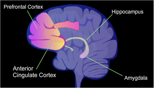
In his most recent research, Carlson collaborated with Dr. Kafui Dzirasa, associate professor of psychiatry and behavioral sciences and assistant professor in neurobiology and neurosurgery, on the effects of stress on the brains of mice, trying to understand the underlying causes of depression.

“What’s happening in neuroscience is the amount of data we’re sorting through is growing rapidly, and it’s really beginning to outstrip our ability to use classical tools,” Carlson says. “A lot of these classical tools made a lot more sense when you had these small data sets, but now we’re talking about this canonically overused word, Big Data”

With machine learning algorithms, it’s easier than ever to find trends in these huge sets of data.  In his most recent study, Carlson and his fellow researchers could find patterns tied to stress and even to how susceptible a mouse was to depression. By continuing this project and looking at new ways to investigate the brain and check their results, Carlson hopes to help improve treatments for depression in the future.

In addition to his ongoing research into depression, Carlson has brought machine learning to a number of other collaborations with the medical center, including research into autism and patient care for diabetes. When there’s too much data for the old ways of data analysis, machine learning can step in, and Carlson sees potential in harnessing this growing technology to improve health and care in the medical field.

“What’s incredibly exciting is the opportunities at the intersection of engineering and medicine,” he said. “I think there’s a lot of opportunities to combine what’s happening in the engineering school and also what’s happening at the medical center to try to create ways of better treating people and coming up with better ways for making people healthier.”

Guest Post by Thomas Yang, a junior at North Carolina School of Math and Science.

Kathleen Pryer: A Passion for the Little-Loved Fern

Most people don’t see in ferns the glory and grandeur of the mighty angiosperms — the flowering plants — but to those who can, ferns may seem like the only thing you could spend your time researching.

Fei-wei Li, Kathleen Pryer

Kathleen Pryer, with former graduate student Fei-Wei Li. (Duke photography)

Kathleen Pryer, a professor of biology at Duke, is an example of one of these people who found their calling in ferns. But she didn’t know it would be ferns from the beginning.

As an undergraduate, she had thought she wanted to be an animal behaviorist, having read books by Jane Goodall, so she enrolled in McGill University in Montreal (she’s Canadian by the way) in the animal behavior program and didn’t end up taking a single botany course until her senior year.  For her final project she worked with snails, a starkly slow endeavor, she thought. Slower even than ferns. After getting her degree in animal behavior, she decided she wanted a masters working with plants, but before jumping right in with only one class’ worth of experience with plants, she worked as a technician for a budding ecologist.  While working there, the ecologist’s wife, who did her masters on ferns, took her on a trip to the annual meeting of the botanical society of America in Blacksburg, VA, a 13-hour trip.

In Virginia, she went on a 2-day field trip through Virginia, led by fern expert Warren Wagner, finding ferns with 107 other people who were mad about ferns.

“It was just serendipity really.”

After that, the idea of ferns stuck, and she’s been working with them ever since.  She’s gotten the chance to name or rename many species of fern, and she created the genus Gaga, named after the singer.  Another new genus she found is soon to be named Mandela by her as well; a nice change from the usual names of “old white guys,” given to new genera, she said.

Through it all though, Pryer is most proud of a paper from 2001, which showed that all modern ferns originated from a central progenitor, showing that they aren’t as archaic as most people think. That paper made the cover of Nature, and has been cited hundreds of times since.

In the end, I guess it’s really hard to tell where you’ll end up.  If an aspiring animal behaviorist can jump to the world of ferns and make a successful career out of it, surely there’s hope for the rest of us too.  In the end, all that matters is if you’re doing what you love, and as for Kathleen Pryer, she’ll keep doing what she loves as long as there’s a “chair and a microscope” for her to sit at.

Isaac PoarchGuest Post by Isaac Poarch, a senior at the North Carolina School of Science and Math

Leonor Corsino: Research and Care Toward Alleviating Diabetes

Dr. Leonor Corsino works to relieve the prevalent issues regarding diabetes and obesity. An endocrinologist and professor at the Duke School of Medicine, her passions lie in understanding the struggles that diabetics face through comprehensive patient care and communication.

Leonor Corsino

Her interests in endocrinology began at a young age. She grew up watching her father and many other members of her family challenged with balancing a normal life alongside diabetes. When she progressed to medical school, she was fascinated by the workings of the hormonal system, one of the most neatly regulated of all the biological systems.

“When it works in harmony, everything is perfect, but when something goes off, it affects many other organs,” she says.

Corsino believes that patient-provider communication is the most important thing for the makings of a good endocrinologist. As the Associate Director for Masters in Biomedical Sciences, she aims to teach students pursuing a career as a healthcare professional to be empathetic. “[A student] can be the smartest person in world, but if [they] don’t know how to communicate with the patient, their ability to provide care gets compromised.”

Another factor that plays a role in providing good patient care is the amount of time available to treat each person, according to Corsino. Although Corsino always aspires to treat her patients to the best of her abilities, occasionally, the limited time she has with each individual can impose difficulties with empathizing and treating patients. However, many regular patients don’t mind when their appointments are delayed because they know that they will receive better care when they are able to get her undivided attention.

Beyond her clinical expertise, Corsino’s research focuses on similar issues. Through her research, she intends to improve the healthcare of minorities in the country, as they are the groups that are most affected by diabetes. In the past 11 years, she has introduced interventions to improve and maintain weight loss and worked with pharmaceutical companies to look at potential drugs to treat diabetes. She intends to answer the questions “How do we motivate people to exercise? What is the reason some people struggle with diabetes and other people don’t?”

Corsino has found that biological factors play an equal role to environmental factors in the risk of getting diabetes. Sometimes, even if a patient strictly adheres to the prescribed treatment, they still don’t see the same results and progress as others do. This distinction can be attributed to things like differences in fat distribution and insulin resistance.

In her work, Dr. Corsino tries to alleviate the stress and difficulties that those with diabetes and obesity encounter. As a doctor and professor, she inspires others to pursue a career in public health and provide healthcare to those who need it.

Sindhu PolavaramGuest Post by Sindhu Polavaram, a senior at North Carolina School of Science and Math

Martin Brooke: Mentoring Students Toward an X Prize for Ocean Robotics

We know less about the ocean floor than the surface of the moon. As one of the most unexplored areas of the world, multiple companies have begun to incentivize ingenuity towards exploring the oceans. Among these organizations are the Gates Foundation, the National Academy of Sciences, and X Prize.

XPrize team at Duke

Martin Brooke, second from left, and the student team with their giant drone.

Martin Brooke, an Associate Professor of Electrical and Computer Engineering at Duke, is presently leading a group of students who are working on mapping the ocean floor in an efficient way for the X Prize challenge.

Brooke said “open ended problems where you don’t know what to do” inspire him to do research about ocean engineering and design.

Martin Brooke

Martin Brooke

Collaborating with professors at the Duke Marine Lab that “strap marine sensors on whales” was a simple lead-in to starting a class about ocean engineering a few years ago. His teaching philosophy includes presenting the students with problems that make them think, “we want to do this, but we have no idea how.”

Before working on a drone that drops sensor pods down into the ocean to map the ocean floor, Brooke and his students built a sensor that could be in the ocean for a month or more and take pH readings every five seconds for a previous X Prize challenge.

Addressing the issues that many fisheries faced, he told me that he met an oyster farmer in Seattle who wished that there were pH sensors in the bay because sometimes tides bring in “waves of high pH water into the sound and kill all of the oysters without warning.” Citing climate change as the cause for this rise in pH, Brooke explained how increased carbon dioxide in the air dissolves into the water and raises the acidity. Emphasizing how “there’s not enough data on it,” it’s clear that knowing more about our oceans is beneficial economically and ecologically.

Guest Post by Sofia Sanchez, a senior at North Carolina School of Math and Science

Anita Layton: A Model of STEM Versatility

Using mathematics to model the kidney and its biological systems is a field of study located at the intersection of two disciplines.

Anita Layton is a math professor at Duke. (Photo by Chris Hildreth, Duke Photography)

But for Duke’s Anita Layton, PhD, the Robert R. and Katherine B. Penn Professor of Mathematics and a professor of biomedical engineering, that just adds to the fun of it.

Growing up, with her father as the head of mathematics at her school, she was always told she was going to be a mathematician just like him. So she knew that was the last thing she wanted to do.

When Layton arrived as an undergraduate at Duke, she began a major in physics, but she seemed rather cursed when it came to getting correct results from her experiments. She settled for a BA in physics, but her academic journey was far from over. She had also taken a computer science course at Duke and fallen in love with it. If an experiment went wrong “things didn’t smell or blow up” and you could fix your mistake and move on, she said.

While pursuing her PhD in computer science at the University of Toronto, Layton was performing very math-oriented computer science, working with and analyzing numbers. However, it would be a while before biology entered the mix

While she was never good at dissections, she told me she was always good at understanding things that ‘flow’ and she came to the realization that blood is something that flows. She thought, “Hey, I can do that.

Anita Layton, Duke

Anita Layton, Ph.D.

Layton began creating programs that could solve the equations that model blood flow quickly, using her background in computer science. She then started learning about physiology, focusing on the renal system, and making models

It was a journey that took her to many different places, with pit stops and U-turns throughout many different fields. Had Layton stuck with just physics or computer science or math, she never would have ventured out and found this field that she is an expert in now.

It’s her interest in many different fields that has set Layton apart from many other people in the STEM field. In learning a wide variety of things, she has gotten better at computer science, mathematics, biology, physics, and more

When asked about what advice she would give her younger self, or any young person going into college, it would be to do just that: “Learn more things that you’re not good at.” She encouraged just taking a chemistry or biology class once in a while, or a philosophy course that makes you think in ways that you don’t normally. It’s often in those classes that you unearth things that can truly set your life in a completely different direction, Layton said, and she’s living proof of that.

Cecilia Poston, NCSSM

Cecilia Poston

Guest Post by Cecilia Poston, a senior at North Carolina School of Science and Math

Glitter and Jell-O Reveal the Science of Oobleck

A black and white image showing a circular disk dropping into a container of oobleck

Mixing black glitter with oobleck allowed researchers to track the movement of individual cornstarch particles after a sudden impact. A computer program locked onto pieces of glitter and illustrated their motion. Credit: Melody Lim.

What do gelatin and glitter have to do with serious science? For some experiments, a lot! Duke alumna Melody Lim used jiggly Jell-O and a just a pinch of glitter to solve a scientific mystery about the curious goo many like to call oobleck.

To the uninitiated, oobleck is almost magic. The simple mixture of cornstarch and water feels solid if you squeeze it, but moments later runs through your fingers like water. You can dance across a bathtub full of oobleck, but stand still for too long and you will be sucked into a goopy mess. Not surprisingly, the stuff is a Youtube favorite.

Oobleck is an example of what scientists call a non-Newtonian fluid, a liquid whose viscosity – how easily it changes shape and flows – depends upon the force that is applied. But exactly how it is that this material switches from solid to liquid and back again has remained a mystery to scientists.

A piece of gelatin being squeezed viewed through a circular polarizer

This blogger mixed up a batch of jello to see the photoelastic effect for herself. When viewed with polarized light – from an iPhone screen and a circular polarizer – the jello changes color when squeezed.

“Water is simple to understand, and so is cornstarch,” said Lim, ’16, who is currently a graduate student at the University of Chicago. “However, a combination of the two produces this ‘liquid’ that ripples and flows, solidifies beneath your feet if you run on it, then turns back into a liquid if you stop running and stand still. I wanted to know why.”

The question beguiling scientists was whether sudden impact causes the cornstarch particles to “jam” into a solid like cement, or whether the suspension remains liquid but simply moves too slowly for its liquid-like properties to be apparent — similar to what happens if you skip a rock off the surface of a lake.

“There are these two opposing pictures,” said Robert Behringer, James B. Duke Professor of Physics at Duke. “Either you squish the material and turn it into cement temporarily, or you simply transmit the stress from the impactor straight to the boundary.”

Lim did two sets of experiments to find out which way oobleck works. In one experiment, she mixed black glitter into a transparent channel filled with oobleck, and then used a high-speed camera to watch how the material responded to the impact. The glitter let her track the motion of individual particles after the disc hit.

A piece of gelatin changes color when you squeeze it.

The photoelastic effect in gelatin.

Her video shows that the particles near the impact site jam and become solid, forming what the researchers call a “mass shock” wave that travels slowly through the suspension.

In a second set of experiments, Lim placed the oobleck in a container lined with gelatin, the main ingredient in Jell-O – besides sugar and food dye, of course. Gelatin is what is called a photoelastic material, which means that applying pressure bends light that travels through it, like a prism.

“Next time you eat Jell-O, get out your sunglasses and get somebody else’s sunglasses and look between them,” Behringer said. “Because if you give it a shake you should see all these stress patterns bouncing around.”

After the metal disc hit the oobleck, the gelatin let Lim see how fast the resulting pressure wave traveled through the material and reached the boundary.

A black and white image showing pressure waves traveling through a transparent material after impact

The researchers poured oobleck into a clear container lined with gelatin, a material that bends light when a pressure is applied to it. They saw that the force of a sudden impact is rapidly transmitted through the oobleck and to the boundary with the gelatin. Credit: Melody Lim.

They found that when the impact is sudden, the pressure wave traveled to the gelatin boundary faster than the “mass shock” wave. This means that the reason oobleck appears solid after a sudden impact is because the force of the collision is quickly transmitted to a solid boundary.

“If you are running across the water, that actually puts you into an impact velocity range where the pressure wave is significantly faster than the mass shock,” Behringer said. “Whereas if you try to walk across it, the impact speeds are slow, and the system actually doesn’t have the ability to transport the momentum quickly through the material and so you just sink in.”

“If you’d told me when I started that I would line a narrow container with Jell-o, add cornstarch, water, and black glitter, drop a piece of metal on it, then publish a paper on the results, I would have laughed at you,” Lim said.

CITATION: “Force and Mass Dynamics in Non-Newtonian Suspensions,” Melody X. Lim, Jonathan Barés, Hu Zheng and Robert P. Behringer. Physical Review Letters, Nov. 3, 2017. DOI: 10.1103/PhysRevLett.119.184501

Post by Kara Manke

Morphogenesis: All Guts and Morning Glories

What is morphogenesis? Morphogenesis examines the development of the living organisms’ forms.

It also is an area of research for Lakshminarayanan Mahadevan, Professor of Applied Mathematics, Organismic and Evolutionary Biology and Physics at Harvard University. On his presentation in the Public Lectures Unveiling Math (PLUM) series here at Duke, he credited the beginnings of morphogenesis to D’Arcy Wentworth Thompson, author of the book On Growth and Form.

Mathematically, morphogenesis focuses on how different rates of growth change the shapes of organisms as they develop. Cell number, cell size, cell shape, and cell position comprise the primary cellular factors of multicellular morphogenesis, which studies larger structures than individual cells and is Mahadevan’s focus.

Effects on tissues appear through changes in sizes, connectivities, and shapes, altering the phenotype, or the outward physical appearance. All these variables change in space and time. Professor Mahadevan presented on morphogenesis studies that have been conducted on plant shoots, guts, and brains.

Research on plant shoots often concentrates on the question, “Why do plant shoots grow in such a wide variety of directions and what determines their shapes?” The picture below shows the different postures appearances of plant shoots from completely straight to leaning to hanging.

Can morphogenesis make sense of these differences? Through mathematical modeling, two stimuli for shoots’ shapes was determined: gravity and itself. Additionally, elasticity as a function of the shoots’ weight plays a role in the mathematical models of plant shoots’ shapes which appear in Mahadevan’s paper co-written with a fellow professor, Raghunath Chelakkot. Mahadevan also explored the formation of flower and leaf shapes with these morphogenesis studies. 

Over twenty feet of guts are coiled up inside you. In order to fit these intestines inside the mammals, they must coil and loop. But what variables determine how these guts loop around? To discover the answer to this question, Mahadevan and other researchers examined chick embryos which increase their gut lengths by a factor greater than twenty over a twelve-day span. They were able to create a physical model using a rubber tube sewn to a sheet that followed the same patterns as the chicks’ guts. Through their observation of not only chicks but also quail and mice, Mahadevan determined that the morphogenesis of the guts has no dependence on genetics or any other microscopic factors.

Mahadevan’s study of how the brain folds occurs through MRI images of human fetal development. Initially, barely any folding exists on fetal brains but eventually the geometry of the surrounding along with local stress forms folds on the brain. By creating a template with gel and treating it to mimic the relationship between the brain’s gray matter and white matter, Mahadevan along with other researchers discovered that they could reproduce the brain’s folds. Because they were able to recreate the folds through only global geometry and local stress, they concluded that morphogenesis evolution does not depend on microscopic factors such as genetics. Further, by examining if folding regions correlate with the activity regions of the brain, questions about the effect of physical form on abilities and the inner functions of the brain.


On a Mission to Increase Exercise

Dr. Zachary Zenko of the Center for Advanced Hindsight at Duke is on a mission to get people to exercise. He shared this mission and his research aimed at achieving this mission at Duke’s Exercise and the Brain Symposium on December 1st.

Dr. Zenko started out with a revealing statistic: Only 1 in 10 people meet the United States activity guidelines for exercise. While I wasn’t completely shocked at this fact as the U.S. is known for its high rates of obesity and easy access to fast-food, I was definitely eager to learn more about how Dr. Zenko planned to fight this daunting statistic.

Next, Dr. Zenko pointed out a common assumption in exercise psychology: if people know how good exercise is for them, they will exercise. However, this hasn’t proven to be true. Most people already know that they should be exercising, but don’t. And those that do often quickly drop out.

Then Dr. Zenko began to break down Dual-Process Theory in Behavioral Economics. Type 1 Processes are those that are fast and non-conscious, while Type 2 Processes are those that are controlled and conscious. While research on exercise usually focuses on Type 2 processes, Zenko believes that we must focus on both.

Ideally, exercise would involve the affect heuristic, which is a mental shortcut in which an emotional response drives an individual. This heuristic involves Type 1 processes. Dr. Zenko’s goal was to shift away from only considering Type 2 processes, and instead focus on using Type 1 processes to make exercise more appealing.

How did he propose doing this? By continually decreasing the difficulty of exercise. By changing the slope of the intensity of a workout and having a continually declining heart rate, exercisers could have a more pleasant experience. In addition, this positive experience could influence memory and make an individual more likely to exercise in the future.

Dr. Zenko put this hypothesis to the test by having unfit adults exercise while continually decreasing the intensity throughout the workout. While test subjects exercised, he measured the amount of pleasure experienced by asking “How do you feel right now?” at certain intervals. This new exercise method  has the most potential when starting at the highest intensity levels because it leaves more room to change the slope of the workout intensity throughout, leading to an overall more pleasurable workout.

Looking forward, this new method of exercise could possibly change the way we think about exercise. It may not only involve doing the right amount of exercise, but also doing the right kind of exercise that leaves us more likely to exercise in the future. Considering that traditional methods of promoting exercise, such as educating people about its benefits, have not been particularly successful thus far, Dr. Zenko’s method is very exciting.

Dr. Zenko wrapped up his talk by suggesting that people consider exercise prescriptions that are safe, effective, pleasant and enjoyable. As exercise has become a huge part of my weekly routine throughout college, I will definitely take this advice to heart. Maybe even look out for me lowering my intensity during a workout soon in a gym near you?

By Nina Cervantes

Exercise is Good for Your Head and Might Fight Alzheimer’s

Recent studies have confirmed that exercising is just about the best thing you can do for your brain health.

Dan Blazer, MD is a psychiatrist who studies aging.

On Dec. 1 during the DIBS event, Exercise and the Brain, Duke psychiatrist Dan Blazer reported findings about the relationship between physical activity and brain health. After lots of research, study groups at the National Academy of Medicine  concluded that their number one recommendation to those experiencing “cognitive aging” is exercise.

Processing speed, memory, and reasoning decline over time in every one of us. But thankfully, simple things like riding a bike or playing pick up basketball can help keep our minds fresh and at their best possible level.

One cool thing a committee conducting the research did to advertise their findings was create keychains saying “take your brain for a walk.” There’s a little image of a brain with legs walking. They wanted to get the word out that physical activity has another benefit than just staying in shape — it can also support your cognitive health.

However, the committees are having a hard time motivating people to exercise in the first place. Even after hearing their findings, it’s not like people everywhere are suddenly going to get off their couches and hit the gym. A world with healthier people — both physically and mentally — sounds nice, but getting there is much more than a matter of publishing these studies.

And, as always, too much of a good thing can make it harmful. While there does seem to appear a potential “biological gradient,” where greater physical activity correlated with better outcomes, you can’t just run a marathon every day of the week and then ~boom~ aging hardly affects your brain anymore. You don’t want to do that to yourself. Just get a healthy amount of exercise and you’ll be keeping your brain young and smart.

One of the best parts about why exercising is so great for you and your brain is because it helps you sleep (and we all know how important sleep is). If you ever have trouble going to bed or are having disrupted sleeps, physical activity could be your savior. It’s a much healthier option for your brain than taking stuff like melatonin, and you’ll get fit in the process.

Regarding exercising and Alzheimer’s, a disease where vital mental functions deteriorate, studies have unfortunately been insufficient to conclude anything. But if getting Alzheimer’s is your worst fear, I’m sure staying active can’t hurt as a preventative. More research on this topic is being conducted as we speak.

When is the best time to start exercising, in order to reap the maximum cognitive benefits, you ask? Well, the sooner the better. As Blazer said, “exercising helps in maintaining or improving cognitive function in later life,” so you better get on that. Go outside and get moving!

Will Sheehan      Post by Will Sheehan


How We Know Where We Are

The brain is a personalized GPS. It can keep track of where you are in time and space without your knowledge.

The hippocampus is a key structure in formation of memories and includes cells that represent a person’s environment.

Daniel Dombeck PhD, and his team of researchers at Northwestern University have been using a technique designed by Dombeck himself to figure out how exactly the brain knows where and when we are. He shared his methods and findings to a group of researchers in neurobiology at Duke on Tuesday.

Domeck and his lab at Northwestern are working at identifying exactly how the brain represents spatial environments.

The apparatus used for these experiments was adapted from a virtual reality system. They position a mouse on a ball-like treadmill that it manipulates to navigate through a virtual reality field or maze projected for the mouse to see. Using water as a reward, Dombeck’s team was able to train mice to traverse their virtual fields in a little over a week.

In order to record data about brain activity in their mice as they navigated virtual hallways, Dombeck and his team designed a specialized microscope that could record activity of single cells in the hippocampus, a deep brain structure previously found to be involved in spatial navigation.

The device allows researchers to observe single cells as a mouse navigates through a simulated hallway.

Previous research has identified hippocampal place cells, specialized cells in the hippocampus that encode information about an individual’s current environment. The representations of the environment that these place cells encode are called place fields.

Dombeck and his colleague Mark Sheffield of the University of Chicago were interested in how we encode new environments in the hippocampus.

Sheffield studied the specific neural mechanisms behind place field formation.

After training the mice to navigate in one virtual environment, Sheffield switched the virtual hallway, thus simulating a new environment for the mouse to navigate.

They found that the formation of these new place cells uses existing neural networks initially, and then requires learning to adapt and strengthen these representations.

After identifying the complex system representing this spatial information, Dombeck and colleagues wondered how the system of representing time compared.

Jim Heys, a colleague of Dombeck, designed a new virtual reality task for the lab mice.

In order to train the mice to rely on an internal representation of passing time, Heys engineered a door-stop task, where a mouse traversing the virtual hallway would encounter an invisible door. If the mouse waited 6 seconds at the door before trying to continue on the track, it would be rewarded with water. After about three months of training the mice, Heys was finally able to collect information about how they encoded the passing of time.

Heys indentified cells in the hippocampus that would become active only after a certain amount of time had passed – one cell would be active after 1 second, then another would become active after 2 seconds, etc. until the 6-second wait time was reached. Then, the mouse knew it was safe to continue down the hallway.

When comparing the cells active in each different task, Dombeck and Heys found that the cells that encode time information are different from the cells that encode spatial information. In other words, the cells that hold information about where we are in time are separate from the ones that tell us where we are in space.

Still these cells work together to create the built-in GPS we share with animals like mice.

By Sarah Haurin

Page 54 of 111

Powered by WordPress & Theme by Anders Norén