Research Blog

Following the people and events that make up the research community at Duke

Students exploring the Innovation Co-Lab

Stretchable, Twistable Wires for Wearable Electronics

A new conductive “felt” carries electricity even when twisted, bent and stretched. Credit: Matthew Catenacci

The exercise-tracking power of a Fitbit may soon jump from your wrist and into your clothing.

Researchers are seeking to embed electronics such as fitness trackers and health monitors into our shirts, hats, and shoes. But no one wants stiff copper wires or silicon transistors deforming their clothing or poking into their skin.

Scientists in Benjamin Wiley’s lab at Duke have created new conductive “felt” that can be easily patterned onto fabrics to create flexible wires. The felt, composed of silver-coated copper nanowires and silicon rubber, carries electricity even when bent, stretched and twisted, over and over again.

“We wanted to create wiring that is stretchable on the body,” said Matthew Catenacci, a graduate student in Wiley’s group.

The conductive felt is made of stacks of interwoven silver-coated copper nanotubes filled with a stretchable silicone rubber (left). When stretched, felt made from more pliable rubber is more resilient to small tears and holes than felts made of stiffer rubber (middle). These tears can be seen in small cavities in the felt (right). Credit: Matthew Catenacci

To create a flexible wire, the team first sucks a solution of copper nanowires and water through a stencil, creating a stack of interwoven nanowires in the desired shape. The material is similar to the interwoven fibers that comprise fabric felt, but on a much smaller scale, said Wiley, an associate professor of chemistry at Duke.

“The way I think about the wires are like tiny sticks of uncooked spaghetti,” Wiley said. “The water passes through, and then you end up with this pile of sticks with a high porosity.”

The interwoven nanowires are heated to 300 F to melt the contacts together, and then silicone rubber is added to fill in the gaps between the wires.

To show the pliability of their new material, Catenacci patterned the nanowire felt into a variety of squiggly, snaking patterns. Stretching and twisting the wires up to 300 times did not degrade the conductivity.

The material maintains its conductivity when twisted and stretched. Credit: Matthew Catenacci

“On a larger scale you could take a whole shirt, put it over a vacuum filter, and with a stencil you could create whatever wire pattern you want,” Catenacci said. “After you add the silicone, so you will just have a patch of fabric that is able to stretch.”

Their felt is not the first conductive material that displays the agility of a gymnast. Flexible wires made of silver microflakes also exhibit this unique set of properties. But the new material has the best performance of any other material so far, and at a much lower cost.

“This material retains its conductivity after stretching better than any other material with this high of an initial conductivity. That is what separates it,” Wiley said.

Stretchable Conductive Composites from Cu-Ag Nanowire Felt,” Matthew J. Catenacci, Christopher Reyes, Mutya A. Cruz and Benjamin J. Wiley. ACS Nano, March 14, 2018. DOI: 10.1021/acsnano.8b00887

Post by Kara Manke

DNA Breakage: What Doesn’t Kill You…

What doesn’t kill you makes you stronger―at least according to Kelly Clarkson’s recovery song for middle school crushes, philosopher Friedrich Nietzsche, and New York University researcher Viji Subramanian.

During the creation of sperm or eggs, DNA molecules exchange genetic material. This increases the differences between offspring and their parents and the overall species diversity and is thought to make an individual and a species stronger.

However, to trade genetic information — through a process called recombination — the DNA molecules must break at points along the chromosomes, risking permanent damage and loss of genomic integrity. In humans, errors during recombination can lead to infertility, fetal loss, and birth defects.

Subramanian, a postdoctoral researcher in the lab of Andreas Hochwagen at NYU, spoke at Duke on February 26. She studies how cells prevent excessive DNA breakage and how they regulate repair.

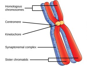
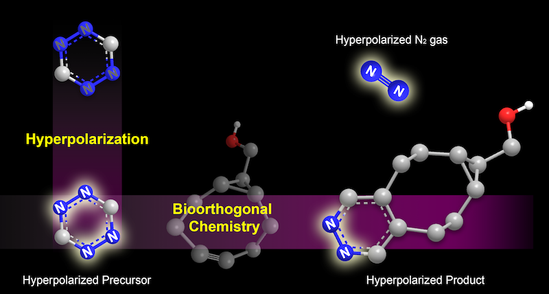
Subramanian uses budding yeast to study the ‘synaptonemal complex,’ a structure that forms between pairing chromosomes as shown in the above image. Over three hundred DNA breakage hotspots exist in the budding yeast’s synaptonemal complex. Normally, double-stranded DNA breaks go from none to some and then return to none.

However, when Subramanian removed the synaptonemal complex, the breaks still appeared, but they did not completely disappear by the end of the process. She  concluded that synaptonemal complex shuts down DNA break formation. The synaptonemal complex therefore is one way cells prevent excessive DNA breakage.

The formation of the synaptonemal complex


During DNA breakage repair, preference must occur between the pairing chromosomes in order for recombination to correctly transpire. A protein called Mek1 promotes this bias by suppressing DNA in select areas. Early in the process of DNA breakage and repair Mek1 levels are high, while synaptonemal complex density is low. Later, the synaptonemal complex increases while the Mek1 decreases.

This led to Subramanian’s conclusion that synaptonemal complex is responsible for removing Mek1, allowing in DNA repair. She then explored if the protein pch2 regulates the removal of Mek1. In pch2-mutant budding yeast cells, DNA breaks were not repaired.

Subramanian showed that at least one aspect of DNA breakage and repair occurs through the Mek1 protein suppression of repair, creating selectivity between chromosomes. The synaptonemal complex then uses pch2 to remove Mek1 allowing DNA breakage repair.

Subramanian had another question about this process though: how is breakage ensured in small chromosomes? Because there are fewer possible breaking points, the chance of recombination seems lower in small chromosomes. However, Subramanian discovered that zones of high DNA break potential exist near the chromosome ends, allowing numerous breaks to form even in smaller chromosomes. This explains why smaller chromosomes actually exhibit a higher density of DNA breaks and recombination since their end zones occupy a larger percentage of their total surface area.

In the future, Subramanian wants to continue studying the specific mechanics behind DNA breaks and repair, including how the chromosomes reorganize during and after this process. She is also curious about how Mek1 suppresses repair and has more than 200 Mek1 mutants in her current study.

Kelly Clarkson may prove that heartbreaks don’t destroy you, but Viji Subramanian proves that DNA breaks create a stronger, more unique genetic code.         

Post by Lydia Goff


Researcher Turns Wood Into Larger-Than-Life Insects

Duke biologist Alejandro Berrio creates larger-than-life insect sculptures. This wooden mantis was exhibited at the Art Science Gallery in Austin, Texas in 2013.

Duke biologist Alejandro Berrio creates larger-than-life insect sculptures. This wooden mantis was exhibited at the Art Science Gallery in Austin, Texas in 2013.

On a recent spring morning, biologist Alejandro Berrio took a break from running genetic analyses on a supercomputer to talk about an unusual passion: creating larger-than-life insect sculptures.

Berrio is a postdoctoral associate in professor Greg Wray’s lab at Duke. He’s also a woodcarver, having exhibited his shoebox-sized models of praying mantises, wasps, crickets and other creatures in museums and galleries in his hometown and in Austin, Texas, where his earned his Ph.D.

The Colombia-born scientist started carving wood in his early teens, when he got interested in model airplanes. He built them out of pieces of lightweight balsa wood that he bought in craft shops.

When he got to college at the University of Antioquia in Medellín, Colombia’s second-largest city, he joined an entomology lab. “One of my first introductions to science was watching insects in the lab and drawing them,” Berrio said. “One day I had an ‘aha’ moment and thought: I can make this. I can make an insect with wings the same way I used to make airplanes.”

Beetle carved by Duke biologist Alejandro Berrio.

His first carvings were of mosquitoes — the main insect in his lab — hand carved from soft balsa wood with an X-Acto knife.

Using photographs for reference, he would sketch the insects from different positions before he started carving.

He worked at his kitchen table, shaping the body from balsa wood or basswood. “I might start with a power saw to make the general form, and then with sandpaper until I started getting the shape I wanted,” Berrio said.

He used metal to join and position the segments in the legs and antennae, then set the joints in place with glue.

“People loved them,” Berrio said. “Scientists were like: Oh, I want a fly. I want a beetle. My professors were giving them to their friends. So I started making them for people and selling them.”

Soon Berrio was carving wooden fungi, dragons, turtles, a snail. “Whatever people wanted me to make,” Berrio said.

He earned just enough money to pay for his lunch, or the bus ride to school.

Duke biologist Alejandro Berrio carved this butterfly using balsa wood for the body and legs, and paper for the wings.

His pieces can take anywhere from a week to two months to complete. “This butterfly was the most time-consuming,” he said, pointing to a model with translucent veined wings.

Since moving to Durham in 2016, he has devoted less time to his hobby than he once did. “Last year I made a crab for a friend who studies crustaceans,” Berrio said. “She got married and that was my wedding gift.”

Still no apes, or finches, or prairie voles — all subjects of his current research. “But I’m planning to restart,” Berrio said. “Every time I go home to Colombia I bring back some wood, or my favorite glue, or one of my carving tools.”

Insect sculptures by Duke biologist Alejandro Berrio.

Insect sculptures by Duke biologist Alejandro Berrio.

Explore more of Berrio’s sculpture and photography at https://www.flickr.com/photos/alejoberrio/.

by Robin Smith

by Robin Smith

Obesity: Do Your Cells Have a Sweet Tooth?

Obesity is a global public health crisis that has doubled since 1980. That is why Damaris N. Lorenzo, a professor of  Cell Biology and Physiology at UNC-Chapel Hill, has devoted her research to this topic.

Specifically, she examines the role of ankyrin-B variants in metabolism. Ankyrins play a role in the movement of substances such as ions into and out of the cell. One of the ways that ankyrins affect this movement is through the glucose transporter protein GLUT4 which is present in the heart, skeletal muscles, and insulin-responsive tissues. GLUT4 plays a large role in glucose levels throughout the entire body.

Through her research, Lorenzo discovered that with modern life spans and high calorie diets, ankyrin-B variants can be a risk factor for metabolic disease. She presented her work for the Duke Developmental & Stem Cell Biology department on March 7th.

Prevalence of Self-Reported Obesity Among U.S. Adults by State, 2016

GLUT4 helps remove glucose from the body’s circulation by moving it into cells. The more GLUT4, the more sugar cells absorb.

Ankyrin-B’s role in regulating GLUT4 therefore proves really important for overall health. Through experiments on mice, Lorenzo discovered that mice manipulated to have ankyrin-B mutations also had high levels of cell surface GLUT4. This led to increased uptake of glucose into cells. Ankyrin-B therefore regulates how quickly glucose enters adipocytes, cells that store fat. These ankyrin-B deficient mice end up with adipocytes that have larger lipid droplets, which are fatty acids.

Lorenzo was able to conclude that ankyrin-B deficiency leads to age-dependent obesity in mutant mice. Age-dependent because young ankyrin-B mutant mice with high fat diets are actually more likely to be affected by this change.

Obese mouse versus a regular mouse

Ankyrin-B has only recently been recognized as part of GLUT4 movement into the cell. As cell sizes grow through increased glucose uptake, not only does the risk of obesity rise but also inflammation is triggered and metabolism becomes impaired, leading to overall poor health.

With obesity becoming a greater problem due to increased calorie consumption, poor dietary habits, physical inactivity, environmental and life stressors, medical conditions, and drug treatments, understanding factors inside of the body can help. Lorenzo seeks to discover how ankyrin-B protein might play a role in the amount of sugar our cells internalize.

Post by Lydia Goff

How a Museum Became a Lab

Encountering and creating art may be some of mankind’s most complex experiences. Art, not just visual but also dancing and singing, requires the brain to understand an object or performance presented to it and then to associate it with memories, facts, and emotions.

A piece in Dario Robleto’s exhibit titled “The Heart’s Knowledge Will Decay” (2014)

In an ongoing experiment, Jose “Pepe” Contreras-Vidal and his team set up in artist Dario Robleto’s exhibit “The Boundary of Life Is Quietly Crossed” at the Menil Collection near downtown Houston. They then asked visitors if they were willing to have their trips through the museum and their brain activities recorded. Robleto’s work was displayed from August 16, 2014 to January 4, 2015. By engaging museum visitors, Contreras-Vidal and Robleto gathered brain activity data while also educating the public, combining research and outreach.

“We need to collect data in a more natural way, beyond the lab” explained Contreras-Vidal, an engineering professor at the University of Houston, during a talk with Robleto sponsored by the Nasher Museum.

More than 3,000 people have participated in this experiment, and the number is growing.

To measure brain activity, the volunteers wear EEG caps which record the electrical impulses that the brain uses for communication. EEG caps are noninvasive because they are just pulled onto the head like swim caps. The caps allow the museum goers to move around freely so Contreras-Vidal can record their natural movements and interactions.

By watching individuals interact with art, Contreras-Vidal and his team can find patterns between their experiences and their brain activity. They also asked the volunteers to reflect on their visit, adding a first person perspective to the experiment. These three sources of data showed them what a young girl’s favorite painting was, how she moved and expressed her reaction to this painting, and how her brain activity reflected this opinion and reaction.

The volunteers can also watch the recordings of their brain signals, giving them an opportunity to ask questions and engage with the science community. For most participants, this is the first time they’ve seen recordings of their brain’s electrical signals. In one trip, these individuals learned about art, science, and how the two can interact. Throughout this entire process, every member of the audience forms a unique opinion and learns something about both the world and themselves as they interact with and make art.

Children with EEG caps explore art.

Contreras-Vidal is especially interested in the gestures people make when exposed to the various stimuli in a museum and hopes to apply this information to robotics. In the future, he wants someone with a robotic arm to not only be able to grab a cup but also to be able to caress it, grip it, or snatch it. For example, you probably can tell if your mom or your best friend is approaching you by their footsteps. Contreras-Vidal wants to restore this level of individuality to people who have prosthetics.

Contreras-Vidal thinks science can benefit art just as much as art can benefit science. Both he and Robleto hope that their research can reduce many artists’ distrust of science and help advance both fields through collaboration.

Post by Lydia Goff

Understanding the Link Between ADHD and Binge Eating Could Point to New Treatments


Binge eating disorder is the most prevalent eating disorder in the United States. Infographic courtesy of Multi-Service Eating Disorders Association

With more than a third of the adult population of the United States meeting criteria for obesity, doctors are becoming increasingly interested in behaviors that contribute to these rates.

Allan Kaplan is interested in improving treatment of binge eating disorder.

Allan Kaplan, MD, of the University of Toronto, is interested in eating disorders, specifically binge eating disorder, which is observed in about 35 percent of people with obesity.

Binge eating disorder (BED) is a pattern of disordered eating characterized by consumption of a large number of calories in a relatively short period of time. In addition to these binges, patients report lack of control and feelings of self-disgust. Because of these patterns of excessive caloric intake, binge eating disorder and obesity go hand-in-hand, and treatment of the disorder could be instrumental in decreasing rates of obesity and improving overall health.

In addition to the health risks associated with obesity, binge eating disorder is associated with anxiety disorders, affective disorders, substance abuse and attention deficit hyperactivity disorder (ADHD) – in fact, about 30 percent of individuals with binge eating disorder also have a history of ADHD.

Binge eating disorder displays a high comorbidity with mood and affective disorders. Infographic courtesy of American Addiction Centers.

ADHD is characterized by inability to focus, hyperactivity, and impulsivity, and substance abuse involves cravings and patterns of losing control followed by regret. These patterns of mental and physiological sympoms resemble those seen in patients with binge eating disorder. Kaplan and other researchers are linking the neurological patterns observed in these disorders to better understand BED.

Researchers have found that the neurological pathways become active when a patient with binge eating disorder is provided with a food-related stimulus. Individuals with the eating disorder are more sensitive to food-related rewards than most people. Researchers have also identified a genetic basis — certain genes make individuals more susceptible to reward and thus more likely to engage in binges.

Because patients with ADHD exhibit similar neurological patterns, doctors are looking to drugs already approved by the FDA to treat ADHD as possible treatments for binge eating disorder. The first of these approved drugs, Vyvanse, has proven not much better than the traditional form of treatment, cognitive behavioral therapy, a form of talk therapy that aims to identify and correct dysfunctions in behavior and thought patterns that lead to disordered behaviors.

Another drug, however, proved promising in a study conducted by Kaplan and his colleagues. The ADHD drug methylphenidate, combined with CBT, led to significant clinical outcomes — pateints engaged in less binges and cravings and body mass index decreased. Kaplan argues that the most effective treatment would reduce binges, treat physiological symptoms like obesity, improve psychological disturbances like low self-esteem, and, of course, be safe. So far, the combination of psychostimulants like methylphenidate and CBT have met these criteria.

Kaplan emphasized a need to make information about binge eating disorder and its treatments more available. Most individuals currently being treated for BED do not obtain treatment knowing they have an eating disorder — they are usually diagnosed only after seeking help with obesity-related health issues or help in weight loss. Making clinicians more familiar with the disorder and its associated behaviors as well as encouraging patients to seek treatment could prove instrumental in combating the current healthcare issue of obesity.

By Sarah Haurin

MRI Tags Stick to Molecules with Chemical “Velcro®”

An extremely close-up view of Velcro

In the new technique, MRI chemical tags attach to a target molecule and nothing else – kind of like how Velcro only sticks to itself. Credit: tanakawho, via Flickr.

Imagine attaching a beacon to a drug molecule and following its journey through our winding innards, tracking just where and how it interacts with the chemicals in our bodies to help treat illnesses.

Duke scientists may be closer to doing just that. They have developed a chemical tag that can be attached to molecules to make them light up under magnetic resonance imaging (MRI).

This tag or “lightbulb” changes its frequency when the molecule interacts with another molecule, potentially allowing researchers to both locate the molecule in the body and see how it is metabolized.

“MRI methods are very sensitive to small changes in the chemical structure, so you can actually use these tags to directly image chemical transformations,” said Thomas Theis, an assistant research professor in the chemistry department at Duke.

Chemical tags that light up under MRI are not new. In 2016, the Duke team of Warren S. Warren’s lab and Qiu Wang’s lab created molecular lightbulbs for MRI that burn brighter and longer than any previously discovered.

A photo of graduate students Junu Bae and Zijian Zhou in front of a bookshelf.

Junu Bae and Zijian Zhou, the co-first authors of the paper. Credit: Qiu Wang, Duke University.

In a study published March 9 in Science Advances, the researchers report a new method for attaching tags to molecules, allowing them to tag molecules indirectly to a broader scope of molecules than they could before.

“The tags are like lightbulbs covered in Velcro,” said Junu Bae, a graduate student in Qiu Wang’s lab at Duke. “We attach the other side of the Velcro to the target molecule, and once they find each other they stick.”

This reaction is what researchers call bioorthogonal, which means that the tag will only stick to the molecular target and won’t react with any other molecules.

And the reaction was designed with another important feature in mind — it generates a rare form of nitrogen gas that also lights up under MRI.

“One could dream up a lot of potential applications for the nitrogen gas, but one that we have been thinking about is lung imaging,” Theis said.

Currently the best way to image the lungs is with xenon gas, but this method has the downside of putting patients to sleep. “Nitrogen gas would be perfectly safe to inhale because it is what you inhale in the air anyways,” Theis said.

A stylized chemical diagram of the hyperpolarization process

In the new technique, a type of molecule called a tetrazine is hyperpolarized, making it “light up” under MRI (illustrated on the left). It is then tagged to a target molecule through a what is called a bioorthogonal reaction. The reaction also generates a rare form of nitrogen gas that can be spotted under MRI (illustrated on the right). Credit: Junu Bae and Seoyoung Cho, Duke University.

Other applications could include watching how air flows through porous materials or studying the nitrogen fixation process in plants.

One downside to the new tags is that they don’t shine as long or as brightly as other MRI molecular lightbulbs, said Zijian Zhou, a graduate student in  Warren’s lab at Duke.

The team is tinkering with the formula for polarizing, or lighting up, the molecule tags to increase their lifetime and brilliance, and to make them more compatible with chemical conditions in the human body.

“We are now developing new techniques and new procedures which may be helpful for driving the polarization levels even higher, so we can have even better signal for these applications,” Zhou said.

15N4-1,2,4,5-tetrazines as potential molecular tags: Integrating bioorthogonal chemistry with hyperpolarization and unearthing para-N2,” Junu Bae, Zijian Zhou, Thomas Theis, Warren S. Warren and Qiu Wang. Science Advances, March 9, 2018. DOI: 10.1126/sciadv.aar2978

Post by Kara Manke

High as a Satellite — Integrating Satellite Data into Science

Professor Tracey Holloway researches air quality at the University of Wisconsin-Madison.

Professor Tracey Holloway researches air quality at the University of Wisconsin-Madison.

Satellite data are contributing more and more to understanding air quality trends, and professor Tracey Holloway wants the world to know.

As a professor of the Department of Atmospheric and Oceanic Science at University of Wisconsin-Madison and the current Team Lead of the NASA Health and Air Quality Applied Sciences Team (HAQAST), she not only helps with the science related to satellites, but also the communication of findings to larger audiences.

Historically, ground-based monitors have provided estimates on changes in concentrations of air pollutants, Holloway explained in her March 2, 2018 seminar, “Connecting Science with Stakeholders,” organized by Duke’s Earth and Ocean Sciences department.

Despite the valuable information ground-based monitors provide, however, factors like high costs limit their widespread use. For example, only about 400 ground-based monitors for nitrogen dioxide currently exist, with many states in the U.S. entirely lacking even a single one. Almost no information on nitrogen dioxide levels had therefore existed before satellites came into the picture.

To close the gap, HAQAST employed earth-observing and polar-orbiting satellites — with fruitful results. Not only have they provided enough data to make more comprehensive maps showing nitrogen dioxide distributions and concentrations, but they also have detected formaldehyde, one of the top causes of cancer, in our atmosphere for the first time.

Satellites have additional long-term benefits. They can help determine potential monitoring sites before actually having to invest large amounts of resources. In the case of formaldehyde, satellite-generated information located areas of higher concentrations — or formaldehyde “hotspots” —  in which HAQAST can now prioritize placing a ground-based monitor. Once established, the site can evaluate air dispersion models, provide air quality information to the public and add to scientific research.

A slide form Holloway’s presentation, in the LSRC A building on March 2, explaining the purposes of a monitoring site.

A slide from Holloway’s presentation, in the LSRC A building on March 2, explaining the purposes of a monitoring site.

Holloway underscored the importance of effectively communicating science. She explained that many policymakers don’t have the strong science backgrounds and therefore need quick and friendly explanations of research from scientists.

Perhaps more significant, though, is the fact that some people don’t even realize that information exists. Specifically, people don’t realize that more satellites are producing new information every day; Holloway has made it a personal goal to have more one-on-one conversations with stakeholders to increase transparency.

Breakthroughs in science aren’t made by individuals: science and change are collaborative. And for Holloway, stakeholders also include the general public. She founded the Earth Science Women’s Network, with one of her goals being to change the vision of what a “scientist” looks like. Through photo campaigns and other communication and engagement activities, she interacted with adults and children to make science more appealing. By making science more sexy, it would be easier to inspire new and continue old discussions, create a more diverse research environment, and make the field more open for all.

Professor Tracey Holloway, air quality researcher at University of Wisconsin-Madison, presented her research at Duke on March 2, 2018.

Professor Tracey Holloway, air quality researcher at University of Wisconsin-Madison, presented her research at Duke on March 2, 2018.

Post by Stella Wang, class of 2019

Post by Stella Wang, class of 2019

How Earth’s Earliest Lifeforms Protected Their Genes

A colorful hot spring in Yellowstone National Park

Heat-loving thermophile bacteria may have been some of the earliest lifeforms on Earth. Researchers are studying their great great great grandchildren, like those living in Yellowstone’s Grand Prismatic Spring, to understand how these early bacteria repaired their DNA.

Think your life is hard? Imagine being a tiny bacterium trying to get a foothold on a young and desolate Earth. The earliest lifeforms on our planet endured searing heat, ultraviolet radiation and an atmosphere devoid of oxygen.

Benjamin Rousseau, a research technician in David Beratan’s lab at Duke, studies one of the molecular machines that helped these bacteria survive their harsh environment. This molecule, called photolyase, fixes DNA damaged by ultraviolet (UV) radiation — the same wavelengths of sunlight that give us sunburn and put us at greater risk of skin cancer.

“Anything under the sun — in both meanings of the phrase — has to have ways to repair itself, and photolyase proteins are one of them,” Rousseau said. “They are one of the most ancient repair proteins.”

Though these proteins have been around for billions of years, scientists are still not quite sure exactly how they work. In a new study, Rousseau and coworkers, working with Professor David Beratan and Assistant Research Professor Agostino Migliore, used computer simulations to study photolyase in thermophiles, the great great great great grandchildren of Earth’s original bacterial pioneers.

The study appeared in the Feb. 28 issue of the Journal of the American Chemical Society.

DNA is built of chains of bases — A, C, G and T — whose order encodes our genetic information. UV light can trigger two adjacent bases to react and latch onto one other, rendering these genetic instructions unreadable.

Photolyase uses a molecular antenna to capture light from the sun and convert it into an electron. It then hands the electron over to the DNA strand, sparking a reaction that splits the two bases apart and restores the genetic information.

A ribbon diagram of a photolyase protein

Photolyase proteins use a molecular antenna (green, blue and red structure on the right) to harvest light and convert it into an electron. The adenine-containing structure in the middle hands the electron to the DNA strand, splitting apart DNA bases. Credit: Benjamin Rousseau, courtesy of the Journal of the American Chemical Society.

Rousseau studied the role of a molecule called adenine in shuttling the electron  from the molecular antenna to the DNA strand. He looked at photolyase in both the heat-loving ancestors of ancient bacteria, called thermophiles, and more modern bacteria like E. Coli that thrive at moderate temperatures, called mesophiles.

He found that in thermophiles, adenine played a role in transferring the electron to the DNA. But in E. coli, the adenine was in a different position, providing mainly structural support.

The results “strongly suggest that mesophiles and thermophiles fundamentally differ in their use of adenine for this electron transfer repair mechanism,” Rousseau said.

He also found that when he cooled E. Coli down to 20 degrees Celsius — about 68 degrees Fahrenheit — the adenine shifted back in place, resuming its transport function.

“It’s like a temperature-controlled switch,” Rousseau said.

Though humans no longer use photolyase for DNA repair, the protein persists in life as diverse as bacteria, fungi and plants — and is even being studied as an ingredient in sunscreens to help repair UV-damaged skin.

Understanding exactly how photolyase works may also help researchers design proteins with a variety of new functions, Rousseau said.

“Photolyase does all of the work on its own — it harvests the light, it transfers the electron over a huge distance to the other site, and then it cleaves the DNA bases,” Rousseau said. “Proteins with that kind of plethora of functions tend to be an attractive target for protein engineering.”

Post by Kara Manke

Using Drones to Feed Billions

A drone flying over an agricultural field

Drones revolutionizing farming

As our population continues its rapid growth, food is becoming increasingly scarce. By the year 2050, we will need to double our current food production to feed the estimated 9.6 million mouths that will inhabit Earth.

A portrait of Maggie Monast

Maggie Monast

Thankfully, introducing drones and other high-tech equipment to farmers could be the solution to keeping our bellies full.

Last week, Dr. Ramon G. Leon of North Carolina State University and Maggie Monast of the Environmental Defense Fund spoke at Duke’s monthly Science & Society Dialogue, sharing their knowledge of what’s known as “precision agriculture.” At its core, precision agriculture is integrating technology with farming in order to maximize production.

It is easy to see that farming has already changed as a result of precision agriculture. The old family-run plot of land with animals and diverse crops has turned into large-scale, single-crop operations. This transition was made possible through the use of new technologies — tractors, irrigation, synthetic fertilizer, GMOs, pesticides — and is no doubt way more productive.

A portrait of Dr. Ramon G. Leon

Dr. Ramon G. Leon

So while the concept of precision agriculture certainly isn’t new, in today’s context it incorporates some particularly advanced and unexpected tools meant to further optimize yield while also conserving resources.

Drones equipped with special cameras and sensors, for example, can be flown over thousands of acres and gather huge amounts of data. This data produces a map of  things like pest damage, crop stress and yield. One image from a drone can easily help a farmer monitor what’s going on: where to cut back on resources, what needs more attention, and where to grow a certain type of crop. Some drones can even plant and water crops for you.

Blue River’s “See & Spray” focuses on cutting back herbicide use. Instead of spraying herbicide over an entire field and wasting most of it, this machine is trained to spray weeds directly, using 10% of the normal amount of herbicide.

Similarly, another machine called the Greenseeker can decide where, when and how much fertilizer should be applied based on the greenness of the crop. Fertilizing efficiently means saving money and emitting less ozone-depleting nitrous oxide.

As you can see, fancy toys like these are extremely beneficial, and there are more out there. They enable farmers to make faster, better decisions and understand their land on an unprecedented level. At the same time, farmers can cut back on their resource usage. This should eventually result in a huge productivity boom while helping out the environment. Nice.

One problem preventing these technologies from really taking off is teaching the farmers how to take advantage of them. As Dr. Leon put it, “we have all these toys, but nobody knows how to play with them.” However, this issue can resolved with enough time. Some older farmers love messing around with the drones, and the next generations of farmers will have more exposure to this kind of technology growing up. Sooner or later, it may be no big deal to spot drones circling above fields of wheat as you road trip through the countryside.

A piece of farm equipment in a field

A Greenseeker mounted on a Boom Sprayer

Precision agriculture is fundamental to the modern agricultural revolution. It increases efficiency and reduces waste, and farming could even become a highly profitable business again as the cost for these technologies goes down. Is it the solution to our environmental and production problems? I guess we’ll know by 2050!

Will Sheehan

Post By Will Sheehan

Page 51 of 111

Powered by WordPress & Theme by Anders Norén