Following the people and events that make up the research community at Duke

Students exploring the Innovation Co-Lab

Category: Neuroscience Page 7 of 15

ECT: Shockingly Safe and Effective

Husain is interested in putting to rest misconceptions about the safety and efficacy of ECT.

Few treatments have proven as controversial and effective as electroconvulsive therapy (ECT), or ‘shock therapy’ in common parlance.

Hippocrates himself saw the therapeutic benefits of inducing seizures in patients with mental illness, observing that convulsions caused by malaria helped attenuate symptoms of mental illness. However, depictions of ECT as a form of medical abuse, as in the infamous scene from One Flew Over the Cuckoo’s Nest, have prevented ECT from becoming a first-line psychiatric treatment.

The Duke Hospital Psychiatry program recently welcomed back Duke Medical School alumnus Mustafa Husain to deliver the 2018 Ewald “Bud” Busse Memorial Lecture, which is held to commemorate a Duke doctor who pioneered the field of geriatric psychiatry.

Husain, from the University of Texas Southwestern, delivered a comprehensive lecture on neuromodulation, a term for the emerging subspecialty of psychiatric medicine that focuses on physiological treatments that are not medication.

The image most people have of ECT is probably the gruesome depiction seen in “One Flew Over the Cuckoo’s Nest.”

Husain began his lecture by stating that ECT is one of the most effective treatments for psychiatric illness. While medication and therapy are helpful for many people with depression, a considerable proportion of patients’ depression can be categorized as “treatment resistant depression” (TRD). In one of the largest controlled experiments of ECT, Husain and colleagues showed that 82 percent of TRD patients treated with ECT were remitted. While this remission rate is impressive, the rate at which remitted individuals experience a relapse into symptoms is also substantial – over 50% of remitted individuals will experience relapse.

Husain’s study continued to test whether a continuation of ECT would be a potentially successful therapy to prevent relapse in the first six months after acute ECT. He found that continuation of ECT worked as well as the current best combination of drugs used.

From this study, Husain made an interesting observation – the people who were doing best in the 6 months after ECT were elderly patients. He then set out to study the best form of treatment for these depressed elderly patients.

Typically, ECT involves stimulation of both sides of the brain (bilateral), but this treatment is associated with adverse cognitive effects like memory loss. Using right unilateral ECT effectively decreased cognitive side effects while maintaining an appreciable remission rate.

After the initial treatment, patients were again assigned to either receive continued drug treatment or continued ECT. In contrast to the previous study, however, the treatment for continued ECT was designed based on the individual patients’ ratings from a commonly used depression scaling system.

The results of this study show the potential that ECT has in becoming a more common treatment for major depressive disorder: maintenance ECT showed a lower relapse rate than drug treatment following initial ECT. If psychiatrists become more flexible in their prescription of ECT, adjusting the treatment plan to accommodate the changing needs of the patients, a disorder that is exceedingly difficult to treat could become more manageable.

In addition to discussing ECT, Husain shared his research into other methods of neuromodulation, including Magnetic Seizure Therapy (MST). MST uses magnetic fields to induce seizures in a more localized region of the brain than available via ECT.

Importantly, MST does not cause the cognitive deficits observed in patients who receive ECT. Husain’s preliminary investigation found that a treatment course relying on MST was comparable in efficacy to ECT. While further research is needed, Husain is hopeful in the possibilities that interventional psychiatry can provide for severely depressed patients.
By Sarah Haurin 

How a Museum Became a Lab

Encountering and creating art may be some of mankind’s most complex experiences. Art, not just visual but also dancing and singing, requires the brain to understand an object or performance presented to it and then to associate it with memories, facts, and emotions.

A piece in Dario Robleto’s exhibit titled “The Heart’s Knowledge Will Decay” (2014)

In an ongoing experiment, Jose “Pepe” Contreras-Vidal and his team set up in artist Dario Robleto’s exhibit “The Boundary of Life Is Quietly Crossed” at the Menil Collection near downtown Houston. They then asked visitors if they were willing to have their trips through the museum and their brain activities recorded. Robleto’s work was displayed from August 16, 2014 to January 4, 2015. By engaging museum visitors, Contreras-Vidal and Robleto gathered brain activity data while also educating the public, combining research and outreach.

“We need to collect data in a more natural way, beyond the lab” explained Contreras-Vidal, an engineering professor at the University of Houston, during a talk with Robleto sponsored by the Nasher Museum.

More than 3,000 people have participated in this experiment, and the number is growing.

To measure brain activity, the volunteers wear EEG caps which record the electrical impulses that the brain uses for communication. EEG caps are noninvasive because they are just pulled onto the head like swim caps. The caps allow the museum goers to move around freely so Contreras-Vidal can record their natural movements and interactions.

By watching individuals interact with art, Contreras-Vidal and his team can find patterns between their experiences and their brain activity. They also asked the volunteers to reflect on their visit, adding a first person perspective to the experiment. These three sources of data showed them what a young girl’s favorite painting was, how she moved and expressed her reaction to this painting, and how her brain activity reflected this opinion and reaction.

The volunteers can also watch the recordings of their brain signals, giving them an opportunity to ask questions and engage with the science community. For most participants, this is the first time they’ve seen recordings of their brain’s electrical signals. In one trip, these individuals learned about art, science, and how the two can interact. Throughout this entire process, every member of the audience forms a unique opinion and learns something about both the world and themselves as they interact with and make art.

Children with EEG caps explore art.

Contreras-Vidal is especially interested in the gestures people make when exposed to the various stimuli in a museum and hopes to apply this information to robotics. In the future, he wants someone with a robotic arm to not only be able to grab a cup but also to be able to caress it, grip it, or snatch it. For example, you probably can tell if your mom or your best friend is approaching you by their footsteps. Contreras-Vidal wants to restore this level of individuality to people who have prosthetics.

Contreras-Vidal thinks science can benefit art just as much as art can benefit science. Both he and Robleto hope that their research can reduce many artists’ distrust of science and help advance both fields through collaboration.

Post by Lydia Goff

Mice, motor learning, and making decisions

Advanced imaging techniques allow neuroscientists to better understand how the motor outputs we observe are created in the brain.

Early understandings of the brain viewed it as a black box that takes sensory input and generates a motor response, with the in-between functioning of the brain as a mystery.

Takaki Komiyama is curious about how the brain produces the stereotypical movements characteristic of motor learning.

Takaki Komiyama of the University of California, San Diego is curious about the relationship between sensory input, motor output, and what happens in between. “What fascinates me the most is the flexibility of this dynamic… this flexibility of the relationship between the environment and the brain is the key element of my research,” Komiyama said to an audience of Duke neuroscience researchers.

Komiyama and his lab have designed experiments to watch how the brain changes as mice learn. Specifically, they train mice to complete a lever-pushing task in response to an auditory stimulus and then use an advanced imaging technique to watch the activity of specific populations of neurons.

Komiyama based his experimental design on a hallmark of motor learning: An “expert” mouse will hear the auditory stimulus and produce a motor response that is exactly the same each time. Komiyama’s team was curious about how these reproducible movements are learned.

Focusing on the primary motor cortex, called M1 for short, Komiyama observed many different neuronal firing patterns as the mouse learned the motion of lever-pushing. As the mouse ventured into “expert” territory, usually after about two weeks of training, this variation was replaced by an activity pattern that is the same from trial to trial. In addition to being consistent, this final pattern starts earlier after the stimulus and takes less time to complete than earlier patterns. In other words, during learning, the brain tries out different pathways for the goal action and then converges on the most efficient way of producing the desired response.

Komiyama then turned his focus to M2, the secondary motor cortex, which he observed to be one of the last areas activated during early learning trials but one of the first activated during late trials. To test M2’s role in learning, Komiyama inactivated the region in trained mice and subjected them to the same stimulus-motor response trial.

The mice with inactivated M2’s missed more trials, took longer to initiate movement, and completed the lever pushing less efficiently. Essentially, the mice behaved as if they had never learned the movement, suggesting that M2 is crucial for coordinating learned motor behavior.

In addition to identifying crucial patterns of motor learning, Komiyama and his team are working to understand decision making. After designing a more complex lever-pushing task that required pushing a joystick in different directions depending on the visual stimulus, Komiyama observed the mice’s accuracy plateaued around 60%.

The mice’s internal biases prevented them from achieving better results in the visual stimuli task.

Komiyama hypothesized that this pattern of inaccuracy could be explained by the mice’s internal biases from previous trials’ outcomes. He designed a statistical model that incorporated the previous trials’ outcomes. With further testing, the model accurately predicted the mice’s wrong choices.

The posterior parietal cortex (PPC) is an area of the brain that has been found to be involved in decision making tasks. Komiyama observed neurons in the PPC that predicted which direction the mice would push the joystick. In addition to being active before the motor response during trials, these neurons were also active in the time between trials.

Seeing this as a neural correlate for internal biases, Komiyama hypothesized that inactivating this region would decrease the influence of bias on the mice’s choices. Sure enough, inactivating the PPC led to more accurate responses in the mice, thus confirming the PPC as a neural source of bias.

 By Sarah Haurin

How A Bat’s Brain Navigates

Most of what we know about how the hippocampus, a region of the brain associated with memory formation and spatial representations, comes from research done on rodents. Rat brains have taught us a lot, but researchers in Israel have found an interesting alternative model to understanding how the hippocampus helps mammals navigate: Bats.

The Egyptian fruit bat proved the perfect subject for studies of mammalian navigation.

Weizmann Institute neurophysiologist Nachum Ulanovsky, PhD, and his team have looked to bats to understand the nuances of navigation through space. While previous research has identified specific cells in the hippocampus, called place cells, that are active when an animal is located in a specific place, there is not much literature describing how animals actually navigate from point A to point B.

Nachum Ulanovsky

Ulanovsky believes that bats are an ingenious model to study mammalian navigation. While bats have the same types of hippocampal neurons found in rats, the patterns of bats’ neurons’ firings more closely match that of humans than rats do.

Ulanovsky sought to test how bats know where they are going. Using GPS tracking equipment, his team found that wild bats that lived in a cave would travel up to 20 kilometers to forage fruit from specific trees. Night after night, these bats followed similar routes past perfectly viable food sources to the same tree over and over again.

The understanding of hippocampal place cells firing at specific locations doesn’t explain the apparent guided travel of the bat night after night, and other explanations like olfactory input do not explain why the bats fly over good food sources to their preferred tree.

The researchers designed an experiment to test how bats encode the 3D information necessary for this navigation. By letting the bats fly around and recording brain activity, Ulanovsky and team found that their 3D models are actually spherical in shape. They also found another type of hippocampal cells that encode the orientation the bat is facing. These head direction cells operate in a coordinate system that allows for a continuity of awareness of its orientation as the animal moves through space.

http://www.cell.com/cms/attachment/2091916945/2076305003/gr1_lrg.jpg

Ulanovsky found bats relied on memory to navigate toward the goal.

To understand how the bats navigate toward a specific goal, the researchers devised another experiment. They constructed a goal with a landing place and a food incentive. The bat would learn where the goal was and find it. In order to test whether the bats’ ability to find the goal was memory-based, or utilized the hippocampus, the researchers then conducted trials where the goal was hidden from the bats’ view.

To test whether the bats’ relied on memory, the Ulvanosky team measured the goal direction angle, or the angle between the bat’s head orientation and the goal. After being familiarized with the location of the goal, the bats tended toward a goal-direction angle of zero, meaning they oriented themselves toward the goal even when the goal was out of sight.

Continued research identified cells that encode information about the distance the bat is from the goal, the final piece allowing bats to navigate to a goal successfully. These hippocampal cells selectively fire when the bat is within specific distances of the goal, allowing for an awareness of location over distance.

While Ulanovsky and his team have met incredible success in identifying new types of cells as well as new functions of known cells in the hippocampus, further research in a more natural setting is required.

“If we study only under these very controlled and sterile environments, we may miss the very thing we are trying to understand, which is behavior,” Ulanovsky concluded.

By Sarah Haurin

Dopamine, Drugs, and Depression

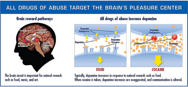
The neurotransmitter dopamine plays a major role in mental illnesses like substance abuse disorders and depressive disorders, as well as a more general role in reward and motivational systems of the brain. But there are still certain aspects of dopamine activity in the brain that we don’t know much about.

Nii Antie Addy and his lab are interested in the role of dopamine in substance abuse and mood disorders.

Duke graduate Nii Antie Addy, PhD, and his lab at Yale School of Medicine have been focusing on dopamine activity in a specific part of the brain that has not been studied: the ventral tegmental area (VTA).

To understand the mechanisms underlying this association, Addy and his team looked at cue-induced drug-seeking behavior. Using classical conditioning, rats can be trained to pair certain cues with the reward of drug administration. When a rat receives an unexpected award, dopamine activity increases. After conditioning, dopamine is released in response to the cue more  than to the drug itself. Looking at the patterns of dopamine release in rats who are forced to undergo detoxification can thus provide insight into how these cues and neurotransmitter activity relate to relapse of substance abuse.

When rats are taught to self-administer cocaine, and each administration of the drug is paired with the cue, after a period of forced detoxification, the rodents continue to try to self-administer the drug, even when the drug is withheld and only the cue is presented. This finding again demonstrates the connection between the cue and drug-seeking behavior.

Studying the activity in the VTA gave additional insights into the regulation of this system. During the period of abstinence, when the rodents are forced to detox, researchers observed an increase in the activity of cholingergic neurons, or neurons in the brain system that respond to the neurotransmitter acetylcholine.

Using these observations, Addy and his team sought to identify which of the various receptors that respond to acetylcholine can be used to regulate the dopamine system behind drug-seeking behaviors. They discovered that a specific type of acetylcholine receptor, the muscarinic receptor, is involved in more general reward-seeking behaviors and thus may be a target for therapies.

Using Isradipine, a drug already approved by the FDA for treatment of high blood pressure, Addy designed an experiment to test the role of these muscarinic receptors. He co-opted the drug to act as a calcium antagonist in the VTA and thus increase dopamine activity in rodents during their forced detox and before returning them to access to cocaine. The outcome was promising: administration of Isradipine was associated with a decrease in the coke-seeking behavior of rodents then placed in the chamber with the cue.

The understanding of the role of cholinergic neurons in regulation of dopamine-related mental illnesses like substance-abuse also contributes insights into depressive and anxiety disorders. If the same pathway implicated in cue-induced drug-seeking were involved in depressive and anxious behaviors, then increasing cholinergic activity should increase pro-depressive behavior.

Addy’s experiment yielded exactly these results, opening up new areas to be further researched to improve the treatment of mood disorders.

Post by Sarah Haurin


Morphogenesis: All Guts and Morning Glories

What is morphogenesis? Morphogenesis examines the development of the living organisms’ forms.

It also is an area of research for Lakshminarayanan Mahadevan, Professor of Applied Mathematics, Organismic and Evolutionary Biology and Physics at Harvard University. On his presentation in the Public Lectures Unveiling Math (PLUM) series here at Duke, he credited the beginnings of morphogenesis to D’Arcy Wentworth Thompson, author of the book On Growth and Form.

Mathematically, morphogenesis focuses on how different rates of growth change the shapes of organisms as they develop. Cell number, cell size, cell shape, and cell position comprise the primary cellular factors of multicellular morphogenesis, which studies larger structures than individual cells and is Mahadevan’s focus.

Effects on tissues appear through changes in sizes, connectivities, and shapes, altering the phenotype, or the outward physical appearance. All these variables change in space and time. Professor Mahadevan presented on morphogenesis studies that have been conducted on plant shoots, guts, and brains.

Research on plant shoots often concentrates on the question, “Why do plant shoots grow in such a wide variety of directions and what determines their shapes?” The picture below shows the different postures appearances of plant shoots from completely straight to leaning to hanging.

Can morphogenesis make sense of these differences? Through mathematical modeling, two stimuli for shoots’ shapes was determined: gravity and itself. Additionally, elasticity as a function of the shoots’ weight plays a role in the mathematical models of plant shoots’ shapes which appear in Mahadevan’s paper co-written with a fellow professor, Raghunath Chelakkot. Mahadevan also explored the formation of flower and leaf shapes with these morphogenesis studies. 
Over twenty feet of guts are coiled up inside you. In order to fit these intestines inside the mammals, they must coil and loop. But what variables determine how these guts loop around? To discover the answer to this question, Mahadevan and other researchers examined chick embryos which increase their gut lengths by a factor greater than twenty over a twelve-day span. They were able to create a physical model using a rubber tube sewn to a sheet that followed the same patterns as the chicks’ guts. Through their observation of not only chicks but also quail and mice, Mahadevan determined that the morphogenesis of the guts has no dependence on genetics or any other microscopic factors.

Mahadevan’s study of how the brain folds occurs through MRI images of human fetal development. Initially, barely any folding exists on fetal brains but eventually the geometry of the surrounding along with local stress forms folds on the brain. By creating a template with gel and treating it to mimic the relationship between the brain’s gray matter and white matter, Mahadevan along with other researchers discovered that they could reproduce the brain’s folds. Because they were able to recreate the folds through only global geometry and local stress, they concluded that morphogenesis evolution does not depend on microscopic factors such as genetics. Further, by examining if folding regions correlate with the activity regions of the brain, questions about the effect of physical form on abilities and the inner functions of the brain.


On a Mission to Increase Exercise

Dr. Zachary Zenko of the Center for Advanced Hindsight at Duke is on a mission to get people to exercise. He shared this mission and his research aimed at achieving this mission at Duke’s Exercise and the Brain Symposium on December 1st.

Dr. Zenko started out with a revealing statistic: Only 1 in 10 people meet the United States activity guidelines for exercise. While I wasn’t completely shocked at this fact as the U.S. is known for its high rates of obesity and easy access to fast-food, I was definitely eager to learn more about how Dr. Zenko planned to fight this daunting statistic.

Next, Dr. Zenko pointed out a common assumption in exercise psychology: if people know how good exercise is for them, they will exercise. However, this hasn’t proven to be true. Most people already know that they should be exercising, but don’t. And those that do often quickly drop out.

Then Dr. Zenko began to break down Dual-Process Theory in Behavioral Economics. Type 1 Processes are those that are fast and non-conscious, while Type 2 Processes are those that are controlled and conscious. While research on exercise usually focuses on Type 2 processes, Zenko believes that we must focus on both.

Ideally, exercise would involve the affect heuristic, which is a mental shortcut in which an emotional response drives an individual. This heuristic involves Type 1 processes. Dr. Zenko’s goal was to shift away from only considering Type 2 processes, and instead focus on using Type 1 processes to make exercise more appealing.

How did he propose doing this? By continually decreasing the difficulty of exercise. By changing the slope of the intensity of a workout and having a continually declining heart rate, exercisers could have a more pleasant experience. In addition, this positive experience could influence memory and make an individual more likely to exercise in the future.

Dr. Zenko put this hypothesis to the test by having unfit adults exercise while continually decreasing the intensity throughout the workout. While test subjects exercised, he measured the amount of pleasure experienced by asking “How do you feel right now?” at certain intervals. This new exercise method  has the most potential when starting at the highest intensity levels because it leaves more room to change the slope of the workout intensity throughout, leading to an overall more pleasurable workout.

Looking forward, this new method of exercise could possibly change the way we think about exercise. It may not only involve doing the right amount of exercise, but also doing the right kind of exercise that leaves us more likely to exercise in the future. Considering that traditional methods of promoting exercise, such as educating people about its benefits, have not been particularly successful thus far, Dr. Zenko’s method is very exciting.

Dr. Zenko wrapped up his talk by suggesting that people consider exercise prescriptions that are safe, effective, pleasant and enjoyable. As exercise has become a huge part of my weekly routine throughout college, I will definitely take this advice to heart. Maybe even look out for me lowering my intensity during a workout soon in a gym near you?

By Nina Cervantes

Exercise is Good for Your Head and Might Fight Alzheimer’s

Recent studies have confirmed that exercising is just about the best thing you can do for your brain health.

Dan Blazer, MD is a psychiatrist who studies aging.

On Dec. 1 during the DIBS event, Exercise and the Brain, Duke psychiatrist Dan Blazer reported findings about the relationship between physical activity and brain health. After lots of research, study groups at the National Academy of Medicine  concluded that their number one recommendation to those experiencing “cognitive aging” is exercise.

Processing speed, memory, and reasoning decline over time in every one of us. But thankfully, simple things like riding a bike or playing pick up basketball can help keep our minds fresh and at their best possible level.

One cool thing a committee conducting the research did to advertise their findings was create keychains saying “take your brain for a walk.” There’s a little image of a brain with legs walking. They wanted to get the word out that physical activity has another benefit than just staying in shape — it can also support your cognitive health.

However, the committees are having a hard time motivating people to exercise in the first place. Even after hearing their findings, it’s not like people everywhere are suddenly going to get off their couches and hit the gym. A world with healthier people — both physically and mentally — sounds nice, but getting there is much more than a matter of publishing these studies.

And, as always, too much of a good thing can make it harmful. While there does seem to appear a potential “biological gradient,” where greater physical activity correlated with better outcomes, you can’t just run a marathon every day of the week and then ~boom~ aging hardly affects your brain anymore. You don’t want to do that to yourself. Just get a healthy amount of exercise and you’ll be keeping your brain young and smart.

One of the best parts about why exercising is so great for you and your brain is because it helps you sleep (and we all know how important sleep is). If you ever have trouble going to bed or are having disrupted sleeps, physical activity could be your savior. It’s a much healthier option for your brain than taking stuff like melatonin, and you’ll get fit in the process.

Regarding exercising and Alzheimer’s, a disease where vital mental functions deteriorate, studies have unfortunately been insufficient to conclude anything. But if getting Alzheimer’s is your worst fear, I’m sure staying active can’t hurt as a preventative. More research on this topic is being conducted as we speak.

When is the best time to start exercising, in order to reap the maximum cognitive benefits, you ask? Well, the sooner the better. As Blazer said, “exercising helps in maintaining or improving cognitive function in later life,” so you better get on that. Go outside and get moving!

Will Sheehan      Post by Will Sheehan


How We Know Where We Are

The brain is a personalized GPS. It can keep track of where you are in time and space without your knowledge.

The hippocampus is a key structure in formation of memories and includes cells that represent a person’s environment.

Daniel Dombeck PhD, and his team of researchers at Northwestern University have been using a technique designed by Dombeck himself to figure out how exactly the brain knows where and when we are. He shared his methods and findings to a group of researchers in neurobiology at Duke on Tuesday.

Domeck and his lab at Northwestern are working at identifying exactly how the brain represents spatial environments.

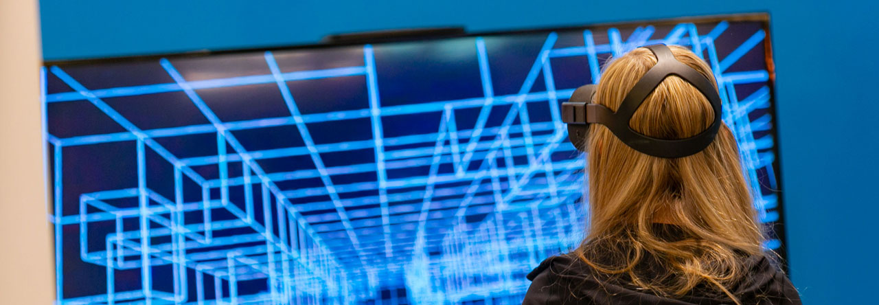
The apparatus used for these experiments was adapted from a virtual reality system. They position a mouse on a ball-like treadmill that it manipulates to navigate through a virtual reality field or maze projected for the mouse to see. Using water as a reward, Dombeck’s team was able to train mice to traverse their virtual fields in a little over a week.

In order to record data about brain activity in their mice as they navigated virtual hallways, Dombeck and his team designed a specialized microscope that could record activity of single cells in the hippocampus, a deep brain structure previously found to be involved in spatial navigation.

The device allows researchers to observe single cells as a mouse navigates through a simulated hallway.

Previous research has identified hippocampal place cells, specialized cells in the hippocampus that encode information about an individual’s current environment. The representations of the environment that these place cells encode are called place fields.

Dombeck and his colleague Mark Sheffield of the University of Chicago were interested in how we encode new environments in the hippocampus.

Sheffield studied the specific neural mechanisms behind place field formation.

After training the mice to navigate in one virtual environment, Sheffield switched the virtual hallway, thus simulating a new environment for the mouse to navigate.

They found that the formation of these new place cells uses existing neural networks initially, and then requires learning to adapt and strengthen these representations.

After identifying the complex system representing this spatial information, Dombeck and colleagues wondered how the system of representing time compared.

Jim Heys, a colleague of Dombeck, designed a new virtual reality task for the lab mice.

In order to train the mice to rely on an internal representation of passing time, Heys engineered a door-stop task, where a mouse traversing the virtual hallway would encounter an invisible door. If the mouse waited 6 seconds at the door before trying to continue on the track, it would be rewarded with water. After about three months of training the mice, Heys was finally able to collect information about how they encoded the passing of time.

Heys indentified cells in the hippocampus that would become active only after a certain amount of time had passed – one cell would be active after 1 second, then another would become active after 2 seconds, etc. until the 6-second wait time was reached. Then, the mouse knew it was safe to continue down the hallway.

When comparing the cells active in each different task, Dombeck and Heys found that the cells that encode time information are different from the cells that encode spatial information. In other words, the cells that hold information about where we are in time are separate from the ones that tell us where we are in space.

Still these cells work together to create the built-in GPS we share with animals like mice.

By Sarah Haurin

New Blogger Nirja Trivedi: Neuroscience Junior with Infinite Curiosity

My name is Nirja Trivedi and I’m a junior from Seattle interested in the intersections between health, technology and business. At Duke, I’m the co-president of P.A.S.H., a writer for the Standard and a member of B.O.W.

Nirja Trivedi blocking the sun with her hand

Nirja Trivedi

During high school, I considered liberal arts and scientific research to be separate disciplines: if technology was my strength then philosophy must be my weakness. In my two years at Duke, I have experienced the duality of these fields through participating in the Global Health Focus Program, developing my own research projects, working with professors and now applying to write for Duke Research. Science truly is for everyone; no matter your field, interests or opinion. Research and discovery are conduits for every mind. Research isn’t just the forefront of innovation, it paves the way for the future.

Growing up with a passion for service and influenced by my family in the medical field, the research I leaned towards combined aspects of community and health. My senior project in high school examined traumatic brain injury (TBI) in youth sports, which provided the research-based approach for designing my own Concussion Prevention Program. After my first semester, I wanted to discover what kinds of research I wanted to fully integrate myself in. I began research with the Duke Institute of Brain Sciences and spent my summer volunteering for the Richman Lab, which examines the effects of psychosocial factors like discrimination, social hierarchies and power. After I declared my Neuroscience major, I spent the year assisting in studies at the Autism Clinic, sparking my interest in technology.

Nirja Trivedi on a mountain top

Nirja Trivedi on a mountain top.

Now going into my third year, my interests in scientific discovery have only grown. From insight into the human psyche and social economic behavior to medical advances, I love the complexity of the human mind and how it fuels innovation.

My unrestricted interests guided me to the Innovation & Entrepreneurship Certificate as well as this writing position, both which foster an environment of curiosity and inspiration. Through writing, I hope to connect with faculty, discover areas of research I never knew existed, widen my breadth of scientific knowledge, and connect students to research opportunities. The threshold of knowledge is where you draw the line – why not make it infinite?

Post by Nirja Trivedi

Page 7 of 15

Powered by WordPress & Theme by Anders Norén