Following the people and events that make up the research community at Duke

Students exploring the Innovation Co-Lab

Category: Biology Page 13 of 32

Tiny Bubbles of Bacterial Mischief

Margarethe (Meta) Kuehn studies vesicles — little bubbles that bud off bacterial membranes. All sorts of things may be tightly packed into these bubbles: viruses, antigens, and information a bacterium will need to make cells vulnerable to infection.

But why do bacteria produce these small membrane vesicles in the first place? Why not spread out to nearby cells themselves?

Jenny and Meta met last month on the Duke campus.

“The short answer is that we don’t know yet,” explains Kuehn, an associate professor of biochemistry at Duke. “But we speculate that it is due to their small size. These vesicles, which serve as delivery ‘bombs,’ can pass through pores that are too small for bacteria to fit through.”

Originally a chemistry major, Kuehn always had an interest in biochemistry. As an undergraduate, she worked in protein purification and then in the infectious disease division of a children’s hospital. There, she learned about pathogenic bacteria and how they secrete proteins to give themselves access to host cells.

Kuehn’s lab studies the mysterious world of bacterial vesicle production,focusing on the genetic, biochemical, and functional features of vesicles. So far, they have identified specific proteins and genes involved in the vesiculation process.

With a fine filter, they showed that vesicles can fit through holes to reach mammalian cells where a bacterium cannot.

Kuehn wonders why the bacteria don’t just use soluble proteins, which are even smaller than vesicles. They must have some reason for preferring the cell’s vesicles. Currently, they believe that vesicles can serve as nice packages — a whole bolus of information delivered together.

Basic anatomy of a vesicle, a bubble-like  membrane-bound package used by cells to move things around.

Not only will this new insight into extracellular vesicles of gram-negative bacteria aid in identifying new medicines, vesicles are also being used for vaccine delivery.

“They are really good antigen vehicles,” reveals Kuehn, “The more we know how they are made, the better we can design effective vaccines for humans.”

According to Kuehn, the amazing part about studying these pathogens is that, “You are never done. You never know it all. Every single pathogen, they each do things differently.” What keeps Kuehn going, she explains, is that the search never ends.

“There is never really a defined end point; you have to come to grips with the fact that you will never know that whole answer.”

Guest Post by Jenny Huang, NCSSM 2019

Biology by the Numbers

Michael C. Reed was trained as a pure mathematician, but from the start, he was, as he explained to me, a “closet physiologist.” He’s a professor of mathematics at Duke, but he’s always wondered how the body works.

Michael Reed in his office

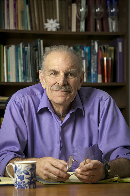
Reed explains an example to me: women have elbows that are bent when their arms are straightened, but men do not. He rationalized his own explanation: women have wide hips and narrow shoulders; their bodies are designed so their arms don’t knock into their sides when they walk. (That basically ended up being the answer.)

Still, Reed never really explored his interest in physiology until he was 40 years old, when he realized that if he wanted to explore something, he should just do it. Why not? He had tenure by that point, so it didn’t really matter what his colleagues thought. He was interested in physiology, but was a mathematician. The obvious answer was mathematical biology.

Now he uses mathematics to find out how various physiological systems work.

In order to decide on a research project, he works with a biologist, Professor Fred Nijhout. They meet for two hours every day and work together. They have lots of projects, but they also just talk science sometimes. That’s how they get their ideas, mainly focusing on things in cell metabolism that have to do with important public health questions.

Reed has been investigating dopamine and serotonin metabolism in the brain, in a collaborative project with Nijhout and Dr. Janet Best, a mathematician at The Ohio State University.

Maybe better math is going to help us understand the human brain

As he explained to me, the brain isn’t like a computer; you don’t know how it works and there are a lot of systems in play. Serotonin is one of them. Low serotonin concentration is thought to be one of the causes of depression. There’s a biochemical network that synthesizes, packages, and transfers serotonin in the brain.

He told me that his work consists of making mathematical models for systems like this that consist of differential equations for concentrations of different chemicals. He then experiments with the system of differential equations to understand how the system works together. It’s not really something you can learn by having it explained to you, he told me. You have to learn through practice.

In a way, biology doesn’t seem like it would be the most compatible science, especially with math. But as Reed explained to me, “Math is easy because it’s very orderly and organized. If you work hard enough, you can understand it.” Biology, on the other hand, “is a mess.”

Everything in biology is linked to everything else in a system of connectedness that ends up all tangled together, and it can be hard to identify how something happens in the human body. But Reed applies math – an organized construct – to understand biological systems.

In the end, Reed does what he does because it’s how we — as human beings — work. He has no regrets about the choices he’s made at all.Mathematical biology seems to be his calling — he’s more interested in understanding how things work, and that’s what he does when he works.

Or rather, he doesn’t really work; because, as he told me,“try to find something to do that you really like, and are passionate about,because if you do, it won’t seem like work.” Reed doesn’t see coming into work as a struggle. He’s excited about it every single day and “it’s because you want to do it, it’s fun.”

Guest Post by Rachel Qu, NCSSM 2019

Gene-Editing Human Embryos: What, How, Why?

Every seat full. Students perched on the aisle stairs and lining the back walls.

What topic could possibly pull so many away from their final exams? Not “How to Stop Procrastinating” nor “How to Pass Life After Failing Your Exams” but rather “Gene-Editing Human Embryos: Unpacking the Current Controversy” on the Duke campus.

Since Chinese researcher He Jiankui announced at the Second International Summit on Human Genome Editing in Hong Kong that he made the world’s first genetically engineered babies, a debate on the ethical implications has raged on social media.

On December 6, the University Program in Genetics and Genomics and the Molecular Genetics and Microbiology department co-hosted a panel responding to He’s claims. Charles A. Gersbach from the Biomedical Engineering department lead the discussion of what exactly happened and then joined the panel which also contained Misha Angrist, a senior fellow in the Science & Society initiative;  Heidi Cope, a genetic counselor; Giny Fouda, an assistant professor in pediatrics; and Vandana Shashi, a genetic counselor.

Dr. He Jiankui announced he had used CRISPR to edit genes in twin embryos that were then born at full term.

But what exactly has He potentially done to these twin girls? Can they fly? Breathe underwater? Photosynthesize? Not exactly. He said he deleted a gene called CCR5 to increase their HIV resistance. Two percent of Northern Europeans naturally have a mutation that removes the CCR5 gene from their DNA and as a result do not display any traits other than increased HIV resistance.

Many researchers have explored blocking CCR5 activity as a potential HIV treatment. Using CRISPR-Cas9, a genetic engineering technology that can cut and paste specific sequences in the DNA, He targeted CCR5 during in vitro fertilization. According to his tests, he successfully removed both copies of the CCR5 gene in one of the girls. However, in the other girl, the CCR5 remained normal on one chromosome and on the other, CRISPR had deleted more than intended.  The effects of that additional deletion are unknown. 
Both the girls are mosaics, meaning the genetic change occurred in some of their cells and not in others, leading to still more uncertainties.

Researchers have conducted genetic engineering experiments on both somatic cells and human embryo cells that were never brought to term. (Somatic cells constitute all parts of the body other than the eggs and sperm.) But because He altered the twin girls as embryos and then they grew to full term, their children could inherit these changes. This alters their family line, not just a single individual, increasing the ethical implications.

According to Shashi, He’s experiment becomes difficult to justify. Additionally, embryos have not consented to these changes in their genetics, unlike a patient undergoing genetic therapy.

Many doctors, scientists, and journalists have also questioned He’s lack of transparency because he hid this work until his grand announcement, which caused China to arrest him. In addition, as Cope explained, “it is not typically the PI who does the informed consent process” as He did with these parents.

While He defends his work by saying that the girls’ father carries HIV and wished to increase the girls’ safety, the twins were not actually at great risk for HIV. Their father’s medical history does not increase their chances of contracting the virus, and the overall risk for HIV in China is low. As Fouda emphasized in the panel, “there was no justification for this experiment.” While He discussed the potential for genetic engineering to help society, for these two individuals, no medical need existed, and that increases the ethical dilemma.

A final concern of researchers is the current inability to ensure technical competency and accuracy. As seen by the additional deletion in one of the girls,  CRISPR-Cas9 still makes errors. Thus using it to alter not only a human being but all of that individual’s progeny would demand a much higher standard, something close to a life-or-death scenario.

But, the panelists also noted, if it hadn’t been He, it would have been somebody else. Perhaps somebody else may have done it more ethically with more transparency and a more traditional consent process, Angrist said.

While He’s claims have yet to be proven, the fact that they could reasonably be true has many concerned. The World Health Organization has announced that they will begin greater oversight of genetic engineering of the human germline.

On campus over the last weeks, I’ve heard mixed reviews on He’s work with some joking about future superhero babies while others have reacted with fear. The technology does live among us; however, the world is working on writing the guidebook and unrolling the yellow tape.

Post by Lydia Goff

Sean Carroll on the Evolution of Snake Venom

What’s in a snake bite?

According to University of Wisconsin-Madison evolutionary biologist Sean Carroll who visited Duke and Durham last week, a snake bite contains a full index of clues.

In his recent research, Carroll has been studying the adaptations of novelties in animal form, such as snake venom. Rattlesnakes, he explains, are the picture of novelty. With traits such as a limbless body, fangs, infrared pits, patterned skin, venom, and the iconic rattle, they represent an amazing incarnation of evolution at work.

Rattlesnakes: the picture of novelty (Photo from USGS)

Snake venoms contain a complex mixture of proteins. This mixture can differ in several ways, but the most interesting difference to Carroll is the presence or absence of neurotoxins. Neurotoxic venom has proven to be a very useful trait, because neurotoxins destroy the nervous tissue of prey, effectively paralyzing the animal’s respiratory system.

Some of today’s rattlesnake species have neurotoxic venom, but some don’t. So how did this happen? That’s what Carroll was wondering too.

Some genes within genomes, such as HOX genes, evolve very slowly from their original position among the chromosomes, and see very few changes in the sequence in millions of years.

But snake venom Pla2 genes are quite the opposite. In recent history, there has been a massive expansion of these genes in the snake genome, Carroll said. When animals evolve new functions or forms, the question always arises: are these changes the result of brand new genes or old genes taking on new functions?

Another important consideration is the concept of regulatory versus structural genes. Regulatory genes control the activity of other genes, such as structural genes, and because of this, duplicates of regulatory genes are generally not going to be a favorable adaptation. In contrast, structural gene activity doesn’t affect other genes, and duplicates are often a positive change. This means it is easier for a new structural gene to evolve than a regulatory one. Carroll explained.

Evolutionary Biologist Sean Carroll (Photo from seanbcarroll.com)

Carroll examined neurotoxic and non-neurotoxic snakes living in overlapping environments. His research showed that the most recent common ancestor of these species was a snake with neurotoxic venom. When comparing the genetic code of neurotoxic snakes to non-neurotoxic ones, he found that the two differed by the presence or absence of 16 genes in the metalloproteinase gene complex. He said this meant that non-neurotoxic venom could not evolve from neurotoxic venom.

So what is the mechanism behind this change? What could be the evolutionary explanation?

When Carroll’s lab compared another pair of neurotoxic and non-neurotoxic species in a different region of the US, they found that the two species differed in exactly the same way, with the same set of genes deleted as had been observed in the first discovery. With this new information, Carroll realized that the differences must have occurred through the mechanism of hybridization, or the interbreeding of neurotoxic and non-neurotoxic species.

Carroll’s lab is now doing the structural work to study if the genes that result in neurotoxic and  non-neurotoxic protein complexes are old genes carrying out new functions or entirely new genes. They are using venom gland organoids to look into the regulatory processes of these genes.

In addition to his research studying the evolution of novelties, Carroll teaches molecular biology and genetics at Madison and has devoted a large portion of his career to  storytelling and science education.

Meet New Blogger Anne Littlewood – Working on Biology and Puppies

My name is Anne Littlewood and I am a sophomore here at Duke. I grew up in San Francisco, spent a brief moment living on the island of Kauai, and finished high school in Pebble Beach, California. I am studying the intersection of biology and psychology here at Duke, in an effort to understand how biological mechanisms inform our interactions with the environment.

Snuggles in Puppy Kindergarten!

Outside the classroom, I can be found frequenting Duke’s beloved Puppy Kindergarten, where I work as a volunteer. Recently, I’ve become an Associate Editor for Duke’s literary magazine, The Archive. I love writing creatively, and it’s been so great to find a community of my literature- loving peers. I’m also participating in a Bass Connections project this year, and working on a team to evaluate the outcomes of different conservation interventions through the synthesis of an evidence gap map for World Wildlife fund.

Me and Cricket on Carmel Beach

Most of all I love to spend time outdoors, whether it’s exploring the mountains of North Carolina on a backpacking trip, lying in my hammock at Eno Quarry, or walking through the gardens each day on my way to class. I’m a huge animal lover, and I’m way too obsessed with my dog, a 12-pound cavalier King Charles spaniel named Cricket.

I’ve always been into science, but I think I really fell in love with Biology my freshman year of high school, when my all time favorite teacher, Mr. Cinti helped me extract my DNA one afternoon, just for fun. Writing is my passion, and I’m excited to explore my skills in a variety of genres this year. This blog is my first ever attempt at journalism/ science writing, and I’m excited to give it a try!

Considerations about AIDS from Brazilian literature

To know what illness is, you have to be ill first.

This was one of the points that post-doctoral student and essayist  Milena Mulatti Magri emphasized in her talk on Oct. 15. She was discussing Brazilian writer Caio Fernando Abreu’s writings about AIDS and its effect on groups who already faced societal prejudice before the breakout of AIDS in the 1980’s and 90’s, when patients were identified mainly as homosexual.

By studying research done by Professor Vladimir Safatle and physician and philosopher Georges Canguilhem, Magri has pieced together that health is seen as a form of normativity and disease as a deviation. Thus, ostracizing and excluding those who contract disease is seen as justifiable because they have deviated from what is seen as normal, even when the disease is not the fault of the patient.

Magri has also analyzed essays about the relationship between disease, metaphor and patient stigma, such as cancer patients who, in addition to combating the illness growing unwelcome inside their own bodies, also have to deal with social stigmas that come with disease, such as cancer as a representation for evil.

Brazilian author and columnist Caio Fernando Abreu. (Image from KD Frases.)

However, while Magri emphasized that social metaphors of different diseases should be deconstructed, she has also found that literature and personal writing can be a way to discuss and otherwise “incommunicable experience.”

During a time when it was seen as shameful to have AIDS, Caio Fernando Abreu began a biweekly publication of his health chronicles in the newspaper O Estado de São Paulo, which was one of the first instances of someone publicly discussing their experience with AIDS from the perspective of the ill person, as opposed to from the perspective of doctors or health experts.

Abreu’s columns confronted the difficulty of living with disease and living in proximity to death, and discuss the increased social prejudice as a result of the disease. Abreu also wrote a play called “O homem e amancha” (in English, “The Man and the Stain”), which is an intertextual reading of the famous Spanish novel Don Quijote de la Mancha. In her talk, Magri explained that “Mancha,” which in English means “stain,” can refer to both the home of Don Quijote before he sets off of on his adventures and the rare skin cancer that often accompanies AIDS called Kaposi Sarcoma, which forms lesions on the skin that resemble stains.

Abreu intended to use his own experiences to question the social prejudice against AIDS, and there Magri highlighted a marked change, even between his own writings at the beginning of his diagnosis compared to those at the end of his life, when he spoke openly and without metaphor about suffering that is amplified by social exclusion. 

Magri believes that Abreu’s writings were pioneering acts of courage, and that from his writing we learn to empathize rather than to judge and stigmatize.

Post by Victoria Priester

Victoria Priester

The Complicated Balance of Predators and Prey

If you knew there was a grizzly bear sitting outside the door, you might wait a while before going to fill up your water bottle, or you might change the way you are communicating with their other people in the room based on your knowledge of the threat.

Ecologists call this “predation risk,” in which animals that could potentially fall prey to a carnivore know this risk is present, and alter their habits and actions accordingly.

A yellow slider turtle.

A yellow slider turtle.

One way in which animals do this is through habitat use, such as a pod of dolphins that changes where they spend most of their time depending on the presence or absence of predators. Animals might also change their feeding habits and diving behavior because of predation risk.

Animals do this all of the time in the wild, but when predators are removed from ecosystems by hunting or over-fishing, the effect of their absence is felt all the way down the food chain.

For example, large amounts of algae growth on coral reefs can be traced back to over-fishing of large ocean predators such as sharks, who then don’t hunt smaller marine mammals like seals. As seal numbers increase, there are more of them to hunt smaller fish that feed on vegetation, which means fewer smaller fish or plankton to keep algal growth in check, and algae begins to grow unchecked.

Meagan Dunphy-Daly

Meagan Dunphy-Daly

This is a “trophic cascade” and it has large effects on ecosystems, Duke Marine Lab instructor Meagan Dunphy-Daly  t0ld the Sustainable Oceans Alliance last Thursday. She has performed research both in labs and in the field to study the effects that removing large predators have on marine ecosystems.

Dunphy-Daly discussed one lab experiment where 10 yellow-bellied slider turtle hatchlings were kept in tanks where they couldn’t see people or anything else on the outside. In real life, blue herons and other large birds prey on these turtle hatchlings, so the researchers made a model skull of a blue heron that they painted and covered with feathers.

Turtles are air-breathing, so each hatchling was given the option to sit where they could be at the surface of their tank and breathe, but this spot was also where the turtle hatchlings thought the bird beak might shoot down at any time to try to “eat” them.

Their options were to get air and risk getting hit by the bird beak, or diving down to the bottom of the tank to get food. During this experiment, Dunphy-Daly found that turtle hatchlings actually decreased their dive time and spent more time at the surface. If the turtles are continuously diving, they are expending lots of energy swimming back and forth between the surface and the bottom, she said, which means if the predator were to actually attack, they would have less energy left to use for a rapid escape.

Even when there is food at the bottom, when a predator is present, these turtles alter their activity by taking deep dives less frequently so as to not max out their aerobic limit before they actually need to escape a predator.

This is one way in which animals alter their behavior due to predation risk.

But let’s say that predators were disappearing in their real habitats, so turtles didn’t feel the need to build up these emergency energy reserves to escape them. They might dive down and feed more frequently, which would then decrease the amount of the vegetation they eat.

This in turn could have an effect on oxygen levels in the water because there would be fewer plants photosynthesizing. Or another species that feeds on the same plant could be out-competed by turtles and run out of food for their own populations.

The absence of large or small predators can have large impacts on ocean ecosystems through these complicated trophic cascades.

Victoria PriesterPost by Victoria Priester

Medicine, Research and HIV

Duke senior Jesse Mangold has had an interest in the intersection of medicine and research since high school. While he took electives in a program called “Science, Medicine, and Research,” it wasn’t until the summer after his first year at Duke that he got to participate in research.

As a member of the inaugural class of Huang fellows, Mangold worked in the lab of Duke assistant professor Christina Meade on the compounding effect of HIV and marijuana use on cognitive abilities like memory and learning.

The following summer, Mangold traveled to Honduras with a group of students to help with collecting data and also meeting the overwhelming need for eye care. Mangold and the other students traveled to schools, administered visual exams, and provided free glasses to the children who needed them. Additionally, the students contributed to a growing research project, and for their part, put together an award-winning poster.

Mangold’s (top right) work in Honduras helped provide countless children with the eye care they so sorely needed.

Returning to school as a junior, Mangold wanted to focus on his greatest research interest: the molecular mechanisms of human immunodeficiency virus (HIV). Mangold found a home in the Permar lab, which investigates mechanisms of mother-to-child transmission of viruses including HIV, Zika, and Cytomegalovirus (CMV).

From co-authoring a book chapter to learning laboratory techniques, he was given “the opportunity to fail, but that was important, because I would learn and come back the next week and fail a little bit less,” Mangold said.

In the absence of any treatment, mothers who are HIV positive transmit the virus to their infants only 30 to 40 percent of the time, suggesting a component of the maternal immune system that provides at least partial protection against transmission.

The immune system functions through the activity of antibodies, or proteins that bind to specific receptors on a microbe and neutralize the threat they pose. The key to an effective HIV vaccine is identifying the most common receptors on the envelope of the virus and engineering a vaccine that can interact with any one of these receptors.

This human T cell (blue) is under attack by HIV (yellow), the virus that causes AIDS. Credit: Seth Pincus, Elizabeth Fischer and Austin Athman, National Institute of Allergy and Infectious Diseases, National Institutes of Health

This human T cell (blue) is under attack by HIV (yellow), the virus that causes AIDS. Credit: Seth Pincus, Elizabeth Fischer and Austin Athman, National Institute of Allergy and Infectious Diseases, National Institutes of Health

Mangold is working with Duke postdoctoral associate Ashley Nelson, Ph.D., to understand the immune response conferred on the infants of HIV positive mothers. To do this, they are using a rhesus macaque model. In order to most closely resemble the disease path as it would progress in humans, they are using a virus called SHIV, which is engineered to have the internal structure of simian immunodeficiency virus (SIV) and the viral envelope of HIV; SHIV can thus serve to naturally infect the macaques but provide insight into antibody response that can be generalized to humans.

The study involves infecting 12 female monkeys with the virus, waiting 12 weeks for the infection to proceed, and treating the monkeys with antiretroviral therapy (ART), which is currently the most effective treatment for HIV. Following the treatment, the level of virus in the blood, or viral load, will drop to undetectable levels. After an additional 12 weeks of treatment and three doses of either a candidate HIV vaccine or a placebo, treatment will be stopped. This design is meant to mirror the gold-standard of treatment for women who are HIV-positive and pregnant.

At this point, because the treatment and vaccine are imperfect, some virus will have survived and will “rebound,” or replicate fast and repopulate the blood. The key to this research is to sequence the virus at this stage, to identify the characteristics of the surviving virus that withstood the best available treatment. This surviving virus is also what is passed from mothers on antiretroviral therapy to their infants, so understanding its properties is vital for preventing mother-to-child transmission.

As a Huang fellow, Mangold had the opportunity to present his research on the compounding effect of HIV and marijuana on cognitive function.

Mangold’s role is looking into the difference in viral diversity before treatment commences and after rebound. This research will prove fundamental in engineering better and more effective treatments.

In addition to working with HIV, Mangold will be working on a project looking into a virus that doesn’t receive the same level of attention as HIV: Cytomegalovirus. CMV is the leading congenital cause of hearing loss, and mother-to-child transmission plays an important role in the transmission of this devastating virus.

Mangold and his mentor, pediatric resident Tiziana Coppola, M.D., are authoring a paper that reviews existing literature on CMV to look for a link between the prevalence of CMV in women of child-bearing age and whether this prevalence is predictive of the number of children suffer CMV-related hearing loss. With this study, Mangold and Coppola are hoping to identify if there is a component of the maternal immune system that confers some immunity to the child, which can then be targeted for vaccine development.

After graduation, Mangold will continue his research in the Permar lab during a gap year while applying to MD/PhD programs. He hopes to continue studying at the intersection of medicine and research in the HIV vaccine field.

Post by undergraduate blogger Sarah Haurin

Post by undergraduate blogger Sarah Haurin


Teaching a Machine to Spot a Crystal

A collection of iridescent crystals grown in space

Not all protein crystals exhibit the colorful iridescence of these crystals grown in space. But no matter their looks, all are important to scientists. Credit: NASA Marshall Space Flight Center (NASA-MSFC).

Protein crystals don’t usually display the glitz and glam of gemstones. But no matter their looks, each and every one is precious to scientists.

Patrick Charbonneau, a professor of chemistry and physics at Duke, along with a worldwide group of scientists, teamed up with researchers at Google Brain to use state-of-the-art machine learning algorithms to spot these rare and valuable crystals. Their work could accelerate drug discovery by making it easier for researchers to map the structures of proteins.

“Every time you miss a protein crystal, because they are so rare, you risk missing on an important biomedical discovery,” Charbonneau said.

Knowing the structure of proteins is key to understanding their function and possibly designing drugs that work with their specific shapes. But the traditional approach to determining these structures, called X-ray crystallography, requires that proteins be crystallized.

Crystallizing proteins is hard — really hard. Unlike the simple atoms and molecules that make up common crystals like salt and sugar, these big, bulky molecules, which can contain tens of thousands of atoms each, struggle to arrange themselves into the ordered arrays that form the basis of crystals.

“What allows an object like a protein to self-assemble into something like a crystal is a bit like magic,” Charbonneau said.

Even after decades of practice, scientists have to rely in part on trial and error to obtain protein crystals. After isolating a protein, they mix it with hundreds of different types of liquid solutions, hoping to find the right recipe that coaxes them to crystallize. They then look at droplets of each mixture under a microscope, hoping to spot the smallest speck of a growing crystal.

“You have to manually say, there is a crystal there, there is none there, there is one there, and usually it is none, none, none,” Charbonneau said. “Not only is it expensive to pay people to do this, but also people fail. They get tired and they get sloppy, and it detracts from their other work.”

Three microscope images of protein crystallization solutions

The machine learning software searches for points and edges (left) to identify crystals in images of droplets of solution. It can also identify when non-crystalline solids have formed (middle) and when no solids have formed (right).

Charbonneau thought perhaps deep learning software, which is now capable of recognizing individual faces in photographs even when they are blurry or caught from the side, should also be able to identify the points and edges that make up a crystal in solution.

Scientists from both academia and industry came together to collect half a million images of protein crystallization experiments into a database called MARCO. The data specify which of these protein cocktails led to crystallization, based on human evaluation.

The team then worked with a group led by Vincent Vanhoucke from Google Brain to apply the latest in artificial intelligence to help identify crystals in the images.

After “training” the deep learning software on a subset of the data, they unleashed it on the full database. The A.I. was able to accurately identify crystals about 95 percent of the time. Estimates show that humans spot crystals correctly only 85 percent of the time.

“And it does remarkably better than humans,” Charbonneau said. “We were a little surprised because most A.I. algorithms are made to recognize cats or dogs, not necessarily geometrical features like the edge of a crystal.”

Other teams of researchers have already asked to use the A.I. model and the MARCO dataset to train their own machine learning algorithms to recognize crystals in protein crystallization experiments, Charbonneau said. These advances should allow researchers to focus more time on biomedical discoveries instead of squinting at samples.

Charbonneau plans to use the data to understand how exactly proteins self-assemble into crystals, so that researchers rely less on chance to get this “magic” to happen.

“We are trying to use this data to see if we can get more insight into the physical chemistry of self-assembly of proteins,” Charbonneau said.

CITATION: “Classification of crystallization outcomes using deep convolutional neural networks,” Andrew E. Bruno, et al. PLOS ONE, June 20, 2018. DOI: 10.1371/journal.pone.0198883


Post by Kara Manke

Better Butterfly Learners Take Longer to Grow Up

Emilie Snell-Rood studies butterflies to understand the factors that influence plasticity.

The ability of animals to vary their phenotypes, or physical expression of their genes, in different environments is a key element to survival in an ever-changing world.

Emilie Snell-Rood, PhD, of the University of Minnesota, is interested in why this phenomena of plasticity varies. Some animals’ phenotypes are relatively stable despite varying environmental pressures, while others display a wide range of behaviors.

Researchers have looked into how the costs of plasticity limit its variability. While many biologists expected that energetic costs should be adequate explanations for the limits to plasticity, only about 30 percent of studies that have looked for plasticity-related costs have found them.

Butterflies’ learning has provided insight into developmental plasticity.

With her model of butterflies, Snell-Rood has worked to understand why these researchers have come up with little results.

Snell-Rood hypothesized that the life history of an animal, or the timing of major developmental events like weaning, should be of vital importance in the constraints on plasticity, specifically on the type of plasticity involved in learning. Much of learning involves trial and error, which is costly – it requires time, energy, and exposure to potential predators while exploring the environment.

Additionally, behavioral flexibility requires an investment in developing brain tissue to accommodate this learning.

Because of these costs, animals that engage in this kind of learning must forgo reproduction until later in life.

To test the costs of learning, Snell-Rood used butterflies as a subject. Butterflies require developmental plasticity to explore their environments and optimize their food finding strategies. Over time, butterflies get more efficient at landing on the best host plants, using color and other visual cues to find the best food sources.

Studying butterfly families shows that families that are better learners have increased volume in the part of the brain associated with sensory integration. Furthermore, experimentally speeding up an organism’s life history leads to a decline in learning ability.

These results support a tradeoff between an organism’s developmental plasticity and life history. While this strategy is more costly in terms of investment in neural development and energy investment, it provides greater efficacy in adaptation to environment. However, further pressures from resource availability can also influence plasticity.

Looking to the butterfly model, Snell-Rood found that quality nutrition increases egg production as well as areas of the brain associated with plasticity.

Understanding factors that influence an animal’s plasticity is becoming increasingly important. Not only does it allow us to understand the role of plasticity in evolution up to this point, but it allows us to predict how organisms will adapt to novel and changing environments, especially those that are changing because of human influence. For the purposes of conservation, these predictions are vital.

By Sarah Haurin

Page 13 of 32

Powered by WordPress & Theme by Anders Norén