My name is Olivia Ares (she/her), and I’d like to provide the opportunity for you to get to know me better. In true blog post fashion, here are some quick facts at the outset:
Those close to me claim I have a “cardigan problem.” (By that, they mean that I own an obscene amount of cardigans. If you ask me, that sounds like the exact opposite of a problem.)
Pictured here is my green three-quarter sleeve cardigan with flower-shaped buttons, which provides a colorful accent.
You may be asking yourself what interest I could possibly have in being a research blogger, since I’m clearly destined for a future in comedy (or cardigan connoisseurship). And especially since, as you’ll soon learn, I’m not a science person.
Like a lot of people during our year of virtual school, I went through a lifetime of hobby phases in a matter of months. I started with baking, which only lasted until the bread flour ran out. I watched a lot of movies that I had always wanted to see (which often disappointed), and I rewatched a lot of movies I loved (which never disappointed). I tried learning the guitar, but I never practiced enough to build up the right callouses, so I never practiced at all. I discovered a love for puzzles and an utter lack of skill for them. I downloaded The Sims 4 on a free trial, spent months building a super cool house, then deleted the whole game.
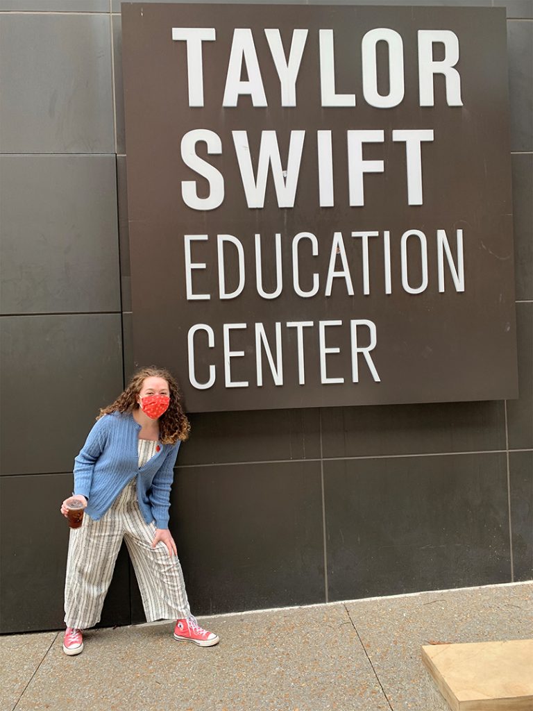
My three favorite things in one picture: lavender cold brew, Taylor Swift, and my blue wool cardigan.
The only thing that’s stuck so far has been reading. In middle school, we used to stay up late with a flashlight under our covers to finish books, then abruptly lost all motivation somewhere between The Giver and The Scarlet Letter. I think we forgot along the way that there are no rules to reading; there’s no one to impress. There’s no one to sample your sourdough or judge your twangy, painful acoustic cover of “Three Blind Mice.” Reading is something you do purely for yourself.
Reading makes information and ideas universally accessible; it connects worlds using only ink on a page. There’s this myth that analytical minds are not creative minds and vice versa, and it alienates people: people who would bring such great perspectives to the table if they hadn’t been defined by a checklist of abilities. Reading is for everyone to find what they love and to love what they find (or hate it; one of the great things about doing things for yourself is that you can just quit whenever you want to).
Scientific research, on the other hand, is something produced for everyone. Humans exploring more and more about the world is something that affects all of us, despite the research being conducted only by a select few of us.
My black long-sleeve cardigan is a personal favorite, as it goes with pretty much everything.
Freshman year of high school, I finished chemistry with a B, which was a miracle considering I was rocking a D around November. I had to change my way of looking at the material; I couldn’t remember the makeup of an atom, but I could remember it if I thought about the stories of individuals who built off of each model in succession. I didn’t understand stoichiometry, but I did understand you have to balance equations just like weights on a scale or kids on a see-saw.
My point is: everyone sees things differently. Exclusivity in different fields is fabricated to make information and education elitist, and it is not reflective of individuals’ ability to understand the world. If you want to read about cool science stuff, you shouldn’t feel left out because you’re more of an art history person. If you want to read about cool art history stuff, you shouldn’t feel left out because you’re an aerospace engineer.
So I like to think that I can be that bridge for some people; at the very least, I can do it for myself.
Engineers, medical students, ecologists, political scientists, ethicists, policymakers — come one, come all to the Duke Space Initiative (DSI), “the interdisciplinary home for all things space at Duke.”
At Duke Polis’ “Perspectives on Space: Introducing the Duke Space Initiative” on Sept. 9, DSI co-founder and undergraduate student Ritika Saligram introduced the initiative and moderated a discussion on the current landscape of space studies both at Duke and beyond.
William R. & Thomas L. Perkins Professor of Law Jonathan Wiener began by expressing his excitement in the amount of interest he’s observed in space at Duke.
One of these interested students was Spencer Kaplan. Kaplan, an undergraduate student studying public policy, couldn’t attend Wiener’s Science & Society Dinner Dialogue about policy and risk in the settlement of Mars. Unwilling to miss the learning opportunity, Kaplan set up a one-on-one conversation with Wiener. One thing led to another: the two created a readings course on space law — Wiener hired Kaplan as a research assistant and they worked together to compile materials for the syllabus — then thought, “Why stop there?”
Wiener and Kaplan, together with Chase Hamilton, Jory Weintraub, Tyler Felgenhauer, Dan Buckland, and Somia Youssef, created the Bass Connections project “Going to Mars: Science, Society, and Sustainability,” through which a highly interdisciplinary team of faculty and students discussed problems ranging from the science and technology of getting to Mars, to the social and political reality of living on another planet.
The team produced a website, research papers, policy memos and recommendations, and a policy report for stakeholders including NASA and some prestigious actors in the private sector. According to Saligram, through their work, the team realized the need for a concerted “space for space” at Duke, and the DSI was born. The Initiative seeks to serve more immediately as a resource center for higher education on space, and eventually as the home of a space studies certificate program for undergraduates at Duke.
Wiener sees space as an “opportunity to reflect on what we’ve learned from being on Earth” — to consider how we could avoid mistakes made here and “try to do better if we settle another planet.” He listed a few of the many problems that the Bass Connections examined.
The economics of space exploration have changed: once, national governments funded space exploration; now, private companies like SpaceX, Blue Origin, and Virgin Galactic seek to run the show. Space debris, satellite and launch junk that could impair future launches, is the tragedy of the commons at work — in space. How would we resolve international disputes on other planets and avoid conflict, especially when settlements have different missions? Can we develop technology to ward off asteroids? What if we unintentionally brought microorganisms from one planet to another? How will we make the rules for the settlement of other planets?
These questions are vast — thereby reflecting the vastness of space, commented Saligram — and weren’t answerable within the hour. However, cutting edge research and thinking around them can be found on the Bass Connections’ website.
Earth and Climate Sciences Senior Lecturer Alexander Glass added to Wiener’s list of problems: “terraforming” — or creating a human habitat — on Mars. According to Glass, oxygen “isn’t a huge issue”: MOXIE can buzz Co2 with electricity to produce it. A greater concern is radiation. Without Earth’s magnetosphere, shielding of some sort will be necessary; it takes sixteen feet of rock to produce the same protection. Humans on Mars might have to live underground.
Glass noted that although “we have the science to solve a lot of these problems, the science we’re lagging in is the human aspects of it: the psychological, of humanity living in conditions like isolation.” The engineering could be rock solid. But the mission “will fail because there will be a sociopath we couldn’t predict beforehand.”
Bass Connections project leader and PhD candidate in political science Somia Youssef discussed the need to examine deeply our laws, systems, and culture. Youssef emphasized that we humans have been on Earth for six million years. Like Wiener, she asked how we will “apply what we’ve learned to space” and what changes we should make. How, she mused, do prevailing ideas about humanity “transform in the confines, the harsh environment of space?” Youssef urged the balancing of unity with protection of the things that make us different, as well as consideration for voices that aren’t being represented.
Material Science Professor, Assistant Professor of Surgery, and NASA Human System Risk Manager Dr. Dan Buckland explained that automation has exciting potential in improving medical care in space. If robots can do the “most dangerous aspects” of mission medical care, humans won’t have to. Offloading onto “repeatable devices” will reduce the amount of accidents and medical capabilities needed in space.
Multiple panelists also discussed the “false dichotomy” between spending resources on space and back home on Earth. Youssef pointed out that many innovations which have benefited (or will benefit) earthly humanity have come from the excitement and passion that comes from investing in space. Saligram stated that space is an “extension of the same social and policy issues as the ones we face on Earth, just in a different context.” This means that solutions we find in our attempt to settle Mars and explore the universe can be “reverse engineered” to help Earth-dwelling humans everywhere.
Saligram opened up the panel for discussion, and one guest asked Buckland how he ended up working for NASA. Buckland said his advice was to “be in rooms you’re not really supposed to be in, and eventually people will start thinking you’re supposed to be there.”
Youssef echoed this view, expressing the need for diverse perspectives in space exploration. She’s most excited by all the people “who are interested in space, but don’t know if there’s enough space for them.”
If this sounds like you, check out the Duke Space Initiative. They’ve got space.
Imagine: you wake on a chilly November morning, alarm blaring, for your 8:30 am class. You toss aside the blankets and grab your phone. Shutting the alarm off reveals a Washington Post notification. But this isn’t your standard election headline. You almost drop your phone in shock. It can’t be, you think. This is too good to be true. It’s not — a second later, you get a text from the SymMon app, notifying you of your upcoming appointment in the Bryan Center.
A vaccine for COVID-19 is finally available, and you’re getting one.
This scenario could be less far-fetched than one might think: the Centers for Disease Control and Prevention has told officials to prepare for a vaccine as soon as November 1st. To a country foundering due to the economic and social effects of COVID-19, this comes as incredible news — a bright spot on a bleak horizon. But to make a vaccine a reality, traditional phase 3 clinical trials may not be enough. What are challenge trials? Should they be used? What’s at stake, and what are the ethical implications of the path we choose?
Dr. Marc Lipsitch, Director of the Center for Communicable Disease Dynamics at the Harvard School of Public Health, began by comparing traditional phase 3 trials and challenge trials.
In both kinds of trials, vaccines are tested for their “safety and ability to provoke an immune response” in phases 1 and 2. In phase 3 trials, large numbers (typically thousands or tens of thousands) of individuals are randomly assigned either the vaccine being tested or a placebo. Scientists observe how many vaccinated individuals become infected compared to participants who received a placebo. This information enables scientists to assess the efficacy — as well as rarer side effects — of the vaccine.
Marc Lipsitch
In challenge trials, instead of random assignment, small numbers of low-risk individuals are deliberately infected in order to more directly study the efficacy of vaccine and treatment candidates. Though none are underway yet, the advocacy group 1Day Sooner has built a list of more than 35,000 volunteers willing to participate.
Dr. Cameron Wolfe, an Infectious Disease Specialist, Associate Professor of Medicine, and Clinical Expert In Respiratory and Infectious Disease at the Duke Medical School, provided an overview of the current vaccine landscape.
Cameron Wolfe
There are currently at least 150 potential vaccine candidates, from preclinical to approved stages of development. Two vaccines, developed by Russia’s Gamelaya Research Institute and China’s CanSinoBIO, have skipped phase 3, but are little more than an idiosyncrasy to Dr. Wolfe, as there is “minimal clarity about their safety and efficacy.” Three more vaccines of interest — Moderna’s mRNA vaccine, Pfizer’s mRNA vaccine, and Oxford and AstraZeneca’s adenovirus vaccine — are all in phase 3 trials with around 30,000 enrollees. Scientists will be watching for a “meaningful infection and a durable immune response.”
Dr. Nir Eyal, the Henry Rutgers Professor of Bioethics and Director of The Center for Population-Level Bioethics at Rutgers University, explained how challenge trials could fit into the vaccine roadmap.
According to Dr. Eyal, challenge trials would most likely be combined with phase 3 trials. One way this could look is the use of challenge trials to weed out vaccine candidates before undergoing more expensive phase 3 trials. Additionally, if phase 3 trials fail to produce meaningful results about efficacy, a challenge trial could be used to obtain information while still collecting safety data from the more comprehensive phase 3 trial.
Nir Eyal
Dr. Eyal emphasized the importance of challenge trials for expediting the arrival of the vaccine. According to his own calculations, getting a vaccine — and making it widely available — just one month sooner would avert the loss of 720,000 years of life and 40 million years of poverty, mostly concentrated in the developing world. (Dr. Eyal stressed that his estimate is extremely conservative as it neglects many factors, including loss of life from avoidance of child vaccines, cancer care, malaria treatment, etc.) Therefore, speed is of “great humanitarian value.”
Dr. Wolfe added that because phase 3 trials rely on a lot of transmission, if the US gets better at mitigating the virus, “the distinction between protective efficacy and simple placebo will take longer to see.” A challenge study, however, is “always a well defined time period… you can anticipate when you’ll get results.”
The panelists then discussed the ethics of challenge trials in the absence of effective treatment — as Krawiec put it, “making people sick without knowing if we can make them better.”
Dr. Wolfe pointed to the flu, citing challenge trialsthat havebeen conducted even though current treatments are not uniformly effective (“tamiflu is no panacea”). He then conceded that the biggest challenge is not a lack of effective therapies, but the current inability to “say to a patient, ‘you will not have a severe outcome.’ It varies so much from person to person, I guess.” (See one troubling example of that variance.)
Dr. Eyal acknowledged the trouble of informed consent when the implications are scarcely known, but argued that “in extraordinary times, business as usual is no longer the standard.” He asserted that if people volunteer with full understanding of what they are committing to, there is no reason to assume they are less informed than when making other decisions where the outcome is as yet unknown.
Dr. Lipsitch compared this to the military: “we are not cheating if we cannot provide a roadmap of future wars because they are not yet known to us.” Rather, we commend brave soldiers (and hope they come home safe).
Furthermore, Dr. Eyal asserted that “informed consent is not a comprehensive understanding of the disease,” lest much of the epidemiological research from the 1970s be called into question too. Instead, volunteers should be considered informed as long as they comprehend questions like, “‘we can’t give you an exact figure yet; do you understand?’”
Agreeing, Dr. Wolfe stated that when critics of challenge trials ask, isn’t your mission to do no harm?, he asks, “Do no harm in regards to whom?” “Who is in front of you matters,” Dr. Wolfe confirmed, “that’s why we put up safeguards. But as clinicians it can be problematic [to stop there]. It’s not just about the patient, but to do no harm in regards to the broader community.”
The experts then discussed what they’d like to see in challenge trials.
Dr. Wolfe said he’d like to see challenge trials carried out with a focus on immunology components, side effect profiles, and a “barrage” of biological safety and health standards for hospitals and facilities.
Dr. Eyal stated the need for exclusion criteria (young adults, perhaps age 20-25, with no risk factors), a “high high high” quality of informed consent ideally involving a third party, and access to therapies and critical care for all volunteers, even those without insurance.
Dr. Lipsitch stressed the scientific importance of assessing participants from a “virological, not symptom bent.” He mused that the issue of viral inoculum was a thorny one — should scientists “titrate down” to where many participants won’t get infected and more volunteers will be needed overall? Or should scientists keep it concentrated, and contend with the increased risk?
Like many questions pondered during the hour — from the ideal viral strain to use to the safest way to collect information about high risk patients — this one remained unanswered.
So don’t mark November 1st on your calendar just yet. But if you do get that life-changing notification, there’s a chance you’ll have human challenge trials to thank.
This squiggly line shows the path taken by a snippet of DNA as it might move around within the soupy interior of a cell. Duke’s Kevin Welsher and colleagues have developed a technique that turns a microscope into a ‘flight tracker’ for molecules, making it possible to follow the paths of viruses and other particles thousands of times smaller than the period at the end of this sentence. Until now, such techniques have required particles to be tethered to make sure they stay within the field of view. But the Welsher lab has developed a way to lock on to freely moving targets and track them for minutes at a time.
Many university labs may have gone quiet amid coronavirus shutdowns, but faculty continue to analyze data, publish papers and write grants. In this guest post from Duke chemistry professor David Beratan and colleagues, the researchers describe a new study showing how water’s ability to shepherd electrons can change with subtle shifts in a water molecule’s 3-D structure:
Water, the humble combination of hydrogen and oxygen, is essential for life. Despite its central place in nature, relatively little is known about the role that single water molecules play in biology.
Researchers at Duke University, in collaboration with Arizona State University, Pennsylvania State University and University of California-Davis have studied how electrons flow though water molecules, a process crucial for the energy-generating machinery of living systems. The team discovered that the way that water molecules cluster on solid surfaces enables the molecules to be either strong or weak mediators of electron transfer, depending on their orientation. The team’s experiments show that water is able to adopt a higher- or a lower-conducting form, much like the electrical switch on your wall. They were able to shift between the two structures using large electric fields.
In a previous paper published fifteen years ago in the journal Science, Duke chemistry professor David Beratan predicted that water’s mediation properties in living systems would depend on how the water molecules are oriented.
Water assemblies and chains occur throughout biological systems. “If you know the conducting properties of the two forms for a single water molecule, then you can predict the conducting properties of a water chain,” said Limin Xiang, a postdoctoral scholar at University of California, Berkeley, and the first author of the paper.
“Just like the piling up of Lego bricks, you could also pile up a water chain with the two forms of water as the building blocks,” Xiang said.
In addition to discovering the two forms of water, the authors also found that water can change its structure at high voltages. Indeed, when the voltage is large, water switches from a high- to a low-conductive form. In fact, it is may be possible that this switching could gate the flow of electron charge in living systems.
This study marks an important first step in establishing water synthetic structures that could assist in making electrical contact between biomolecules and electrodes. In addition, the research may help reveal nature’s strategies for maintaining appropriate electron transport through water molecules and could shed light on diseases linked to oxidative damage processes.
The researchers dedicate this study to the memory of Prof. Nongjian (NJ) Tao.
Science
is increasingly asking artificial intelligence machines to help us search and interpret
huge collections of data, and it’s making a difference.
But unfortunately, polymer chemistry — the study of large, complex molecules — has been hampered in this effort because it lacks a crisp, coherent language to describe molecules that are not tidy and orderly.
Think nylon. Teflon. Silicone. Polyester. These and other polymers are what the chemists call “stochastic,” they’re assembled from predictable building blocks and follow a finite set of attachment rules, but can be very different in the details from one strand to the next, even within the same polymer formulation.
Plastics, love ’em or hate ’em, they’re here to stay. Foto: Mathias Cramer/temporealfoto.com
Chemistry’s
old stick and ball models and shorthand chemical notations aren’t adequate for a
long molecule that can best be described as a series of probabilities that one
kind of piece might be in a given spot, or not.
Polymer chemists searching for new materials for medical treatments or plastics that won’t become an environmental burden have been somewhat hampered by using a written language that looks like long strings of consonants, equal signs, brackets, carets and parentheses. It’s also somewhat equivocal, so the polymer Nylon-6-6 ends up written like this:
{<C(=O)CCCCC(=O)<,>NCCCCCCN>}
Or this,
{<C(=O)CCCCC(=O)NCCCCCCN>}
And when we get to something called ‘concatenation
syntax,’ matters only get worse.
Stephen Craig, the William T. Miller Professor of Chemistry, has been a polymer chemist for almost two decades and he says the notation language above has some utility for polymers. But Craig, who now heads the National Science Foundation’s Center for the Chemistry of Molecularly Optimized Networks (MONET), and his MONET colleagues thought they could do better.
Stephen Craig
“Once you have that insight about how a polymer is grown,
you need to define some symbols that say there’s a probability of this kind of
structure occurring here, or some other structure occurring at that spot,”
Craig says. “And then it’s reducing that to practice and sort of defining a set
of symbols.”
Now he and his MONET colleagues at MIT and Northwestern University have done just that, resulting in a new language – BigSMILES – that’s an adaptation of the existing language called SMILES (simplified molecular-input line-entry system). They they think it can reduce this hugely combinatorial problem of describing polymers down to something even a dumb computer can understand.
And that, Craig says, should enable computers to do all the stuff they’re good at – searching huge datasets for patterns and finding needles in haystacks.
The initial heavy lifting was done by MONET members Prof. Brad Olsen and his co-worker Tzyy-Shyang Lin at MIT who conceived of the idea and developed the set of symbols and the syntax together. Now polymers and their constituent building blocks and variety of linkages might be described like this:
Examples of bigSMILES symbols from the recent paper
It’s
certainly not the best reading material for us and it would be terribly difficult
to read aloud, but it becomes child’s play for a computer.
Members
of MONET spent a couple of weeks trying to stump the new language with the
weirdest polymers they could imagine, which turned up the need for a few more
parts to the ‘alphabet.’ But by and large, it holds up, Craig says. They also threw
a huge database of polymers at it and it translated them with ease.
“One of the things I’m excited about
is how the data entry might eventually be tied directly to the synthetic
methods used to make a particular polymer,” Craig says. “There’s an opportunity
to actually capture and process more information about the molecules than is
typically available from standard characterizations. If that can be done,
it will enable all sorts of discoveries.”
BigSMILES was introduced to the
polymer community by an article in ACS Central Science
last week, and the MONET team is eager to see the response.
“Can other people use it and does it work for
everything?” Craig asks. “Because polymer structure space is effectively
infinite.” Which is just the kind of thing you need Big Data and machine
learning to address.
“This is an area where the
intersection of chemistry and data science can have a huge impact,” Craig says.
Many people turn to the Internet to find a Mr. or Ms. Right. But lemurs don’t have to cyberstalk potential love interests to find a good match — they just give them a sniff.
A study of lemur scents finds that an individual’s distinctive body odor reflects genetic differences in their immune system, and that other lemurs can detect these differences by smell.
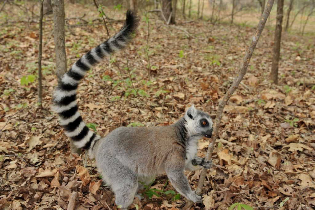
Smell check: Fritz the ring-tailed lemur sniffs a tree for traces of other lemurs’ scents. Photo by David Haring, Duke Lemur Center.
From just one whiff, these primates are able to tell which prospective partners have immune genes different from their own. The ability to sniff out mates with different immune genes could make their offspring’s immune systems more diverse and able to fight more pathogens, said first author Kathleen Grogan, who did the research while working on her Ph.D. with professor Christine Drea at Duke University.
The results appeared online August 22 in the journal BMC Evolutionary Biology.
Lemurs advertise their presence by scent marking — rubbing stinky glands against trees to broadcast information about their sex, kin, and whether they are ready to mate.
Lemurs can tell whether a mate’s immune genes are a good genetic match by the scents they leave behind. Photo by David Haring, Duke Lemur Center
For the study, Grogan, Drea and colleagues collected scent secretions from roughly 60 lemurs at the Duke Lemur Center, the Indianapolis Zoo, and the Cincinnati Zoo. The team used a technique called gas chromatography-mass spectrometry to tease out the hundreds of compounds that make up each animal’s signature scent.
They also analyzed the lemurs’ DNA, looking for differences within a cluster of genes called MHC that help trigger the body’s defenses against foreign invaders such as bacteria and viruses.
Their tests reveal that the chemical cocktail lemurs emit varies depending on which MHC types they carry.
To see if potential mates can smell
the difference, the researchers presented lemurs with pairs of wooden rods
smeared with the bodily secretions of two unfamiliar mates and observed their
responses. Within seconds, the animals were drawn to the smells wafting from
the rods, engaging in a frenzy of licking, sniffing, or rubbing their own
scents on top.
In 300 trials, the team found that
females paid more attention to the scents of males whose immune genes differed
from their own.
MHC genes code for proteins that help the immune system recognize foreign invaders and distinguish “friend” from “foe.” Since different genetic versions respond to different sets of foreign substances, Grogan said, sniffing out genetically dissimilar mates produces offspring more capable of fighting a broad range of pathogens.
Just because females spent more time
checking out the scents of dissimilar males doesn’t necessarily make them more
likely to have kids together, Grogan said. Moving forward, she and her
colleagues plan to use maternity and paternity DNA test results from wild
lemurs living in Beza Mahafaly Reserve in Madagascar to see if lemur couples
are more different in their MHC type than would be expected by chance.
Similar results have been found in humans, but this is the first time the ability to sniff out partners based on their immune genes has been shown in such distant primate kin, said Grogan, who is currently a postdoctoral fellow at Pennsylvania State University.
“Growing evidence suggests that
primates rely on olfactory cues way more than we thought they did,” Grogan
said. “It’s possible that all primates can do this.”
This research was supported by the National Science Foundation (BCS #0409367, IOS #0719003), the National Institutes of Health (F32 GM123634–01), and the Duke University Center for Science Education.
CITATION: “Genetic Variation at MHC class II Loci Influences Both Olfactory Signals and Scent Discrimination in Ring-Tailed Lemurs,” Kathleen E. Grogan, Rachel L. Harris, Marylène Boulet, and Christine M. Drea. BMC Evolutionary Biology, August 22, 2019. DOI: 10.1186/s12862-019-1486-0
The technical-sounding category of “light-driven
charge-transfer reactions,” becomes more familiar to non-physicists when you just
call it photosynthesis or solar electricity.
When a molecule (in a leaf or solar cell) is hit by an
energetic photon of light, it first absorbs the little meteor’s energy, generating
what chemists call an excited state. This excited state then almost immediately
(like trillionths of a second) shuttles an electron away to a charge acceptor
to lower its energy. That transference of charge is what drives plant life and
photovoltaic current.
A 20 Megawatt solar farm ( Aerial Innovations via wikimedia commons)
The energy of the excited state plays an important role in
determining solar energy conversion efficiency. That is, the more of that
photon’s energy that can be retained in the charge-separated state, the better.
For most solar-electric devices, the excited state rapidly loses energy,
resulting in less efficient devices.
But what if there were a way to create even more energetic
excited states from that incoming photon?
Using a very efficient photosynthesizing bacterium as their inspiration,
a team of Duke chemists that included graduate students Nick Polizzi and Ting
Jiang, and faculty members David Beratan and Michael Therien, synthesized a
“supermolecule” to help address this question.
“Nick and Ting discovered a really cool trick about electron
transfer that we might be able to adapt to improving solar cells,” said Michael
Therien, the William R. Kenan, Jr. Professor of Chemistry. “Biology figured
this out eons ago,” he said.
“When molecules absorb light, they have more energy,” Therien said. “One of the things that these molecular excited states do is that they move charge. Generally speaking, most solar energy conversion structures that chemists design feature molecules that push electron density in the direction they want charge to move when a photon is absorbed. The solar-fueled microbe, Rhodobacter sphaeroides, however, does the opposite. What Nick and Ting demonstrated is that this could also be a winning strategy for solar cells.”
Ting Jiang
Nick Polizzi
The chemists devised a clever synthetic molecule that shows the advantages of an excited state that pushes electron density in the direction opposite to where charge flows. In effect, this allows more of the energy harvested from a photon to be used in a solar cell.
“Nick and Ting’s work shows that there are huge advantages
to pushing electron density in the exact opposite direction where you want
charge to flow,” Therien said in his top-floor office of the French Family
Science Center. “The biggest advantage of an excited state that pushes charge the
wrong way is it stops a really critical pathway for excited state relaxation.”
“So, in many ways it’s a Rube Goldberg Like conception,”
Therien said. “It is a design strategy that’s been maybe staring us in the face
for several years, but no one’s connected the dots like Nick and Ting have
here.”
In a July 2
commentary for the Proceedings of the National Academy of Sciences, Bowling
Green State University chemist and photoscientist Malcom D.E. Forbes calls this
work “a great leap forward,” and says it “should be regarded as one of the most
beautiful experiments in physical chemistry in the 21st century.”
Here’s a schematic from the paper. (Image by Nick Polizzi)
CITATION: “Engineering Opposite Electronic Polarization of
Singlet and Triplet States Increases the Yield of High-Energy Photoproducts,”
Nicholas Polizzi, Ting Jiang, David Beratan, Michael Therien. Proceedings of
the National Academy of Sciences, June 10, 2019. DOI: 10.1073/pnas.1901752116
Online: https://www.pnas.org/content/early/2019/07/01/1908872116
Have you ever questioned the quality of the water you drink every day? Or worried that cooking with tap water might be dangerous? For most of us, the answer to these questions is probably no. However, students from a Bass Connections team at Duke say we may want to think otherwise.
From bottle refilling
stations to the tap, drinking water is so habitual and commonplace that we often
take it for granted. Only in moments of crisis do we start worrying about what’s
in the water we drink daily. The reality is that safe drinking water isn’t accessible
for a lot of people.
Pig waste discoloring lagoon water
Images like this hog farm motivated the Bass Connections project team DECIPHER to take a closer look at the quality of water in North Carolina. On April 16 they presented their concerning findings from three case studies looking at lead contamination, coal ash impoundments, and aging infrastructure at the Motorco Music Hall.
Motorco in Durham. The talk was inside, though.
Nadratun Chowdhury, a Ph.D. student in Civil and Environmental Engineering, investigated lead contamination in water. Lead is an abundant and corrosion-resistant material, making it appealing for use in things like paint, batteries, faucets and pipes. While we’ve successfully removed lead from paint and gasoline, a lot of old water pipes in use today are still fashioned from lead. That’s not good – lead is very toxic and can leach into the water.
Just how toxic is it? Anything over a blood-lead level concentration of fifty parts per billion – fifty drops of water in a giant Olympic swimming pool – is considered dangerous. According to Duke graduate student Aaron Reuben, this much lead in one’s blood is correlated with downward social mobility, serious health concerns, diminished capacity to regulate thoughts and emotions, and hyperactivity. Lower income and minority areas are more at risk due to the higher likelihood of owning contaminated older homes.
Rupanjali Karthik, a Master of Laws student, conducted research on the intersection of water and aging infrastructure in Orange County. Breaks in water pipes are common and can result in serious consequences, like the loss of 9 million gallons of drinkable water. Sometimes it takes 8 or 9 months just to find the location of a broken pipe. In 2018, the UNC-Chapel Hill water main break caused a huge shortage on campus and at the medical center.
Excess fluoridation is also
an issue caused by aging infrastructure. In February 2017, a combination of
human and machine error caused an excessive fluoride concentration coming out
of an Orange County Water Treatment Plant. People were advised not to use their
water even to shower. A UNC basketball game had to move locations, and stores
were completely swept of bottled water.
Another issue is that arsenic, a known carcinogen, is often used as the fluoridation agent. We definitely don’t want that in our drinking water. Fluoridation isn’t even that necessary these days when we have toothpaste and mouthwash that supports our dental health.
Tommy Lin, an undergraduate
studying Chemistry and Computer Science, topped off the group’s presentation with
findings surrounding coal ash in Belmont, NC. Coal ash, the residue after coal
is burned in power plants, can pollute rivers and seep into ground water,
affecting domestic wells of neighboring communities. This creates a cocktail of
highly concentrated heavy metals and carcinogens. Drinking it can cause damage
to your nervous system, cancer, and birth defects, among other things. Not so great.
The group’s presentation.
Forty-five plastic water bottles.
That’s how much water it takes Laura, a Belmont resident, to cook her middle-sized
family Thanksgiving. She knows that number because it’s been her family’s
tradition the past three years. The Allen Plant Steam Station is a big culprit
of polluting water with coal ash. Tons of homes nearby the station, like Laura’s,
are told not to use the tap water. You can find these homes excessively
stockpiled with cases on cases of plastic water bottles.
These issues aren’t that apparent to people unless they have been directly impacted. Lead, aging infrastructure, and coal ash all pose real threats but are also very invisible problems. Kathleen Burns, a Ph.D. student in English, notes that only in moments of crisis will people start to care, but by then it may be too late.
So, what can people do? Not much, according to the Bass Connections team. They noted that providing clean water is very much a structural issue which will require some complex steps to be solved. So, for now, you may want to go buy a Brita.
Protein crystals don’t usually display the glitz and glam of gemstones. But no matter their looks, each and every one is precious to scientists.
Patrick Charbonneau, a professor of chemistry and physics at Duke, along with a worldwide group of scientists, teamed up with researchers at Google Brain to use state-of-the-art machine learning algorithms to spot these rare and valuable crystals. Their work could accelerate drug discovery by making it easier for researchers to map the structures of proteins.
“Every time you miss a protein crystal, because they are so rare, you risk missing on an important biomedical discovery,” Charbonneau said.
Knowing the structure of proteins is key to understanding their function and possibly designing drugs that work with their specific shapes. But the traditional approach to determining these structures, called X-ray crystallography, requires that proteins be crystallized.
Crystallizing proteins is hard — really hard. Unlike the simple atoms and molecules that make up common crystals like salt and sugar, these big, bulky molecules, which can contain tens of thousands of atoms each, struggle to arrange themselves into the ordered arrays that form the basis of crystals.
“What allows an object like a protein to self-assemble into something like a crystal is a bit like magic,” Charbonneau said.
Even after decades of practice, scientists have to rely in part on trial and error to obtain protein crystals. After isolating a protein, they mix it with hundreds of different types of liquid solutions, hoping to find the right recipe that coaxes them to crystallize. They then look at droplets of each mixture under a microscope, hoping to spot the smallest speck of a growing crystal.
“You have to manually say, there is a crystal there, there is none there, there is one there, and usually it is none, none, none,” Charbonneau said. “Not only is it expensive to pay people to do this, but also people fail. They get tired and they get sloppy, and it detracts from their other work.”
The machine learning software searches for points and edges (left) to identify crystals in images of droplets of solution. It can also identify when non-crystalline solids have formed (middle) and when no solids have formed (right).
Charbonneau thought perhaps deep learning software, which is now capable of recognizing individual faces in photographs even when they are blurry or caught from the side, should also be able to identify the points and edges that make up a crystal in solution.
Scientists from both academia and industry came together to collect half a million images of protein crystallization experiments into a database called MARCO. The data specify which of these protein cocktails led to crystallization, based on human evaluation.
The team then worked with a group led by Vincent Vanhoucke from Google Brain to apply the latest in artificial intelligence to help identify crystals in the images.
After “training” the deep learning software on a subset of the data, they unleashed it on the full database. The A.I. was able to accurately identify crystals about 95 percent of the time. Estimates show that humans spot crystals correctly only 85 percent of the time.
“And it does remarkably better than humans,” Charbonneau said. “We were a little surprised because most A.I. algorithms are made to recognize cats or dogs, not necessarily geometrical features like the edge of a crystal.”
Other teams of researchers have already asked to use the A.I. model and the MARCO dataset to train their own machine learning algorithms to recognize crystals in protein crystallization experiments, Charbonneau said. These advances should allow researchers to focus more time on biomedical discoveries instead of squinting at samples.
Charbonneau plans to use the data to understand how exactly proteins self-assemble into crystals, so that researchers rely less on chance to get this “magic” to happen.
“We are trying to use this data to see if we can get more insight into the physical chemistry of self-assembly of proteins,” Charbonneau said.
CITATION: “Classification of crystallization outcomes using deep convolutional neural networks,” Andrew E. Bruno, et al. PLOS ONE, June 20, 2018. DOI: 10.1371/journal.pone.0198883


 Post by Robin A. Smith


