Following the people and events that make up the research community at Duke

Students exploring the Innovation Co-Lab

Category: Biology Page 17 of 32

Closing the Funding Gap for Minority Scientists

DURHAM, N.C. — The barriers to minority students in science, technology, engineering and math (STEM) don’t go away once they’ve finished school and landed a job, studies show. But one nationwide initiative aims to level the playing field once they get there.

With support from a 3-year, $500,0000 grant from the National Science Foundation, assistant professors and postdoctoral fellows who come from underrepresented minorities are encouraged to apply by May 5 for a free grant writing workshop to be held June 22-24 in Washington, D.C..

It’s no secret that STEM has a diversity problem. In 2015, African-Americans and Latinos made up 29 percent of the U.S. workforce, but only 11 percent of scientists and engineers.

A study published in the journal Science in 2011 revealed that minority scientists also were less likely to win grants from the National Institutes of Health, the largest source of research funding to universities.

Based on an analysis of 83,000 grant applications from 2000 to 2006, the study authors found that applications from black researchers were 13 percent less likely to succeed than applications from their white peers. Applications from Asian and Hispanic scientists were 5 and 3 percent less likely to be awarded, respectively.

Even when the study authors made sure they were comparing applicants with similar educational backgrounds, training, employers and publication records, the funding gap persisted — particularly for African-Americans.

Competition for federal research dollars is already tough. But white scientists won 29 percent of the time, and black scientists succeeded only 16 percent of the time.

Pennsylvania State University chemistry professor Squire Booker is co-principal investigator of a $500,000 initiative funded by the National Science Foundation to help underrepresented minority scientists write winning research grants.

“That report sent a shock wave through the scientific community,” said Squire Booker, a Howard Hughes Medical Institute investigator and chemistry professor at Pennsylvania State University. Speaking last week in the Nanaline H. Duke building on Duke’s Research Drive, Booker outlined a mentoring initiative that aims to close the gap.

In 2013, Booker and colleagues on the Minority Affairs Committee of the American Society for Biochemistry and Molecular Biology decided to host a workshop to demystify the grant application process and help minority scientists write winning grants.

Grant success is key to making it in academia. Even at universities that don’t make funding a formal requirement for tenure and promotion, research is expensive. Outside funding is often required to keep a lab going, and research productivity — generating data and publishing results — is critical.

To insure underrepresented minorities have every chance to compete for increasingly tight federal research dollars, Booker and colleagues developed the Interactive Mentoring Activities for Grantsmanship Enhancement program, known as IMAGE. Program officers from NIH and NSF offer tips on navigating the funding process, crafting a successful proposal, decoding reviews and revising and resubmitting. The organizers also stage a mock review panel, and participants receive real-time, constructive feedback on potential research proposals.

Participants include researchers in biology, biophysics, biochemistry and molecular biology. More than half of the program’s 130 alumni have been awarded NSF or NIH grants since the workshop series started in 2013.

Booker anticipates this year’s program will include more postdoctoral fellows. “Now we’re trying to expand the program to intervene at an earlier stage,” Booker said.

To apply for the 2017 workshop visit http://www.asbmb.org/grantwriting/.  The application deadline is May 5.

s200_robin.smith

Post by Robin Smith

The Road to a Tastier Tomato

This week, I discovered that I’ve lived life deprived of a good tomato.

As a tomato-lover, I was surprised to learn from Professor Harry Klee of the University of Florida that the supermarket tomatoes I’ve enjoyed throughout my 18-year existence are all flavorless compared to the tomatoes of the past. He spoke at Duke as a guest of the University Program in Genetics and Genomics on Feb. 28.

It turns out that commercial tomato growers, by breeding more profitable (i.e. higher-yield, redder-color, larger-fruit) tomato varieties over the past 50 years, inadvertently excluded what Klee believes is the most important tomato trait of all:

Commercial tomato growers have bred larger, redder tomatoes that are less flavorful than heirloom and older varieties. Image courtesy of Harry Klee.

Flavor.

Apparently, I was one of very few people unaware of this issue. The public outcry in response to the increasing flavorlessness of commercial tomatoes began over a decade ago, when Klee first began to study tomato genetics.

From his research, Klee has drawn several important, unexpected conclusions, chief among them:

1: Flavor has more to do with smell than taste;

2: Lesser-known biochemical compounds called “volatiles” influence the flavor of tomatoes more than sugars, acids, and other well-known, larger compounds;

3: These “volatiles” are less present in modern tomato varieties than in tastier, older, and heirloom varieties;

But fear not—

4: Tomatoes can be back-bred to regain the genes that code for volatile compounds.

In other words, Klee has mapped the way back to the flavorful tomatoes of the past. His work culminated in a cover story of the Jan. 27 issue of Science. The corresponding paper describing the analysis of over 300 tomato strains to identify the chemicals associated with “good” and “bad” tomatoes.

Dr. Harry Klee and collaborators in his lab at the University of Florida. Image courtesy of Harry Klee.

To prove that modern tomatoes have less of the compounds that make them tasty, Klee and his team recruited a panel of 100 taste-testers to rank 160 representative tomato varieties. According to Klee, the team “developed statistical models to explain the chemistry of ‘liking’ [tomatoes],” then narrowed down the list of compounds that correlated with “liking” from 400 to 26. After tracing these 26 compounds to genetic loci, they used whole-genome sequencing to show that these loci are less expressed in modern tomatoes than in “cerasiforme” (i.e. old) and heirloom tomato varieties.

Further studies showed that tomato weight is inversely correlated with sugar content—in other words, “a gigantic fruit doesn’t taste as good,” Klee said.

If Klee can convince tomato growers that consumers value flavor over size, color, and quantity, then he might just single-handedly put flavorful tomatoes back on the shelves. Nevertheless—and despite the publicity surrounding his work—Klee understands it make take a while before commercial tomato growers see the light.

Klee and his team of scientists have genetically mapped the way back to the tasty tomatoes of the past. Image courtesy of Harry Klee.

“Growers get no more money if the tomato tastes good or bad; they’re paid for how many pounds of red objects they put in a box…[but] we can’t just blame the modern breeders. We’ve been selecting bigger and bigger fruit for millennia, and that has come at the cost of reducing flavor,” Klee said.

Post by Maya Iskandarani

Cells Need Their Personal Space

One of the body’s first lines of defense against harmful pathogens is the skin. The constant maintenance of this epithelial cell layer which serves as a barrier to infection  is essential to fighting off disease.

Jody Rosenblatt, an Associate Professor in the Department of Oncological Sciences at the University of Utah School of Medicine, has made it her lab’s mission to study the function of epithelia as a barrier, how this barrier is maintained, and what happens when it goes awry.

Jody Rosenblatt, PhD is an investigator for the Huntsman Cancer Institute at the University of Utah School of Medicine and a Howard Hughes Medical Institute Faculty Scholar

Rosenblatt recently spoke at Duke’s Developmental & Stem Cell Biology Colloquium where she presented some extraordinary findings about how epithelia can squeeze out  both healthy and dying cells  to preserve the protective barrier.

Some c cells commit suicide via programed cell death and are forced out of the cell layer because they are no longer functional. But in the case of forcing out living cells, “cell extrusion is more like a homicide” said Rosenblatt. The fact that perfectly functional living cells are pushed out of a cell layer perplexed her group until they discovered it was happening as a response to cell overcrowding.

Rosenblatt explained that like people, cells tend to like their personal space, so when this is compromised, live cells are actively pushed out of the cell layer, restoring balanced cell numbers.

Rosenblatt’s lab took this discovery a step farther and pinpointed the pathway that likely induces the extrusion of live cells.

Piezo1, a stretch-activated calcium ion channel present in epithelial cells, senses crowding and activates sphingosine-1-phosphate (S1P), the driver of epithelial cell extrusion. When Piezo1 channels are inhibited and don’t sense stretching, cells cannot extrude.

Using zebrafish, Rosenblatt showed that when extrusion was blocked by compromising the S1P2 pathway, epidermal cells form masses that are resistant to chemotherapy drugs and signals for programmed cell death.

Rosenblatt explains the importance of regulating cell extrusion in the epithelium to maintain the tissue’s function as a protective barrier for our organs. Misregulation of this function can result in diseases such as metastatic cancers.

This finding lead them to examine samples of human pancreatic, lung, colon, and breast tumors. They found that in all of these cancers, S1P2 is significantly reduced. But if they restored S1P2 activity in cell lines of these cancers, the extrusion pathway was rescued and tumor size and metastases were greatly decreased!

Rosenblatt and her colleagues have shown that the importance of cell extrusion cannot be overstated. If extrusion is compromised, cells can begin to pile up and move beneath the cell layer, which can lead to invasion of the tissues beneath the epithelium and metastasis to other sites in the body.

Now that we are uncovering more of the pathways involved in tumor formation and metastasis, we can develop new drugs that may be the key to fighting these devastating diseases.

Guest Post by Amanda Cox, PhD candidate in biology


Young Scientists, Making the Rounds

“Can you make a photosynthetic human?!” an 8th grader enthusiastically asks me while staring at a tiny fern in a jar.

He’s not the only one who asked me that either — another student asked if Superman was a plant, since he gets his power from the sun.

These aren’t the normal questions I get about my research as a Biology PhD candidate studying how plants get nutrients, but they were perfect for the day’s activity –A science round robin with Durham eighth-graders.

Biology grad student Leslie Slota showing Durham 8th graders some fun science.

After seeing a post under #scicomm on Twitter describing a public engagement activity for scientists, I put together a group of Duke graduate scientists to visit local middle schools and share our science with kids. We had students from biomedical engineering, physics, developmental biology, statistics, and many others — a pretty diverse range of sciences.

With help from David Stein at the Duke-Durham Neighborhood Partnership, we made connections with science teachers at the Durham School of the Arts and Lakewood Montessori school, and the event was in motion!

The outreach activity we developed works like speed dating, where people pair up, talk for 3-5 mins, and then rotate. We started out calling it “Science Speed Dating,” but for a middle school audience, we thought “Science Round-Robin” was more appropriate. Typically, a round-robin is a tournament where every team plays each of the other teams. So, every middle schooler got to meet each of us graduate students and talk to us about what we do.

The topics ranged from growing back limbs and mapping the brain, to using math to choose medicines and manipulating the different states of matter.

The kids were really excited for our visit, and kept asking their teachers for the inside scoop on what we did.

After much anticipation, and a little training and practice with Jory Weintraub from the Science & Society Initiative, two groups of 7-12 graduate students armed themselves with photos, animals, plants, and activities related to our work and went to visit these science classes full of eager students.

First-year MGM grad student Tulika Singh (top right) brought cardboard props to show students how antibodies match up with cell receptors.

“The kids really enjoyed it!” said Alex LeMay, middle- and high-school science teacher at the Durham School of the Arts. “They also mentioned that the grad students were really good at explaining ideas in a simple way, while still not talking down to them.”

That’s the ultimate trick with science communication: simplifying what we do, but not talking to people like they’re stupid.

I’m sure you’ve heard the old saying, “dumb it down.” But it really doesn’t work that way. These kids were bright, and often we found them asking questions we’re actively researching in our work. We don’t need to talk down to them, we just need to talk to them without all of the exclusive trappings of science. That was one thing the grad students picked up on too.

“It’s really useful to take a step back from the minutia of our projects and look at the big picture,” said Shannon McNulty, a PhD candidate in Molecular Genetics and Microbiology.

The kids also loved the enthusiasm we showed for our work! That made a big difference in whether they were interested in learning more and asking questions. Take note, fellow scientists: share your enthusiasm for what you do, it’s contagious!

Another thing that worked really well was connecting with the students in a personal way. According to Ms. LeMay, “if the person seemed to like them, they wanted to learn more.” Several of the grad students would ask each student their names and what they were passionate about, or even talk about their own passions outside of their research, and these simple questions allowed the students to connect as people.

There was one girl who shared with me that she didn’t know what she wanted to do when she grew up, and I told her that’s exactly where I was when I was in 8th grade too. We then bonded over our mutual love of baking, and through that interaction she saw herself reflected in me a little bit; making a career in science seem like a possibility, which is especially important for a young girl with a growing interest in science.

Making the rounds in these science classrooms, we learned just as much from the students we spoke to as they did from us. Our lesson being: science outreach is a really rewarding way to spend our time, and who knows, maybe we’ll even spark someone who loves Superman to figure out how to make the first photosynthesizing super-person!

Guest post by Ariana Eily , PhD Candidate in Biology, shown sharing her floating ferns at left.


Seeing Nano

Take pictures at more than 300,000 times magnification with electron microscopes at Duke

Sewer gnat head

An image of a sewer gnat’s head taken through a scanning electron microscope. Courtesy of Fred Nijhout.

The sewer gnat is a common nuisance around kitchen and bathroom drains that’s no bigger than a pea. But magnified thousands of times, its compound eyes and bushy antennae resemble a first place winner in a Movember mustache contest.

Sewer gnats’ larger cousins, horseflies are known for their painful bite. Zoom in and it’s easy to see how they hold onto their furry livestock prey:  the tiny hooked hairs on their feet look like Velcro.

Students in professor Fred Nijhout’s entomology class photograph these and other specimens at more than 300,000 times magnification at Duke’s Shared Material & Instrumentation Facility (SMIF).

There the insects are dried, coated in gold and palladium, and then bombarded with a beam of electrons from a scanning electron microscope, which can resolve structures tens of thousands of times smaller than the width of a human hair.

From a ladybug’s leg to a weevil’s suit of armor, the bristly, bumpy, pitted surfaces of insects are surprisingly beautiful when viewed up close.

“The students have come to treat travels across the surface of an insect as the exploration of a different planet,” Nijhout said.

Horsefly foot

The foot of a horsefly is equipped with menacing claws and Velcro-like hairs that help them hang onto fur. Photo by Valerie Tornini.

Weevil

The hard outer skeleton of a weevil looks smooth and shiny from afar, but up close it’s covered with scales and bristles. Courtesy of Fred Nijhout.

fruit fly wing

Magnified 500 times, the rippled edges of this fruit fly wing are the result of changes in the insect’s genetic code. Courtesy of Eric Spana.

You, too, can gaze at alien worlds too small to see with the naked eye. Students and instructors across campus can use the SMIF’s high-powered microscopes and other state of the art research equipment at no charge with support from the Class-Based Explorations Program.

Biologist Eric Spana’s experimental genetics class uses the microscopes to study fruit flies that carry genetic mutations that alter the shape of their wings.

Students in professor Hadley Cocks’ mechanical engineering 415L class take lessons from objects that break. A scanning electron micrograph of a cracked cymbal once used by the Duke pep band reveals grooves and ridges consistent with the wear and tear from repeated banging.

cracked cymbal

Magnified 3000 times, the surface of this broken cymbal once used by the Duke Pep Band reveals signs of fatigue cracking. Courtesy of Hadley Cocks.

These students are among more than 200 undergraduates in eight classes who benefitted from the program last year, thanks to a grant from the Donald Alstadt Foundation.

You don’t have to be a scientist, either. Historians and art conservators have used scanning electron microscopes to study the surfaces of Bronze Age pottery, the composition of ancient paints and even dust from Egyptian mummies and the Shroud of Turin.

Instructors and undergraduates are invited to find out how they could use the microscopes and other nanotech equipement in the SMIF in their teaching and research. Queries should be directed to Dr. Mark Walters, Director of SMIF, via email at mark.walters@duke.edu.

Located on Duke’s West Campus in the Fitzpatrick Building, the SMIF is a shared use facility available to Duke researchers and educators as well as external users from other universities, government laboratories or industry through a partnership called the Research Triangle Nanotechnology Network. For more info visit http://smif.pratt.duke.edu/.

Scanning electron microscope

This scanning electron microscope could easily be mistaken for equipment from a dentist’s office.

s200_robin.smith

Post by Robin Smith

Totally Tubular! Fluid forces that affect the development of biological tubes

Have you ever wondered how something as simple as fluid can impact the development of a large organism? How about the way tubes form in relation to each other? Or maybe you’ve wondered how it is possible for something as rigid as a spine to be formed from fluid?

Zebrafish embryos are relatively transparent, making them easier to study.

Zebrafish embryos are relatively transparent, making them easier to study.

Dr. Michel Bagnat and his lab work to analyze each of these questions and more in their research about how biological tubes are formed and how pressure exerted by these fluids affects the formation of these tubes.

Dr. Bagnat, an associate professor of cell biology, uses ‘forward genetics,’ a process by which genes are modified in order to see the effect and function of each gene in the organism. The technique enables them to identify and analyze the role of fluid secretion in zebrafish. Fluid secretion also plays a role in many human diseases, including cystic fibrosis and polycystic kidney disease.

The void in a blood vessel is called the lumen. Bagnat studies the cells lining the lumen.

The void in a blood vessel is called the lumen. Bagnat studies the cells lining the lumen.

One of the most interesting aspects of tubal formation is that biological tubes often form in relation to each other. Dr. Bagnat and his lab study this type of tubal formation through studying the lumen, or the thin membrane lining the intestinal tubes of zebrafish. There are many cellular mechanisms that can affect the formation of the lumen, and extensive research is conducted in order to better understand these mechanisms.

These same sorts of forces can even help build a structure as complex as the spine. Dr. Bagnat’s research covers this specific field. The notochord of zebrafish, or the scaffold which will develop into a spine, is heavily affected by the growth of vacuoles, or fluid-filled sacs in the cell. Dr. Bagnat’s research explores the deeper mechanisms behind the filling of these fluid vacuoles in cells and how each cell’s vacuole stops and starts filling with fluid.

This image of fluid-filled sacs forming a fish notochord was on the cover of a journal.

This image of fluid-filled sacs forming a fish notochord was on the cover of a journal.

Overall, Dr. Bagnat’s research holds strong implications for how we understand the development and formation of biological tubes not just in zebrafish but in our own human bodies.

Guest Post by Vaishnavi Siripurapu, North Carolina School of Math and Science, Class of 2018

bagnatselfie


Life Lessons from a Neuroscientist

I recently had the privilege of sitting down with Dr. Anne Buckley, a professor and  neuropathologist working in Dr. Chay Kuo’s cell biology lab at Duke. I got a first-hand account of her research on neuron development and function in mice. But just as fascinating to me were the life lessons she had learned during her time as a researcher.

Anne Buckley, M.D. Ph.D., is an assistant professor of pathology

Anne Buckley, M.D. Ph.D., is an assistant professor of pathology

Buckley’s research looks at brain tumors in mice. She recently found that some of the mice developed the tumors in an area full of neurons, the roof of the fourth ventricle, which is of particular interest because humans have developed tumors in the same location. This discovery could show how neurological pathways affect tumor formation and progression.

Buckley also gave me some critical words of advice, cautioning me that research isn’t for everyone.

“Research is not glamorous, and not always rewarding,” she warned me. When she first started research, Buckley learned a hard lesson: work doesn’t necessarily lead to results. “For every question I went after, I found ten more unresolved,” she said. “To be a researcher, it takes a lot of perseverance and resilience. A lot of long nights.”

But that’s also the beauty of research. Buckley says that she’s learned to find happiness in the small successes, and that she “enjoys the process, enjoys the challenge.”

And when discoveries happen?

“When I look at data, and I see something unexpected, I get really excited,” she says. “I know something that no one else knows. Tomorrow, everyone will know. But tonight, I’m the only person in the world who knows.”

kendra_zhong_headshotGuest Post by Kendra Zhong, North Carolina School of Science and Math, Class of 2017

Evolutionary Genetics Shaping Health and Behavior

Dr. Jenny Tung is interested in the connections between genes and behavior: How does behavior influence genetic variation and regulation and how do genetic differences influence behavior?

A young Amboseli baboon hitches a ride with its mother. (Photo by Noah Snyder-Mackler)

A young Amboseli baboon hitches a ride with its mother. (Photo by Noah Snyder-Mackler)

An assistant professor in the Departments of Evolutionary Anthropology and Biology at Duke, Tung is interested in evolution because it gives us a window into why the living world is the way it is. It explains how organisms relate to one another and their environment. Genetics explains the actual molecular foundation for evolutionary change, and it gives part of the answer for trait variation. Tung was drawn as an undergrad towards the combination of evolution and genetics to explain every living thing we see around us; she loves the explanatory power and elegance to it.

Tung’s longest collaborative project is the Amboseli Baboon Research Project (ABRP), located in the Amboseli ecosystem of East Africa. She co-leads it with Susan Alberts, chair of evolutionary anthropology at Duke, Jeanne Altmann at Princeton, and Beth Archie at Notre Dame.

Tung has spent months at a time on the savannah next to Mount Kilimanjaro for this project. The ABRP monitors hundreds of baboons in several social groups and studies social processes at several levels. Recently the project has begun to include genetics and other aspects of baboon biology, including the social behaviors within the social groups and populations, and how these behaviors have changed along with the changing Amboseli ecosystem. Tung enjoys different aspects of all of her projects, but is incredibly grateful to be a part of the long-term Amboseli study.

Jenny Tung

Jenny Tung is an assistant professor in evolutionary anthropology and biology.

The process of discovery excites Tung. It is hard for her to pin down a single thing that makes research worth it, but “new analyses, discussions with students who teach me something new, seeing a great talk that makes you think in a different way or gives you new research directions to pursue” are all very exciting, she said.

Depending on the project, the fun part varies for her; watching a student develop as a scientist through their own project is rewarding, and she loves collaborating with extraordinary scientists. Specific sets of collaborators make the research worth it. “When collaborations work, you really push each other to be better scientists and researchers,” Tung said.

Raechel ZellerGuest post by Raechel Zeller, North Carolina School of Science and Math, Class of 2017
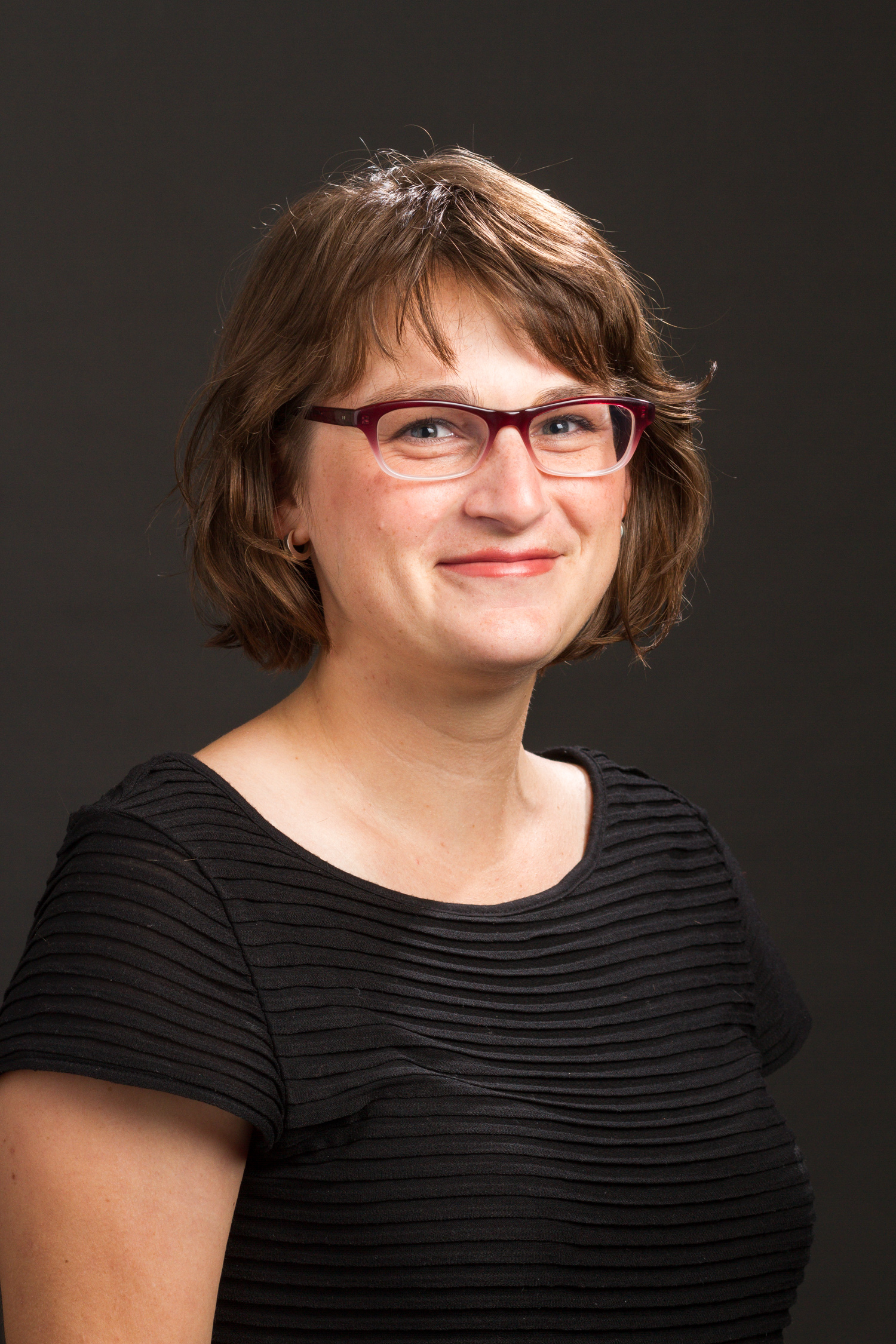
Diabetes — and Privacy — Meet 'Big Data'

“Click here to consent forever.”

If consent to participate in medical research were that simple, Joanna Radin of Yale University would have to find a new focus for her research, and I would never have found the Trent Center for Bioethics, Humanities & History of Medicine.

Luckily for us both, this is not the case. Medical consent is a very complex issue that can, as Radin’s research attests, traverse generations.

joanna-radin-headshot

Joanna Radin’s reserach focuses on the intersection of medical history, anthropology and ethics at Yale University. Source: Yale School of Medicine

Radin is an Associate Professor of Medical History at Yale, the perfect fit for the Humanities in Medicine Lecture Series taking place this month at the Trent Center. Her research nails the narrow intersection of medical history, anthropology, bioethics and data analytics. In fact, Radin’s appeal is so broad that her visit to Duke was sponsored by no less than six Duke departments, including the Departments of Computer Science, History, Electrical and Computer Engineering, Cultural Anthropology and Statistical Science.

Radin’s lecture honed in on a well-known case in the realm of bioethics and medical history: the Pima Native American tribe in Arizona, which is known for unusually high rates of diabetes and obesity. The Pima were the first Native American tribe to be granted a reservation in Arizona—30,000 acres—at the beginning of the California Gold Rush. In 1963, following nearly half a century of mass famine among the Pima, the National Institute of Health (NIH) conducted a survey for rheumatoid arthritis in the Pima tribe, instead discovering a frighteningly high frequency of diabetes.

In 1965, the NIH initiated a long-term observational study of the Pima that continued for about 40 years, though it was meant to last no more than 10. The goal of the study was to learn about diabetes in the “natural laboratory” of sorts that the Pima reservation unwittingly provided. The data collected in this study came to be known as the Pima Indian Diabetes Data set (PIDD).

Machine learning enters the story around 1987, when David Aha and colleagues at the University of California, Irvine (UCI) created the UCI Machine Learning Repository, an archive containing thousands of data sets, databases and data generators. The repository is still active today, virtually a gold mine for researchers in machine learning to test their algorithms. The PIDD is one of the oldest data sets on file in the UCI archive, “a standard for testing data mining algorithms for accuracy in predicting diabetes,” according to Radin.

pima_indian_man_miguel_a_farmer_pima_arizona_ca-1900_chs-3625

A Pima farmer in Pima, Arizona, circa 1900. Source: Wikimedia Commons

Generations’ worth of data on the Pima tribe have been publicly accessible in the UCI archive for over two decades, creating ethical controversy around the accessibility of information as personal as blood pressure, body mass index (BMI) and number of pregnancies of Pima Native Americans. Though the PIDD can help refine machine learning algorithms that could accurately predict—and prevent—diabetes, the privacy issues provoked by the publicness of the data are impossible to ignore.

This is where “eternal” medical consent enters the equation: no researcher can realistically inform a study participant of what their medical data will be used for 40 years in the future.

These are the interdisciplinary questions that Radin brought forth in her lecture, weaving together seemingly opposite fields of study in an engaging, thought-provoking presentation. No one who left that room will look at the Apple Terms & Conditions the same way again.


Post by Maya Iskandarani iskandarani_maya_100hed

Meet the Newbie: Maya Iskandarani

Hello!

My name is Maya Iskandarani, and I’m a freshman at Duke from Miami, Florida—meaning I’ll probably be in trouble once the temperature in Durham drops below 50°.

Messing around with an electric circuit at the Center for Sustainable Development (CSD) at the Island School. Source: Island School Communications Team

Messing around with an electric circuit in the Center for Sustainable Development (CSD) at The Island School. Source: The Island School Communications Team.

Though I might be poorly prepared for “real” seasons, I’m no less excited to start my adventure at Duke. For now, my academic interests lie in Biology (particularly Genetics and Marine Science), Earth and Ocean Sciences, Neuroscience, Spanish, Arabic, and French. I’m also in the Genetics and Genomics cluster of the Duke Focus Program. As you can see, I have some intense self-reflection to do before I declare a major.

I’m a walk-on to the Duke Women’s Rowing team, and feel very lucky to start fresh in an awesome sport with fantastic coaches and teammates. I also participated in Project WILD this summer, camping for two weeks in Pisgah National Forest without much prior camping, hiking, or backpacking experience. Otherwise, I’ve fought my instinct to jump into a million cool extracurricular clubs and programs, so I can get a handle on this “college” thing before I bite off more than I can chew.

In high school, I was an editor for the school newsmagazine (s/o highlights), president of the International Baccalaureate Honor Society (IBHS), vice president of the environmental club, Gables Earth, and a scooper at Whip ‘n Dip, an ice cream shop down the street from my house. I trained with the club swim team Miami Swimming for six years, and competed for my high school swim team for four.

My first taste of research was volunteering with Dr. Claire Paris of the University of Miami in my junior year of high school. I spent a few weeks helping then-PhD-candidate Dr. Erica Staaterman (a Duke alumna!) complete her dissertation on the relationship between marine soundscapes and biodiversity. My role was small, but fascinating: I used a computer program to manually filter hydrophone recordings from coral reefs in the Florida Keys for boat noise.

Preparing for a boat dive off of Cape Eleuthera, with underwater slate in hand to take notes while observing the coral reef.

Preparing for a boat dive off of Cape Eleuthera, with underwater slate in hand to take notes on coral reef life. Source: The Island School Communications Team.

My experience in Dr. Paris’s lab spurred me to further explore marine science, so I applied to attend The Island School over the summer entering my senior year. I spent a month in the blazing heat of the Bahamas, freediving through the same crystal-clear waters in the mornings that I returned to study on SCUBA in the afternoons. Although I’m not quite set on Marine Science as a course of study at Duke, I absolutely intend to spend a semester or two at the Duke Marine Lab to figure it out.

My curiosity in nearly all academic subjects pulls me in a hundred different directions, a few of which I hope to follow through with as part of the Duke Research Blog team. I can’t wait to meet researchers who are passionate about their work, and, perhaps, discover a research passion of my own.

Page 17 of 32

Powered by WordPress & Theme by Anders Norén