Take pictures at more than 300,000 times magnification with electron microscopes at Duke
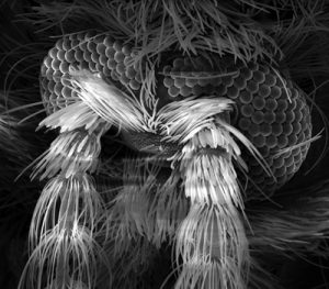
An image of a sewer gnat’s head taken through a scanning electron microscope. Courtesy of Fred Nijhout.
The sewer gnat is a common nuisance around kitchen and bathroom drains that’s no bigger than a pea. But magnified thousands of times, its compound eyes and bushy antennae resemble a first place winner in a Movember mustache contest.
Sewer gnats’ larger cousins, horseflies are known for their painful bite. Zoom in and it’s easy to see how they hold onto their furry livestock prey: the tiny hooked hairs on their feet look like Velcro.
Students in professor Fred Nijhout’s entomology class photograph these and other specimens at more than 300,000 times magnification at Duke’s Shared Material & Instrumentation Facility (SMIF).
There the insects are dried, coated in gold and palladium, and then bombarded with a beam of electrons from a scanning electron microscope, which can resolve structures tens of thousands of times smaller than the width of a human hair.
From a ladybug’s leg to a weevil’s suit of armor, the bristly, bumpy, pitted surfaces of insects are surprisingly beautiful when viewed up close.
“The students have come to treat travels across the surface of an insect as the exploration of a different planet,” Nijhout said.
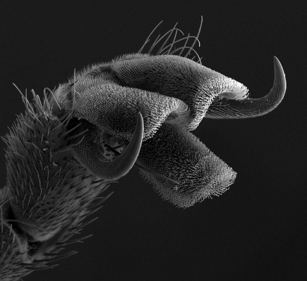
The foot of a horsefly is equipped with menacing claws and Velcro-like hairs that help them hang onto fur. Photo by Valerie Tornini.
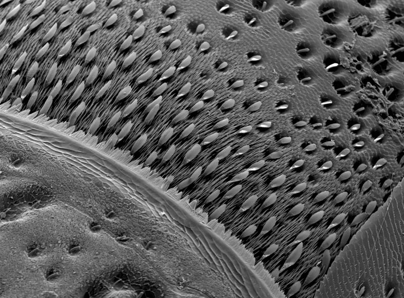
The hard outer skeleton of a weevil looks smooth and shiny from afar, but up close it’s covered with scales and bristles. Courtesy of Fred Nijhout.
Magnified 500 times, the rippled edges of this fruit fly wing are the result of changes in the insect’s genetic code. Courtesy of Eric Spana.
You, too, can gaze at alien worlds too small to see with the naked eye. Students and instructors across campus can use the SMIF’s high-powered microscopes and other state of the art research equipment at no charge with support from the Class-Based Explorations Program.
Biologist Eric Spana’s experimental genetics class uses the microscopes to study fruit flies that carry genetic mutations that alter the shape of their wings.
Students in professor Hadley Cocks’ mechanical engineering 415L class take lessons from objects that break. A scanning electron micrograph of a cracked cymbal once used by the Duke pep band reveals grooves and ridges consistent with the wear and tear from repeated banging.
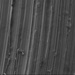
Magnified 3000 times, the surface of this broken cymbal once used by the Duke Pep Band reveals signs of fatigue cracking. Courtesy of Hadley Cocks.
These students are among more than 200 undergraduates in eight classes who benefitted from the program last year, thanks to a grant from the Donald Alstadt Foundation.
You don’t have to be a scientist, either. Historians and art conservators have used scanning electron microscopes to study the surfaces of Bronze Age pottery, the composition of ancient paints and even dust from Egyptian mummies and the Shroud of Turin.
Instructors and undergraduates are invited to find out how they could use the microscopes and other nanotech equipement in the SMIF in their teaching and research. Queries should be directed to Dr. Mark Walters, Director of SMIF, via email at mark.walters@duke.edu.
Located on Duke’s West Campus in the Fitzpatrick Building, the SMIF is a shared use facility available to Duke researchers and educators as well as external users from other universities, government laboratories or industry through a partnership called the Research Triangle Nanotechnology Network. For more info visit http://smif.pratt.duke.edu/.
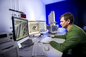
This scanning electron microscope could easily be mistaken for equipment from a dentist’s office.

Post by Robin Smith
