Research Blog

Following the people and events that make up the research community at Duke

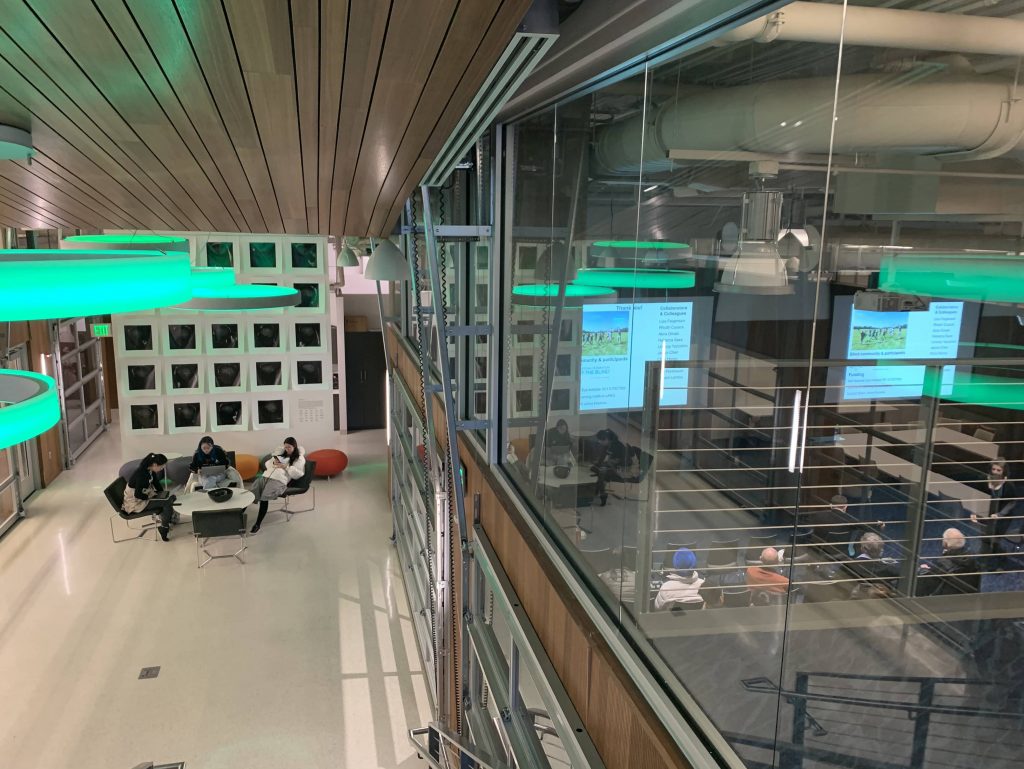
Students exploring the Innovation Co-Lab

Brain networks change with age

Graph theory allows researchers to model the structural and functional connection between regions of the brain. Image courtesy of Shu-Hsien Chu et al.

As we age, our bodies change, and these changes extend into our brains and cognition. Although research has identified many changes to the brain with age, like decreases in gray matter volume or delayed recall from memory, researchers like Shivangi Jain, PhD, are interested in a deeper look at how the brain changes with age.

Shivangi Jain uses graph theory to study how the brain changes with age.

As a post-doctoral associate in the David Madden Lab at Duke, Jain is interested in how structural and functional connectivity in the brain change with age. Jain relies on the increasingly popular method of graph theory, which is a way of modeling the brain as a set of nodes or brain regions that are interconnected. Studying the brain in this way allows researchers to make connections between the physical layout of the brain and how these regions interact when they are active. Structural connectivity represents actual anatomical connections between regions in the brain, while functional connectivity refers to correlated activity between brain regions.

Jain’s studies use a series of tasks that test speed, executive function, and memory, each of which decline with age. Using fMRI data, Jain observed a decline in functional connectivity, where functional modules become less segregated with age.  In terms of structural connectivity, aging was associated with a decline in the strength of white matter connections and global efficiency, which represents the length between modules with shorter paths being more efficient. Thus, the aging brain shows changes at the anatomical, activational, and behavioral levels.

Jain then examined how these network-level changes played a role in the observed behavioral changes. Using statistical modeling, she found that the decline in performance in tasks for executive control could be explained by the observed changes in functional connectivity. Furthermore, Jain found that the changes in structural connectivity caused the change in functional connectivity. Taken together, these results indicate that the physical connections between areas in the brain deteriorate with age, which in turn causes a decrease in functional connectedness and a decline in cognitive ability.

Research like Jain’s can help explain the complicated relationships between brain structure and function, and how these relationships affect behavioral output.

Post by undergraduate blogger Sarah Haurin
Post by Sarah Haurin

Man’s Best Friend, Our Relationship to Dogs

The average dog costs its human owner $10,000-20,000 over the course of its lifetime, from vet care and grooming to treats and toys to the new fad of doggie DNA testing. But what’s in it for us? Researcher Kerri Rodriguez – a Duke alum of evolutionary anthropology and current grad student with Purdue University’s College of Veterinary Medicine – explores just that.

Rodriguez is a member of the OHAIRE Lab at Purdue, which stands for the Organization for Human-Animal Interaction Research and Education. Continuing her work from undergrad, Rodriguez researches the dynamic duo between humans and dogs – a relationship some 15,000 to 40,000 years in the evolutionary making. Rodriguez returned to Duke to speak on February 12th, honoring both Darwin Day and Duke’s second annual Dog Day.

It’s well-known that dogs are man’s best friend, but they do much more than just hang out with us. Dogs provide emotional support when we are stressed or anxious and are highly attentive to us and our emotional states.

In a study of 975 adult dog owners, dogs ranked closely to romantic partners and above best friends, children, parents, and siblings when their owners were asked who they turn to when feeling a variety of ways. Dogs provide non-judgmental support in a unique way. They have also been found to reduce levels of the stress hormone cortisol, lower perceived stress in individuals, improve mood, and improve energy up to 10 hours after interactions. Therapy dogs are prevalent on many college campuses now due to these impacts and are found in hospitals for the same reasons, having been found to reduce subjective pain, increase good hormones and dampen bad ones, causing some patients to require less pain medications.

(Creative Commons)

 Along with reduced stress, dogs make us healthier in other ways, from making us exercise to reducing risk of cardiovascular disease. A study of 424 heart attack survivors found that non-dog owners were four times more likely to be deceased one year after the attack than victims who owned dogs.

The increased social interaction that dogs offer their human companions is also quite amazing due to the social facilitation effect they provide by offering a neutral way to start conversations. One study with people who have intellectual disabilities found that they received 30% more smiles along with increased social interactions when out in public with a dog. Similar studies with people who use wheelchairs have produced similar results, offering that dogs decreased their loneliness in public spaces and led to more social engagements.

Rodriguez also shared results from a study dubbed Pet Wingman. Using dating platforms Tinder and Bumble, researchers found that after one month, simulated profiles containing pictures with dogs received 38% more matches, 58% more messages, and 46% more interactions than simulated profiles without. Even just having a dog in photos makes you appear more likable, happier, relaxed, and approachable – it’s science!

 A large bulk of Rodriguez’s own work is focused on dogs in working roles, particularly the roles of a service dog. She explained that unlike therapy or emotional support dogs, service dogs are trained for one person, to do work and perform tasks to help with a disability, and are the only dogs granted public access by the American Disability Association. Rodriguez is particularly interested in the work of dogs who help American veterans with post-traumatic stress disorder (PTSD).

(Creative Commons)

 Around one out of five post-9/11 military veterans have PTSD and the disorder is difficult to treat. Service dogs are becoming increasingly popular to help combat effects of PTSD, ranking at the third highest placed type of service dog in the United States. PTSD service dogs are able to use their body weight as a grounding method, provide tactile interruption, reduce hypervigilance, and prevent crowding of their veterans. However, because of the lack of research for the practice, the Veterans Association doesn’t support the use of the dogs as a therapy option. This is an issue Rodriguez is currently trying to address.           

 Working with a group called K9s for Warriors, Rodriguez’s research evaluated the mental health, social health, quality of life, and cortisol levels of veterans who have received service dogs and those who were on the wait list for dogs. Veterans with service dogs had lower PTSD symptoms, better mental health, and better social health. Rodriguez is now working on a modification to this study using both veterans and their spouses that will be able to measure these changes to their well-being and health over time, as well as assessing the dog’s health too. Unlike other organizations, K9s for Warriors uses 90% shelter dogs, most of which are mutts. Each dog is as unique as the human it is placed with, but no bond is any less special.

By Cydney Livingston

Contaminated Drinking Water in Our Backyard

About 70% of the human body is made up of water. Water is something we consume on a daily basis. Therefore, when a community’s water source is threatened or contaminated it can be extremely detrimental. 

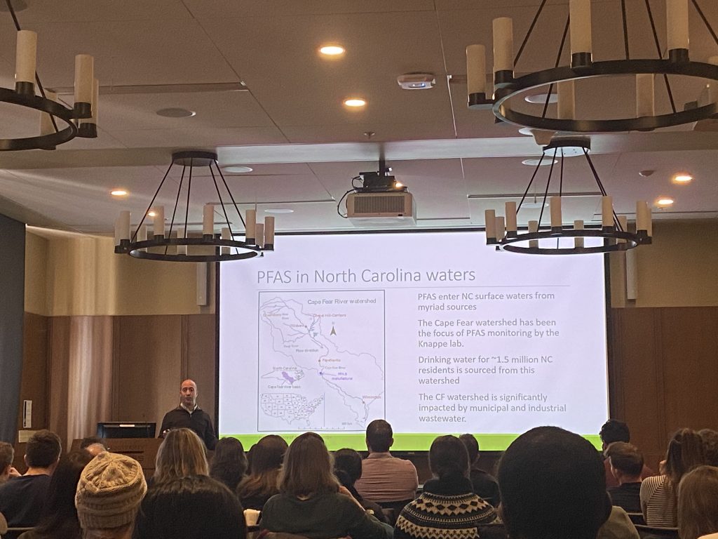
In 2017, it became apparent that there was water contamination in eastern North Carolina. Specifically, PFAS or per- and polyfluoroalkyl Alkyl chemicals were found in the water supply. As a result, several legislative mandates were issued in 2018 establishing a PFAS Testing Network to investigate the contamination.

Lee Ferguson, an Associate Professor of Civil and Environmental Engineering at Duke and Kathleen Gray, a professor at UNC’s Institute for the Environment, are testing PFAS water contamination and communicating any risks to the public. 

Gray is part of the network’s risk communication team. She explained that PFASs are hard to address because the health effects are unknown and they have yet to determine a standard or guideline for these substances. However, because this water contamination affects the lives of everyone connected to the water supply it is extremely important to communicate risk to the affected community but without eliciting panic. 

Gray explained that people often ask, “Are my family and I safe?” “What can I do to protect myself and my family?” “Why did this happen?” and “Why wasn’t it prevented?”

In the last year Ferguson and his research team have tested 409 sites in North Carolina for PFAS compounds.

He explained that PFAS substances are particularly dangerous because they are non-degradable, potentially toxic and constantly changing. Long-chain PFASs are being replaced by fluorinated alternatives.

Ferguson described this phenomenon as “playing environmental ‘whack-a-mole’ with different substances.”

Ferguson and his testing team have found two contaminated water supply sites in North Carolina. Dangerous contamination is based on the EPA health advisory level of 70ng/Liter. The exceedances were found in Maysville and Orange Water and Sewer Authority. Maysville was able to switch to the Jones County water source once the problem was identified.

New data that came in within the last couple weeks found high month-to-month variability in PFAS in the Haw River near Pittsboro. Ferguson and his team predict that it is coming downstream from a waste treatment plant. 

Brunswick County is shown having the worst PFAS concentrations. However, Dr. Ferguson and his team have recently found that the contamination in Haw River is even worse.

While all of this information may seem very alarming, Gray and Ferguson both reiterated that it is not necessary to panic. Instead, people should make sure they are drinking filtered water or invest in a water filter. 

Ferguson added, “The best choice is reverse osmosis.”

Gray and Ferguson presented their work at a SciComm Lunch-and-Learn, a monthly event sponsored by Duke Science & Society Initiative that explores interesting and innovative aspects of science communication. The event is free and open to anyone in the Duke community.

By Anna Gotskind

Origami-inspired robots that could fit in a cell?

Imagine robots that can move, sense and respond to stimuli, but that are smaller than a hair’s width. This is the project that Cornell professor and biophysicist Itai Cohen, who gave a talk on Wednesday, January 29 as a part of Duke’s Physics Colloquium, has been working on with and his team. His project is inspired by the microscopic robots in Paul McEuen’s book Spiral. Building robots at such a small scale involves a lot more innovation than simply shrinking all of the parts of a normal robot. At low Reynolds number, fluids are viscous instead of inertial, Van der Waals forces come into play, as well as other factors that affect how the robot can move and function. 

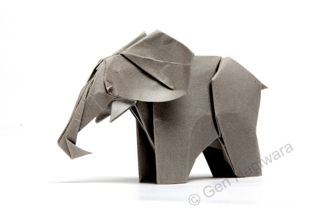
Cohen’s team designs robots that fold similar to origami creatures. Image from Origami.me

To resolve this issue, Cohen and his team decided to build and pattern their micro robots in 2D. Then, inspired by origami, a computer would print the 2D pattern of a robot that can fold itself into a 3D structure. Because paper origami is scale invariant, mechanisms built at one scale will work at another, so the idea is to build robot patterns than can be printed and then walk off of the page or out of a petri dish. However, as Cohen said in his talk last Wednesday, “an origami artist is only as good as their origami paper.” And to build robots at a microscopic scale, one would need some pretty thin paper. Cohen’s team uses graphene, a single sheet of which is only one atom thick. Atomic layer deposition films also behave very similarly to paper, and can be cut up, stretch locally and adopt a 3D shape. Some key steps to making sure the robot self-folds include making elements that bend, and putting additional stiff pads that localize bends in the pattern of the robot. This is what allows them to produce what they call “graphene bimorphs.” 

Cilia on the surface of a cell. Image from MedicalXpress.

Cohen and his team are looking to use microscopic robots in making artificial cilia, which are small leg-like protrusions in cells. Cilia can be sensory or used for locomotion. In the brain, there are cavities where neurotransmitters are redirected based on cilial beatings, so if one can control the individual beating of cilia, they can control where neurotransmitters are directed. This could potentially have biomedical implications for detecting and resolving neurological disorders. 

Right now, Cohen and his lab have microscopic robots made of graphene, which have photovoltaics attached to their legs. When a light shines on the photovoltaic receptor, it activates the robot’s arm movement, and it can wave hello. The advantage of using photovoltaics is that to control the robot, scientists can shine light instead of supplying voltage through a probe—the robot doesn’t need any tethers. During his presentation, Cohen showed the audience a video of his “Brobot,” a robot that flexes its arms when a light shines on it. His team has also successfully made microscopic robots with front and back legs that can walk off a petri dish. Their dimensions are 70 microns long, 40 microns wide and two microns thick. 

Cohen wants to think critically about what problems are important to use technology to solve; he wants make projects that can predict the behavior of people in crowds, predict the direction people will go in response to political issues, and help resolve water crises. Cohen’s research has the potential to find solutions for a wide variety of current issues. Using science fiction and origami as the inspiration for his projects reminds us that the ideas we dream of can become tangible realities. 

By Victoria Priester

Undergraduate Research in Duke’s Wired! Lab

Meet Jules Nasco, a sophomore studying Political Science and Philosophy, Politics, and Economics.

Jules is intrigued by the theories behind “how and why people form governments.” Yet, beyond her participation in various theatrical performances, commitment to several social and living-learning communities, and multiple campus jobs — from being a tour guide to editing Twitter and the Medium blog for DukeStudents — Jules also brandishes the role of undergraduate researcher in the Wired! Lab.

Duke’s Wired Lab is dedicated to digital art history and visual culture. The group – facilitated by Olga Grlic and Bill Broom and comprised of three current undergraduates – works in conjunction with the University of Catania in Italy and senior researchers around the world. Jules works specifically on the Medieval Kingdom of Sicily database, “a collection of historic images of the medieval monuments and cities in the Kingdom of Sicily, available as an open-source resource for travelers, researchers, academics, and anyone curious about the history of this part of the world!”

Since the spring semester of her first year at Duke, Jules has been searching high and low through public and private “collections, museums, archives, libraries, and publications in search of relevant paintings, drawing, etchings, photographs, or other images for the database.” She says that this can be as straightforward and easy as checking the permissions of a digital photo and downloading it or as complicated as contacting persons about image rights or scanning and editing photos from old books. Jules also collects metadata about the images she compiles such as artist or photographer, the date it was produced, the reason for production, or any relevant notes about the work. This data is then reviewed and added onto by senior researchers before being added to the public database.

The work can lead to “super-duper cool discoveries.” Earlier this year, Jules found an illustration of Salerno in a book that was drawn over 500 years ago, which led the team to a collection containing another illustration – likely by the same unknown author – likely drawn solely to depict the event of someone’s execution. However, the execution drawing is now the oldest depiction collected by the Wired! Lab of Castel Nuovo in Naples, which is one of the most prominent monuments studied by the lab.

The photo of Castel Nuovo in Naples that undergraduate researcher Jules found.

Though she admits that more career-focused endeavors may eventually take precedence over her work in the database, it’s her passion for art history that initially drew Jules into the research. Knowing that other pursuits would fill her time at Duke, she wanted to keep her interests alive in other ways. After participating in the Medieval and Renaissance Europe FOCUS program, Jules’ professor introduced her to Olga and Bill and the project. “The rest is art history!”

Jules’ favorite part of the work is the feeling that she is “meaningfully contributing to a community of interested travelers, researchers, and academics.”

Jules is able to provide people globally with information about a part of the world that she believes may otherwise be too hard to find. Her work facilitates and spreads knowledge in an interactive way, which she says makes the sometimes-tedious parts all worth it. In their data review at the end of each semester, Jules is able to see where in the world the database has been accessed and finds it awesome to know that people in Africa, Asia, and Australia use the information she has helped provide.

Post by Cydney Livingston

Visual Perception in Congenitally Blind Adults

Vision provides a rich source of information that most people’s lives revolve around. Yet, for blind people, how do they conceive of visual intake and what happens to regions of the brain dedicated to vision if a person doesn’t have typical visual input? These are questions that drive Marina Bedny PhD, an Assistant Professor of Psychological and Brain Sciences and principal investigator of a neuroplasticity and development lab at John Hopkins University.

Bedny spoke at Duke’s Institute for Brain Sciences on Friday, January 17th, about her work with congenitally blind adults. Her lab explores similarities and distinctions of visual perceptions between blind and seeing people and seeks to understand how nuanced, natural variation in experience shapes the human mind and brain.

Many of the studies Bedny discussed have very important linguistic components. In one trial, she investigated the meaning of verbs pertaining to light events and visual perception as compared to touch, amodal, auditory, and motion verbs.

Both blind and sighted people displayed nearly identical results when comparing the different types of verbs used in the study. This showed that there were no differences in what blind people knew about the terms. Analysis of the verbs revealed that linguistic dimensions of intensity and instability were used to evaluate the words’ comparative meanings. Blind people agreed more on the comparison of sound emission and touch perception words. This shows that blind participants have more aligned comprehension of the meanings of other sensory terms compared to sighted people.

In other cases, Bedny’s lab assessed what blind individuals know about color. One study used three object types – natural kinds, functional artifacts, and non-functional artifacts. These categories were used to evaluate agreeance not only on color, but the relevancy of color to certain objects’ functions as well.

Another crucial question of Bedny’s work looks at how the innate structure of the brain constrains cortical function. The findings show that the visual system in blind participants has been repurposed for higher cognitive functions and that portions of the visual system connected to high cognitive abilities are invaded by the visual systems. Along with repurposing visual regions for linguistic use, Bedny’s lab found that visual regions of the brain are active during numerical processing tasks too.

Blind people display additional activity in the visual centers of their brain in numerous studies beyond having the same regional brain responsiveness as sighted people. Though further research is necessary, Bedny proposes that there is a sensitive period during development that is critical to the specialization of the brain. Study participants who have adult-onset blindness do not show the same sensitivity and patterned responses in visual cortices repurposed for different functions as congenitally blind subjects.

At birth, the human cortex is pluripotent – providing the best of both worlds, Bedny said. The brain is prepared but highly flexible. Her studies have repeatedly shown that the brain is built for and transformed by language, and they underscore the importance of nature and nurture in human development.

Post by Cydney Livingston

The evolution of a tumor

The results of evolution are often awe-inspiring — from the long neck of the giraffe to the majestic colors of a peacock — but evolution does not always create structures of function and beauty.

In the case of cancer, the growth of a population of malignant cells from a single cell reflects a process of evolution too, but with much more harrowing results.

Johannes Reiter uses mathematical models to understand the evolution of cancer

Researchers like Johannes Reiter, PhD, of Stanford University’s Translational Cancer Evolution Laboratory, are examining the path of cancer from a single sell to many metastatic tumors. By using this perspective and simple mathematical models, Reiter interrogates the current practices in cancer treatment. He spoke at Duke’s mathematical biology seminar on Jan. 17.

 The evolutionary process of cancer begins with a single cell. At each division, a cell acquires a few mutations to its genetic code, most of which are inconsequential. However, if the mutations occur in certain genes called driver genes, the cell lineage can follow a different path of rapid growth. If these mutations can survive, cells continue to divide at a rate faster than normal, and the result is a tumor.

As cells divide, they acquire mutations that can drive abnormal growth and form tumors. Tumors and their metastases can consist of diverse cell populations, complicating treatment plans out patient outcomes. Image courtesy of Reiter Lab

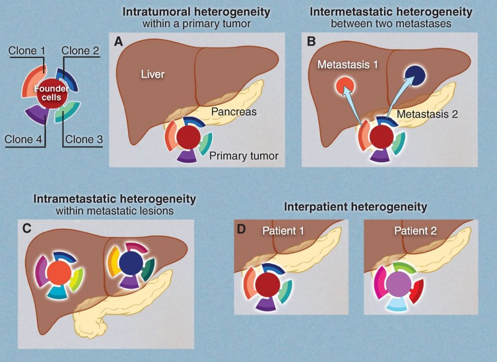
With each additional division, the cell continues to acquire mutations. The result is that a single tumor can consist of a variety of unique cell populations; this diversity is called intratumoral heterogeneity (ITH). As tumors metastasize, or spread to other locations throughout the body, the possibility for diversity grows.

Intratumoral heterogeneity can exist within primary tumors, within metastases, or between metastases. Vogelstein et al., Science, 2013

Reiter describes three flavors of ITH. Intra-primary heterogeneity describes the diversity of cell types within the initial tumor. Intrametastatic heterogeneity describes the diversity of cell types within a single metastasis. Finally, inter-metastatic heterogeneity describes diversity between metastases from the same primary tumor.

For Reiter, inter-metastatic heterogeneity presents a particularly compelling problem. If treatment plans are made based on biopsy of the primary tumor but the metastases differ from each other and from the primary tumor, the efficacy of treatment will be greatly limited.

With this in mind, Reiter developed a mathematical model to predict whether a cell sample collected by biopsy of just the primary tumor would provide adequate information for treatment.

Using genetic sequence data from patients who had at least two untreated metastases and a primary tumor, Reiter found that metastases and primary tumors overwhelmingly share a single driver gene. Reiter said this confirmed that a biopsy of the primary tumor should be sufficient to plan targeted therapies, because the risk of missing driver genes that are functional in the metastases proved to be negligible.

 In his next endeavors as a new member of the Canary Center for Cancer Early Detection, Reiter plans to use his knack for mathematical modeling to tackle problems of identifying cancer while still in its most treatable stage.  

Post by undergraduate blogger Sarah Haurin

Post by Sarah Haurin

Curating a New Portrait of Black America

It’s been over three years since the National Museum of African American History & Culture (NMAAHC) opened in D.C. in September 2016, but the excitement around it doesn’t seem to have dimmed much. Chances are, you’re going to have to get your tickets three months in advance if you want to visit. Infants need their own timed pass, too.

The National Museum of African American History and Culture.
Photo courtesy of Prabal Tiwari

On Friday, January 17, Duke’s From Slavery to Freedom Lab hosted a panel in conjunction with the Franklin Humanities Institute on the topic of contemporary Black arts and icons. The panel, “New Black Aesthetics,” featured speakers Rhea L. Combs, curator at the National Museum of African American & Culture, and Richard J. Powell, John Spencer Bassett Professor of Art & Art History at Duke, and was one half of a two-panel conference titled “Black Images, Black Histories.”

According to Combs and Powell, the reason for the unprecedented popularity of works like the NMAAHC by contemporary Black artists is likely because they do something that other pieces and people rarely do: allow African Americans to tell the African American story.

As a museum curator, Combs doesn’t simply curate cohesive mixed-media exhibitions that shed light on the Black experience. In order to create those exhibitions, she must also dig through and analyze a wide range of old archival materials.

20180925-Rhea Resized.jpg
Rhea L. Combs, Curator at the NMAAHC.
Photo courtesy of the Smithsonian

However, these archival materials at the NMAAHC aren’t necessarily just historical artifacts and records associated with figures like Rosa Parks or the Obamas; the Museum wants people to shuffle through their own attics to find things to donate. It demystifies the question of who belongs in a museum, according to Combs. “We create agency in terms of who gets to tell everyday stories,” she said.

She’s especially interested in the role of photography and film in African American studies. “We use cameras to culturally agitate the ways in which African Americans are understood,” she explained; the camera is a pathway into self-representation.

Captured in the Museum’s photos and moving images are stories of duplicity, or “celebrations that happened in the midst of tragedies.” Combs often finds themes of faith and activism as well as education and uplift, but she says that there’s plenty of variety within those overarching ideas. A photo of boys playing basketball on unicycles, for example.

“Art creates social understanding of who we are,” Combs said. Like hip-hop remixes and re-envisions things that are already understood in one way, so too does the NMAAHC.

On a similar vein, Powell’s presentation focused on the famous Obama portraits, and I’m guessing you might already know which ones I’m referring to. A fully-suited Barack Obama, seated in a wooden chair against a lush green background of flora and fauna; Michelle Obama in a flowing black-and-white colorblock dress, her chin resting on the back of her hand.

Powell examines how these portraits, simply titled “President Barack Obama” and “First Lady Michelle Obama,” manage to blend visual elements with socio-historical allusions and contexts to become world-famous 21st-century icons.

Richard J. Powell, Professor of Art and Art History at Duke.

While the portraits are visually exceptional, Powell said their context is what envelops. These images of the first Black U.S. president and first lady do allude to the old, white traditions of portraiture, “but they dismantle the genre’s conventional outcomes” for something new, he explained.

The portrait of Barack Obama is, visually, extremely similar to those of Abraham Lincoln and Franklin Delano Roosevelt. Likewise, Michelle Obama’s portrait quite closely resembles that of Madame Moitessier, for example. But unlike these representations of pre-21st-century white men and women, the Obama portraits finally depict people of color. According to Powell, portraits elevate status, and it isn’t very often that you see Black individuals portrayed.

And yet there’s also a sad irony involved, Powell explained. Especially for other similar contemporary works of portraiture that depict Black people, there’s a decorative, incongruous grandeur that highlights the tension between social realities and the manner of portrayal. For instance, “saintly” portraits exist of Black men wearing urban clothing, but despite whatever “saintliness” might be visually depicted, the realities of Blackness in the inner cities of America is often far from positive.

One of the most striking features of the Barack Obama portrait is the blooming greenery behind the former president. It’s a metaphor of sorts, Powell said: social and historical context isn’t absent from art. Or, in other words, “The world can never be left out of the garden.”

By Irene Park

First-Year Students Designing Real-World Solutions

In the first week of fall semester, four first-year engineering students, Sean Burrell, Teya Evans, Adam Kramer, and Eloise Sinwell, had a brainstorming session to determine how to create a set of physical therapy stairs designed for children with disabilities. Their goal was to construct something that provided motivation through reward, had variable step height, and could physically support the students. 

Evans explained, “The one they were using before did not have handrails and the kids were feeling really unstable.”

,
Teya Evans is pictured stepping on the staircase her team designed and built. With each step, the lightbox displays different colors.

The team was extremely successful and the staircase they designed met all of the goals set out by their client, physical therapists. It provided motivation through the multi-colored lightbox, included an additional smaller step that could be pulled out to adjust step height, had a handrail to physically support the students and could even be taken apart for easy transportation.

This is a part of the Engineering 101 course all Pratt students are required to take. Teams are paired with a real client and work together throughout the semester to design and create a deliverable solution to the problem they are presented with. At the end of the semester, they present their products at a poster presentation that I attended. It was pretty incredible to see what first-year undergraduates were able to create in just a few months.

The next poster I visited focused on designing a device to stabilize hand tremors. The team’s client, Kate, has Ataxia, a neurological disorder that causes her to have uncontrollable tremors in her arms and hands. She wanted a device that would enable her to use her iPad independently, because she currently needs a caregiver to stabilize her arm to use it. This team, Mohanapriya Cumaran, Richard Sheng, Jolie Mason, and Tess Foote, needed to design something that would allow Kate to access the entire screen while stabilizing tremors, being comfortable, easy to set up and durable.

The team was able to accomplish its task by developing a device that allowed Kate to stabilize her tremors by gripping a 3D printed handlebar. The handlebar was then attached to two rods that rested on springs allowing for vertical motion and a drawer slide allowing for horizontal motion.

“We had her [Kate] touch apps in all areas of the iPad and she could do it.” Foote said. “Future plans are to make it comfier.”

The team plans to improve the product by adding a foam grip to the handlebar, attaching a ball and socket joint for index finger support, and adding a waterproof layer to the wooden pieces in their design. 

The last project I visited created a “Fly Flipping Device.” The team, C. Fischer, E. Song, L. Tarman, and S. Gorbaly, were paired with the Mohamed Noor Lab in the Duke Biology Department as their client. 

Tarman explained, “We were asked to design a device that would expedite the process of transferring fruit flies from one vial to another.”

The Noor lab frequently uses fruit flies to study genetics and currently fly flipping has to be done by hand, which can take a lot of time. The goal was to increase the efficiency of lab experiments by creating a device that would last for more than a year, avoid damaging the vials or flies, was portable and fit within a desk space. 

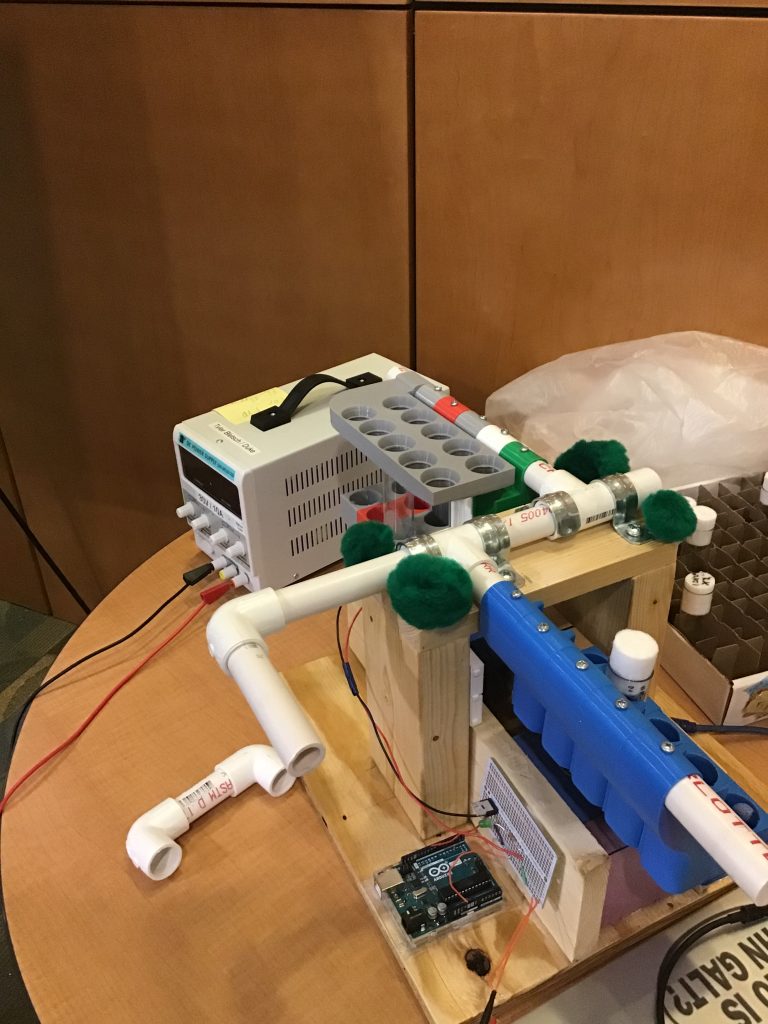
The team came up with over 50 ideas on how to accomplish this task that they narrowed down to one that they would build. The product they created comprised of two arms made of PVC pipe resting on a wooden base. Attached to the arms were “sleeves” 3D printed to hold the vials containing flies. In order to efficiently flip the flies, one of the arms moves about the axis allowing for multiple vials to be flipped that the time it would normally take to flip one vial. The team was very successful and their creation will contribute to important genetic research.

The Fly Flipping Device

It was mind-blowing to see what first-year students were able to create in their first few months at Duke and I think it is a great concept to begin student education in engineering through a hands-on design process that allows them to develop a solution to a problem and take it from idea to implementation. I am excited about what else other EGR 101 students will design in the future.

By Anna Gotskind


Teens Have the Feels About Their Family’s Standing

A study of British twins appearing this week in the Proceedings of the National Academy of Sciences shows that an adolescent’s sense of their own family’s social and economic standing is closely linked to that child’s physical and cognitive health.

In fact, the adolescent’s perception of status was a more powerful predictor of their well-being and readiness for further education than their family’s actual status. The study sample represented the full range of socioeconomic conditions in the U.K.

“Testing how young people’s perceptions related to well-being among twins provided a rare opportunity to control for poverty status as well as environmental and genetic factors shared by children within the same family,” said lead author Joshua Rivenbark, an MD/PhD student in Duke’s Medical School and Sanford School of Public Policy.

Joshua Rivenbark is an MD/PhD student in medicine and policy

“Siblings grew up with equal access to objective resources, but many differed in where they placed their family on the social ladder – which then signaled how well each twin was doing,” Rivenbark said.

Researchers followed 2,232 same-sex twins born in England and Wales in 1994-95 who were part of the Environmental Risk (E-Risk) Longitudinal Twin Study based at King’s College London. Adolescents assessed their family’s social ranking at ages 12 and 18. By late adolescence, these beliefs signaled how well the teen was doing, independent of the family’s access to financial resources, healthcare, adequate nutrition and educational opportunities. This pattern was not seen at age 12.

“The amount of financial resources children have access to is one of the most reliable predictors of their health and life chances,” said Candice Odgers, a professor of psychological science at the University of California, Irvine, who is the senior author of the report. “But these findings show that how young people see their family’s place in a hierarchical system also matters. Their perceptions of social status were an equally good, and often stronger, indicator of how well they were going to do with respect to mental health and social outcomes.”

Study findings also showed that despite growing up in the same family, the twins’ views were not always identical. By age 18, the twin who rated the family’s standing as higher was less likely to be convicted of a crime; was more often educated, employed or in training; and had fewer mental health problems than his or her sibling.

“Studies that experimentally manipulate how young people see their social position would be needed to sort out cause from effect,” Rivenbark said.

The E-Risk study was founded and is co-directed by Duke professors Avshalom Caspi and Terrie Moffitt at King’s College London.

Guest Post by Pat Harriman, UC-Irvine News @UCIPat

Page 37 of 111

Powered by WordPress & Theme by Anders Norén