Following the people and events that make up the research community at Duke

Students exploring the Innovation Co-Lab

Category: Neuroscience Page 11 of 15

Imagining Alternate Realities: Is Brian Williams in the Clear?

By Duncan Dodson

When I go home and reminisce with family about road trips we took or embarrassing moments they facilitated, eventually we’ll disagree on “what actually happened.” We’re all so certain—our memories unfold vividly yet contrarily. It’s clear the past can be subjective, but why is this so?

As part of Duke University’s Brain Awareness Week, I went to a talk at Fullsteam Brewery on imagining alternate realities by Dr. Felipe De Brigard, Assistant Professor of Philosophy and member of Center for Cognitive Neuroscience. De Brigard began by discussing studies of patients with hippocampal atrophy (as in amnesia, PTSD, and severe depression) struggling to place themselves in both the future and the past. Their impoverished answers contrast with those of healthy controls, suggesting a link between areas of the brain accessed for recalling the past and picturing the future.

Dr. Felipe De Brigard presents his recent studies on the relationship between the neural default network and autobiographical thoughts at Fullsteam Brewery 3/19.

Dr. Felipe De Brigard presents his recent studies on the relationship between the neural default network and autobiographical thoughts at Fullsteam Brewery 3/19.

De Brigard buttressed this by displaying fMRI neural images of parts of the brain used when imagining future events and evoking memories. These parts encompass the default network: a system of functions and firings executed when the brain is not engaged in a specific task. Evidence shows the default network allows engagement in “mental time travel” or the projection of oneself into the future or onto the singular, objective past. This assumption leads to temporal asymmetry: only one past exists with which the imagination can corroborate yet it can visualize limitless possibilities.

De Brigard challenged this view: what if the default network works in both directions? He argues that the parts of the brain used for imagining possible futures also allow us to conceive potential outcomes in our past that did not occur, the process of counterfactual thinking. He has found that when contemplating an alternative reality considered likely to have occurred, the brain behaves as if it were remembering. Memory is not haphazard reproduction but probabilistic reconstruction — our memory is constantly rebuilding the past with both fact and what are likely facts, and frequently the distinction is blurred.

A fascinated and packed Fullsteam, many audience members were at their second or third event for Brain Awareness week at Duke.

A fascinated and packed Fullsteam; many audience members were at their second or third event for Brain Awareness week at Duke.

“Perhaps we should cut Brian Williams a little slack?” De Brigard chuckled. Ample evidence shows that engagement in especially rich and detailed counterfactual thinking can increase the probability of constructing — and believing the authenticity of — false memories.

More intriguing than pardoning Williams are potential contributions to treatment of anxiety, depression, and PTSD. A common debilitating trigger among these disorders is repetitive counterfactual thinking, “I shouldn’t have said that, I shouldn’t have said that.” Perhaps with further study on the default network and its relationship to autobiographical contemplations, neuroscientists might develop tools to alter the pathways or functionality of the default network.

As for my family, they have some major counterfactual thinking patterns to alter; my memory is immaculate.

How To Get Your Foot In The Door At A Research Lab

By: Thabit Pulak

So now you are at Duke — one of the world’s best research universities — but now what? You might be taking cool classes, but how can you take advantage of the world-class research happening here? Roughly 50 percent of Duke undergrads do so at some point. Getting involved in research as a freshman might sound intimidating (I know it did to me!), but a little luck and perseverance can get you off to a strong start.

Alan working in Dr. Eroglu's laboratory.

Alan working in Dr. Eroglu’s laboratory.

I had the opportunity to talk with Duke freshman Alan Kong about his experiences trying to get into research labs, and how he successfully ended up finding one to join. Alan is considering majoring in biology whilst on the premed track.

He initially started to look into labs within a month of  starting classes at Duke. He spent about two months sending out emails to professors who were working on interesting projects.

“It was a very frustrating search, and initially difficult. I emailed five professors, and emailed each many times,” Alan said. “But perseverance ultimately paid off.”

Alan now works in the lab of Dr. Eroglu who is an assistant professor of cell biology, associated with the Duke School of Medicine. According to the Duke Institute of Brain Science website description, Eroglu’s laboratory “is interested in understanding how central nervous system (CNS) synapses are formed.”

Alan was also accepted into two others labs, but ultimately felt Dr. Eroglu was the best fit. “I picked Eroglu because her research was very interesting, and relevant to my interests,” Alan says. “I felt I could learn more interesting techniques in research, such as working with live animals.”

Now, Alan has been working in Dr. Eroglu’s lab for a month. When I asked him how it was going, with a smile he exclaimed, “It’s great!”

“Right now I am learning techniques such as genotyping, western blot. I even took out the retina of a rat!” Alan said. “I am learning the ropes of the lab, and my mentor said that down the road, if I learn properly, I can eventually work on my own independent project!”

When asked for any advice for other students thinking of getting into research, Alan said “Persistence is key — don’t give up! It’s a difficult process; don’t let small things get in the way. Keep trying until you find one.”

Alan

“Persistence is key – don’t give up!” Alan says

Learn more:

More information regarding Dr. Eroglu and her research: http://www.dibs.duke.edu/research/profiles/46-agla-eroglu

List of all Duke Faculty affiliated with Cell Biology with contact details: http://www.cellbio.duke.edu/all-faculty/

Summer research opportunities in math and statistics: http://bigdata.duke.edu/research/?field_project_topics_tid=19

Research opportunities in biology: http://cubr.biology.duke.edu/projects

For other research and summer opportunities visit https://biology.duke.edu/undergraduate/current-students/research-independent-study/summer-opportunities

For a list of research opportunities across the sciences, arts, humanities and social sciences visit: http://undergraduateresearch.duke.edu/uploads/media_items/summer-2015-funding-opps.original.pdf


RISK: The Adolescent Mind

By Anika Radiya-Dixit

Have you ever been labeled an out-of-control teenager? A risky driver? An impulsive troublemaker? Here’s the bad news: That’s partially correct. The good news? It’s not your fault: blame the brain.

On November 18, the department of Psychology and Neuroscience introduced students to “The Origins of Heightened Risk Behavior in Adolescence.” The presenter, Dustin Albert, is a PhD research scientist at the Center for Child and Family Policy here at Duke University, who is interested in cognitive neuroscience, problem behaviors, and peer influence.

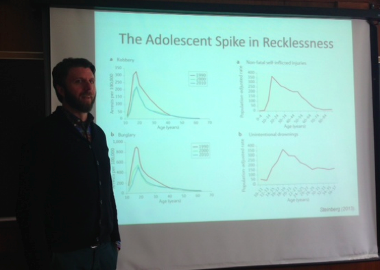
Researchers have identified the stage of adolescence as the peak time of health and performance, but at the same time, they noticed a jump in morbidity and mortality as children approached teen years, as seen in the graphs below. Specifically, adolescents show increased rates of risky behavior, alcohol use, homicide, suicide, and sexually transmitted diseases. However, as Allen tells the audience, “These are only the consequences.” In other words, what teenagers are stereotypically ridiculed for is actually the result of something else. If that’s the case, then what are the causes?

Professor Albert

Professor Albert explaining the spike in risky behavior during teenage years.

Psychologically speaking, researchers believed that these behaviors are caused by a lack of rational decision, perhaps because adolescents “are unable to see their own vulnerability” to the outcomes, meaning that teens are apparently inept at identifying consequences to their actions. However, the studies they took demonstrated that adolescents are not only able to see their own vulnerability, but are also able to intelligently evaluate costs and effects to a certain decision. If teenagers are so smart, then what is actually causing this “risky behavior”?

One important reason Professor Albert discussed is brain activity and maturation before, during, and after adolescence. As a child ages from early to middle adolescence, fast maturation of incentive processing circuitry drives sensation seeking – in other words, the willingness to take risks in order to gain a reward increases as the child approaches teen years. In the brain, this occurs due to increased dopamine availability in reward paths as well as heightened sensitivity to monetary and social reward cues. In one interesting study, adolescents were instructed to press a button only when they saw an angry face. However, the researchers noticed that when the teens saw a happy face, they had a “particularly difficult time restraining themselves” to not press the button. Essentially, the happy-angry face study demonstrates that adolescents have more struggle in restraining themselves against impulsive actions, which often translates into responses during driving, alcohol use, and the other aforementioned risky behaviors.

Later in their life, there is a slower maturation of cognitive control circuitry that leaves a window of imbalance in the teen’s life. In the brain, this period is noted by thinning of gray matter and increasingly efficient cortical activation during inhibition tasks. In other words, older people “use smaller parts of [their] cortex to stop inappropriate responses.” Essentially, due to the way the physical and hormonal brain matures, adolescents are more prone to impulsive behavior. The take away: it’s not your fault.

Another influence on teens’ risky behavior is called the peer presence effect, commonly known as “peer pressure.” Based on arrest records, “adolescents, but not adults, [are] riskier in the presence of peers,” pointing out that the percentage of co-offenders arrested for the top eight crimes decreased with age after teenage years (Gardner & Steinberg, 2005). Perhaps the need to “establish their status,” Albert speculated, decreases with age as they gain more experience about living in the real world.

The test to evaluate the result of peer presence simulates the effect of teens taking a driving exam when in the car alone as compared to when with peers. In terms of peer influence, the study shows that adolescents ran more intersections when sitting with a peer than when sitting alone. In terms of risky behavior compared with adults, adolescents when watched by peers showed over 20% increase in risky behavior of running through intersections, as opposed to the 5-10% increase seen for adults in peer presence. Albert partially attributed this effect to the fact that “teens driving the first time could assess the probability of crashing less than adults do,” but he doesn’t have specific evidence for this claim.

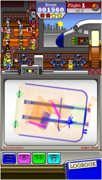
While Albert claimed that the study was valid because the adolescents participating were made aware of the outcome of driving recklessly – damage to the car, injury, time it would take to get a new car, insurance problems – I believe that the study should have taken into account the fact that the teens may have subconsciously known the simulated driving test wasn’t real – viewing it as a mere video game – and so may have succumbed more into peer pressure as the true fear of dying in a crash would not have been present.

Albert ended his talk by giving one last piece of advice to people working with teens: It’s “not enough to [simply] increase their knowledge,” but rather to “understand and work towards developing impulse control and reward sensitivity.”

Below are some of the thought-provoking questions raised by audience members during the Q&A session:

Q: What would be the result of peer presence effect for same-sex peers as compared to peers of the opposite sex?

A: While Albert admitted that this particular situation has not been tested yet, he believes it may be based on personal perceptions of what the peer thinks, and what the opposite person likes.

Q: What would be the result of risky behavior for the simulated driving test if the participant’s parent(s) and peer(s) were both present in the car?

A: On one hand, the participant might drive more carefully due to the presence of an authoritative figure. However, if the participant opinionates the peer as a stronger influence, he / she would effectively neutralize the effect the parent has and drive more recklessly. Other audience members claimed that they would drive more cautiously irrespective of who was sitting with them in the car because they are aware there is another life at stake for every decision they made behind the wheel. “It would be interesting to see the [results of the study] based on this internal conflict,” the audience member who posed this question said. Overall, Albert said the results would be primarily influenced by the type of person participating – whether they would “take the small amount of money or be willing to wait for the big amount” in front of peers – that would determine whether the parent or peer becomes a stronger influence in risky behavior.

Q: How could someone going into education help keep high school students away from risky behaviors?

A: Albert noted that these behaviors are more the result of personal experience rather than something that can be quickly taught. In a school setting, teachers could introduce the practice of challenging situations to help the kids acting ‘in-the-moment’ recognize and understand “changes in their own thought patterns for decision making,” but simply giving them a “lesson in health class is not necessarily going to translate into the Friday night situation.”

If you are interested in these type of topics, Professor Albert is teaching PUBPOL 241: METHODS SOCIAL POLICY RESEARCH  this Spring (2015).

More details about the presenter can be read at: http://fds.duke.edu/db/Sanford/ccfp/william.albert

Why Airport Scanners Catch the Water Bottle but Miss the Dynamite

Guest post by Caroline Drucker

A screenshot of the Airport Scanner game, with a suitcase containing two dynamite sticks. Courtesy of the Mitroff Lab and the Kedlin Company.

A screenshot of the Airport Scanner game, with a suitcase containing two dynamite sticks. Courtesy of the Mitroff Lab and the Kedlin Company.

You’re at the airport waiting to pass through security and board your flight. The security agent stops the person in line ahead of you: there was a full water bottle in his carry-on bag. He throws out the bottle and proceeds through the airport. Later that evening, you see that person’s face on the news, for having pulled out dynamite on their flight. Why did the TSA agent overlook the dynamite?

A team of researchers at Duke led by Dr. Stephen Mitroff is using a cell phone game to provide answers to this and other questions about airport baggage screenings. Last year, they reported that luggage screeners are likely to miss extremely rare illegal items. In a new study which will appear in the Journal of Experimental Psychology: Human Perception and Performance, Dr. Mitroff’s team once again leverages the power of big data to address a different issue: what happens when a carry-on suitcase contains multiple illicit items?

Baggage screening is an example of what cognitive scientists refer to as visual search: attempting to locate a target among a crowded visual display. Many laboratory studies have demonstrated that when more than one target is present in the display, people are less likely to find additional targets once they have spotted a first target. One possible reason for this “subsequent search misses” phenomenon is that people become biased toward searching for targets that match the first target. That is, a baggage screener who finds a water bottle might enter “water bottle” mode and be unprepared to see dynamite. This theory is most likely to hold in situations with large and unpredictable sets of targets – which is precisely why it has been difficult to test in the lab, where the relatively small amount of trials that subjects can perform has limited the possible target numbers and frequencies.

A smartphone game called Airport Scanner circumvents this problem. In the game, players act as TSA agents and view X-ray images of carry-on luggage, earning points for correctly tapping illegal items. More than 200 possible illegal items can appear, and each bag can contain between zero and three illegal items among up to twenty legal items. The game is available as a free download from the Kedlin Company, who share the data with the Mitroff lab. They have now collected close to two billion trials (bags searched) from over seven million people, which would take centuries to collect in the lab.

Using these data, Dr. Mitroff and his colleagues were able to make an important discovery about subsequent search misses. When two identical targets are present in a bag, it is more likely that both will be found than when two different targets are present. In other words, if someone first spots a water bottle, it is more likely that they will also find a second item if it is a water bottle than if it is dynamite.

This result supports the theory that finding a visual target biases a person’s perceptions. We become better prepared to find another instance of the same item, rather than a different item. According to Dr. Mitroff, “Knowing this fact can help create search environments and standard operating procedures to overcome this priming effect.”

The Science of Self-Agency: Dr. Nicolelis and the Walk Again Project

By Olivia Zhu

Screen grab from Univision of Juliano's robo-kick at the World Cup opening ceremony.

Screen grab from Univision of Juliano’s robo-kick at the World Cup opening ceremony.

Over the course of his 20-year career, Dr. Miguel Nicolelis has restored movement and self-agency to paraplegic patients.

On November 11th, as part of the Grand Challenge Seminar Series, Dr. Nicolelis captivated his audience by explaining the extensive process that culminated in Juliano, a Brazilian 29-year-old paralyzed from the chest downward in a car accident, performing the opening kick of the World Cup simply by using his mind.

Dr. Nicolelis has several faculty appointments in the Duke School of Medicine, Department of Psychology and Neuroscience, Institute for Brain Sciences, and Center for Neuroengineering. He has also written a book, Beyond Boundaries, about his work. His program, Walk Again, is supported by the Edmond and Lily Safia International Institute of Neuroscience in Brazil.

Dr. Nicolelis began making progress in 1999-2000 at Duke by developing electrodes that could record firing from multiple neurons. Using this technology, he determined which neurons were necessary for a monkey to move a joystick during a video game. Then, Dr. Nicolelis focused on creating a bypass that would bridge the mind directly to a computer, essentially removing the body as an intermediary.

He called this bypass a “Brain-Machine Interface,” or BMI, a term he coined at a cheese steak joint outside of Philadelphia. With the BMI, Dr. Nicolelis’s monkeys could play the video game without moving their arms or the joystick—they simply imagined themselves moving the joystick. The monkeys could even use their arms to do other tasks like eat or scratch themselves, creating a “third arm.”

Since then, with an extensive team of engineers, Dr. Nicolelis has implemented this technology by creating a IMG_1941hydraulically-powered exoskeleton that interprets a patient’s firing neurons and moves a patient’s legs accordingly.

He has also created artificial “skin,” which provides tactile feedback of movement to a patient’s upper body or, eventually, through an implant directly to the tactile cortex of the brain.

The technology is so accurate that patients report feeling “ghost limbs”—they believe that their legs are actually walking. The legendary Brazilian soccer player, Ronaldo, reportedly exclaimed “I’m moving!” with incredulity, when he was strapped to a chair testing Nicolelis’s technology.

Training with the exoskeleton also improves patients’ cardiovascular circulation, mental health, gastrointestinal health, and sensitivity in previously paralyzed areas.

Dr. Nicolelis is truly using science to stretch the boundaries of the human body.

NIH Getting Serious about the BRAIN

brain landing on moon

The NIH’s mapping initiative called BRAIN has been likened to a moon shot.

By Kelly Rae Chi

The federal government’s  BRAIN Initiative to chart the neural connections in the human brain and explain how its diverse and ever-changing cells make us who we are has been compared to landing a person on the moon.

Last summer the National Institutes of Health (NIH) described its vision as a 100-plus-item list of deliverables, proposed budgets and milestones. Last week, the NIH awarded the first round of seed money toward those goals — $46 million — some it for Duke scientists.

And this week, Gregory Farber, director of the National Institute of Mental Health’s Office of Technology Development and Coordination, visited Duke to talk about the timeline for the decade-long initiative.

Duke scientists in the audience peppered him with questions about how he sees it evolving.

“What I learned in Greg Farber’s talk is that the BRAIN Initiative offers a serious –and I mean serious — ten-year plan to catalyze game-changing discoveries in understanding the human brain, and in doing so, provide new treatments for disorders, like Alzheimer’s disease, that can rob us of our very humanity,” said Michael Platt, director of the Duke Institute for Brain Sciences.

BRAIN stands for ‘Brain Research through Advancing Innovative Neurotechnologies.’ It’s all about technology, and it will cost a pretty penny for its public and private partners. Fiscal Year 2014’s  $46 will develop a “parts list” for the brain and probe neural circuitry in a variety of ways. And that’s only the start.

The initiative is expected to begin in earnest in 2016, take five years for tool development and five more to apply those tools to study humans wherever possible.

“We have a strong sense that we want to see these tools have clinical applications in the not-too-distant future, but I’m being careful not to define ‘not-too-distant’,” Farber told a room full of neuroscientists, many of them working on human brain imaging.

Allen Song is a professor of Radiology, Neurobiology, Psychiatry and Biomedical Engineering.

Allen Song is a professor of Radiology, Neurobiology, Psychiatry and Biomedical Engineering.

In the audience was Allen Song, professor and director of the Duke-UNC Brain Imaging Analysis Center and among the first scientists awarded NIH BRAIN funds. He is leading a team that will further develop and validate a human brain imaging technique dubbed ‘NEMO,’ for Neuro-Electro-Magnetic Oscillations.

The hope is that NEMO, and other ‘next-generation’ brain imaging advances supported with the initiative, will help solve some of the limitations of today’s technologies. Functional magnetic resonance imaging (fMRI), for example, measures changes in the levels of oxygenated blood in the brain. It’s an indirect way of seeing neural activity, and it comes with a several-second delay.

Used with traditional MRI scanners, NEMO will more directly tune into neurons, which naturally create waves of electrical activity in the brain at specific frequencies.

For example, “if this technology works, we can tune our machine to listen to the 10-Hertz oscillation in the brain as a result of neuron firing,” Song said. Then, by driving neurons into specific oscillations at different times and during different tasks, the scientists may be able to resolve the brain in better spatial and temporal detail.

Song said that although he’s excited and confident, he already feels the pressure of a tight timeline for the project. It won’t be possible to finish it in the three-year timeframe. Even with continued funding, at the end of 12 years, “we don’t know where we will be,” he said.

Still, Song and his colleagues were all smiles as they filed in for Farber’s talk. “It’s a thrill to see Allen Song and his colleagues win support in the first round of BRAIN grants to develop the next generation in human brain imaging technology,” Platt said. “I’m confident Duke neuroscience will figure prominently in the BRAIN Initiative, given our focus on interdisciplinary innovation and collaboration.”

No actual cartoon fish will be used in the NEMO project.

No actual cartoon fish will be used in the NEMO project.

Duke Shaking up Basal Ganglia Research

By Kelly Rae Chi

Duke brain scientists are shaking up their field’s understanding of a part of the brain called the basal ganglia that’s sort of a crossroads for many important functions.

A simplified map of the pathways domamine and serotonin travel to the basal ganglia, the snail-shaped structure in the middle of the human brain.

A simplified map of the pathways dopamine (blue) and serotonin travel to the basal ganglia, the snail-shaped structure in the middle of the human brain.

Basal ganglia signaling is involved in movement, learning, language, attention, and motivation.  But this centrality also makes it  challenging to figure out how it works, said Henry Yin,  an assistant professor of psychology and neuroscience at Duke, and a member of the Duke Institute for Brain Sciences.

As healthy mice collected food pellets delivered into a cup once per minute every minute for two hours. Yin’s team was recording the electrical activity of  neurons projecting to and from the basal ganglia.

Henry Yin is an assistant professor in Psychology and Neuroscience.

Henry Yin is an assistant professor in Psychology and Neuroscience.

Naturally, the mice picked up food less often as they became full and some of the cells that use dopamine to signal reward showed less activity.

But other dopamine cells became more active.

In a paper describing these experiments , Yin’s group proposed that the cells’ activity reflected not reward but what the animals are physically doing.

This was new and Yin became curious. Was there a direct relationship between movement and dopamine activity?

Using a different experimental setup with cameras and pressure pads, Yin’s group quantified mouse movements while recording neural activity. “What happens is that whenever there’s movement, (there are phases of) dopamine activity,” Yin said to a room full of fellow neuroscientists during a recent seminar at Duke.

Putting the mice on top of a “shaker,” a piece of lab equipment  normally used to gently shake tubes and dishes full of liquids,  they found individual dopamine neurons responded to specific directions the mouse was tilted on the shaker. The same was true for nearby neurons that signal using GABA, an inhibitory chemical in the brain.

Using additional methods for tracking motions of freely moving mice, the group has discovered specific sub-populations of neurons that respond to different aspects of movement, especially movement speed and acceleration.

The researchers have also created transgenic mice whose dopamine neurons can be stimulated using light. Turning on these neurons makes the mice move.

Yin is working on publishing these results, but he said there’s a lot of resistance in the field.  His work appears to be directly challenging the dogma that  dopamine is linked to reward. He says it might actually be involved in generating movements.

“Let’s say you’re drinking coffee and that’s a reward,” Yin said. “I record your neural activity, and it’s correlated with coffee. You might say it’s a coffee neuron. But that’s not true unless you can measure the movement kinematics and rule out other possible correlations. What we’re seeing is that, with no exceptions, the phasic activity of DA neurons is always correlated with movement.”

Yin’s work also challenges theories about why people with Parkinson’s disease, whose dopamine cells degenerate, often have trouble initiating movement, or they move more slowly than they mean to.

“If you’re a doctor, a neurologist, what you study is the rate model. That’s the textbook description,” Yin said. The gist of the rate model is that the basal ganglia is constantly putting the “brakes” on behavior, and when its neurons settle down, that allows for movement.  Parkinson’s patients can’t initiate movements, it’s thought, because their basal ganglia output (more specifically, the rate of firing in the inhibitory output neurons) is too high, producing excessive braking.

In contrast, according to Yin’s work, at least four different types of basal ganglia output neurons are adjusting behavior dynamically and continuously, to shape the speed and direction of movement.

When the activity of these neurons is constant, it reflects a stable posture, Yin said.  So he argues that the problem with Parkinson’s patients is not that their basal ganglia output is too high, but that this output is stuck in firing mode. The downstream brain areas required for postural control don’t get the right commands.

Nicole Calakos is an associate professor of neurology.

Nicole Calakos is an associate professor of neurology.

“Henry’s studies are really exciting because we’ve thought about this circuitry in one way for a very long time and his findings really cast a new light on those interpretations,” said Nicole Calakos,  M.D., Ph.D., an associate professor of neurology. “I treat patients with Parkinson’s disease and other diseases that involve this circuitry. It is interesting to consider this alternate view to explain the problems my patients face in doing their day-to-day activities.”

Calakos’ own research focuses on how learning alters signal processing by the basal ganglia, and how the signaling goes awry in brain diseases such as obsessive-compulsive disorder. Duke researchers are finding compelling links between different behavioral states and specific long-lasting patterns of activity in the basal ganglia.


Duke's ALS Challenge is Conventional Wisdom

[youtube http://www.youtube.com/watch?v=w1IH9fbXFIs?rel=0]

By Kelly Rae Chi

What all those folks dumping ice water over their heads to raise money for the ALS Association may not realize is that a small number of patients with the degenerative neurological disease might sometimes get better.

In a new patient-funded program called ALS Reversals, Duke researchers are trying to find out why.

“Any time you have a patient with ALS who’s getting better, no matter what it is that they’re doing, I think you should try to put a lot of effort into understanding that patient,” said Richard Bedlack, M.D., Ph.D., associate professor of neurology at Duke University School of Medicine and director of the Duke ALS Clinic.

Richard Bedlack, M.D. Ph.D.

Richard Bedlack heads Duke’s ALS Clinic.

Not everyone believes that the reversal of ALS is real. And if it is, then some skeptics say that the number of examples might be too few to learn anything of value from, Bedlack said.  “I would say the only way you’ll know the answer to that is to try,” he added.

Bedlack said there may be three possible explanations for why some people with ALS stop progressing or get better. First, the person may never have had ALS in the first place; he or she may instead have an unusual form of myasthenia gravis, for example.

Second, something unique about that person’s body might be helping them fight the disease. “The [first and second] possibilities can be teased apart. We can get these folks to send their records and come to Duke, and we can study them,” Bedlack said. The researchers would like to compare these patients’ gene sequences, gene expression data or antibody profiles to those of more typical ALS patients.

The third, and perhaps most controversial, explanation for ALS reversal is that the patient tried a treatment that worked.

The idea for the ALSReversals came to Bedlack as he was reviewing alternative and off-label therapies for ALS. In an effort to quash misinformation floating around the web about such therapies, Bedlack had started ALSUntangled, a group of scientists and clinicians who systematically study any available evidence behind a given therapy — elected by the public via social media — and publish an open review about it.

To Bedlack’s surprise, the ALSUntangled team found that some alternative ALS therapies might show some promise, and probably need more study. One of those was lunasin, a peptide derived from soybeans that is sold as a nutritional supplement. The group is still finishing their review of lunasin and Bedlack plans to carry out a pilot study of it through the ALS Reversals program.

In the past decade, new research has already helped ALS patients by improving their quality of life and functioning. This has made the work of the Duke ALS Clinic more about helping people live with the disease rather than just diagnosing them.

And just for good measure, the clinic team also participated in the Ice Bucket Challenge.

Bedlack is excited by the new attention to ALS. “I’ve never seen ALS being talked about by so many people,” he said, adding that he looks forward to seeing what comes of the boost in funds.

The viral  Ice Bucket Challenge has so far raised more than $94 million in donations to the ALS Association as of Aug. 27.

An estimated 30,000 Americans are living with ALS. One in 500 people will develop ALS in his or her lifetime. It’s incurable, and terrible. But maybe you have already learned that through the Ice Bucket Challenge.

Duke students present Alzheimer's research at Montana conference

By Sonal Gagrani

The Federation of American Societies for Experimental Biology (FASEB) brought together neuroimmunologists from all over the world to Big Sky, Montana in July to discuss their current and upcoming research on mechanisms and therapeutics in neuroimmunology.

They covered a plethora of topics in the field from multiple sclerosis, a neurodegenerative brain disorder, to neuroprotection by microglia, the resident immune cells of the brain, to the effects that intestinal imbalances can have on the brain via the blood brain barrier.

My primary focus at the meeting was to expand my knowledge on Alzheimer’s disease (AD), a dementia-causing neurodegenerative disease of the central nervous system that I am currently researching.

photo

Lauren Kane with her poster at the FASEB conference

We were fortunate to have Lauren Kane, a rising senior in Dr. Carol Colton’s lab at Duke and the only undergraduate student with a poster at the conference, be able to present her work on her Alzheimer’s mouse model.

It is known as a CVN-AD model and has many pathologies found in AD such as β-amyloid plaque formation, neuron loss, tau protein defects, and behavioral deficits. Lauren is studying the changes in myelin, the primary make-up of white matter in the brain. Myelin wraps around axons in order to allow faster communication between neurons. She has found that there is some breakdown in myelin in the CVN-AD model, and this could lead us to find treatments for AD that promote remyelination in the brain.

Matthew Kan, an MD/PhD student in Dr. Michael Gunn’s lab at Duke, also presented his work on Alzheimer’s at the conference. He showed in the CVN-AD mouse model that a possible mechanism of neuronal death may be decreased arginine, an essential amino acid in the brain. Microglia produce arginase-1, an enzyme that breaks down arginine, and Matthew found that blocking arginase-1 activity reversed some neurodegeneration found in the CVN-AD mice. This arginine depletion pathway is known to suppress the brain’s immune system rather than cause inflammation, which many people thought was the mechanism for AD pathology in the past. These results may shift some focus to arginine in looking for AD treatments.

The conference strived to integrate and improve neuroimmunology research by providing a venue for creating connections with the experts in the field. There are many therapeutics for brain disorders in progress that key in on the importance of the brain’s immune system in regulating pathology.

Messenger of Pain Identified

Pain researcher Ru-Rong Ji is a distinguished professor of anesthesiology and professor of neurobiology.

Pain researcher Ru-Rong Ji is a distinguished professor of anesthesiology and professor of neurobiology.

By Karl Leif Bates

In their pursuit of understanding how pain works at the molecular level, a research team lead by Ru-Rong Ji of anesthesiology and neurobiology has found a new function for MicroRNAs, short stretches of genetic material that signal genes to turn on or off.

In a paper appearing online April 2 in the journal Neuron, Ji and his colleagues in the Pain Signaling and Plasticity Lab describe one MicroRNA called “let-7b” that is found floating outside cells and can bind specifically to pain-sensing neurons.

Let-7b rapidly excites these neurons through the toll-like receptor-7 (TLR7) and its associated ion channel, TRPA1, which leads to a rapid inward flow of ions to the neurons.

Injecting the 22-basepair RNA molecule into the feet of mice induced a sensation of pain within minutes. (The mice are seen lifting the affected paw or licking it.)

The MicroRNA let-7b is the 22 red nucleotides in this diagram. (Image from MiRNAMap site, Institute of Bioinformatics National Chiao Tung University, Hsinchu, Taiwan.)

The MicroRNA let-7b is the 22 red nucleotides in this diagram. (Image from MiRNAMap site, Institute of Bioinformatics National Chiao Tung University, Hsinchu, Taiwan.)

Mutant mice missing the genes for TLR7 and TRPA1 were found to be less susceptible to the signaling molecule or even unaffected by it.

Pain is notoriously difficult to measure, but Ji said the new molecule may serve as a biomarker for pain. “We’re also interested to know if targeting this miRNA would be a way to alleviate pain.”

The study was supported by NIH grants R01-DE17794, R01-DE22743, and NS67686 to Ru-Rong Ji and R21-NS82985 to Zhen-Zhong Xu.

CITATION: “Extracellular MicroRNAs Activate Nociceptor Neurons to Elicit Pain via TLR7 and TRPA1,” Park, Xu, Berta, Han, Chen, Liu and Ji. Neuron, Online April 2, 2014. DOI: 10.1016/j.neuron.2014.02.011


Page 11 of 15

Powered by WordPress & Theme by Anders Norén