Following the people and events that make up the research community at Duke

Students exploring the Innovation Co-Lab

Category: Neuroscience Page 9 of 15

Mapping the Brain With Stories

alex-huth_

Dr. Alex Huth. Image courtesy of The Gallant Lab.

On October 15, I attended a presentation on “Using Stories to Understand How The Brain Represents Words,” sponsored by the Franklin Humanities Institute and Neurohumanities Research Group and presented by Dr. Alex Huth. Dr. Huth is a neuroscience postdoc who works in the Gallant Lab at UC Berkeley and was here on behalf of Dr. Jack Gallant.

Dr. Huth started off the lecture by discussing how semantic tasks activate huge swaths of the cortex. The semantic system places importance on stories. The issue was in understanding “how the brain represents words.”

To investigate this, the Gallant Lab designed a natural language experiment. Subjects lay in an fMRI scanner and listened to 72 hours’ worth of ten naturally spoken narratives, or stories. They heard many different words and concepts. Using an imaging technique called GE-EPI fMRI, the researchers were able to record BOLD responses from the whole brain.

Dr. Huth explaining the process of obtaining the new colored models that revealed semantic "maps are consistent across subjects."

Dr. Huth explaining the process of obtaining the new colored models that revealed semantic “maps are consistent across subjects.”

Dr. Huth showed a scan and said, “So looking…at this volume of 3D space, which is what you get from an fMRI scan…is actually not that useful to understanding how things are related across the surface of the cortex.” This limitation led the researchers to improve upon their methods by reconstructing the cortical surface and manipulating it to create a 2D image that reveals what is going on throughout the brain.  This approach would allow them to see where in the brain the relationship between what the subject was hearing and what was happening was occurring.

A model was then created that would require voxel interpretation, which “is hard and lots of work,” said Dr. Huth, “There’s a lot of subjectivity that goes into this.” In order to simplify voxel interpretation, the researchers simplified the dimensional subspace to find the classes of voxels using principal components analysis. This meant that they took data, found the important factors that were similar across the subjects, and interpreted the meaning of the components. To visualize these components, researchers sorted words into twelve different categories.

img_2431

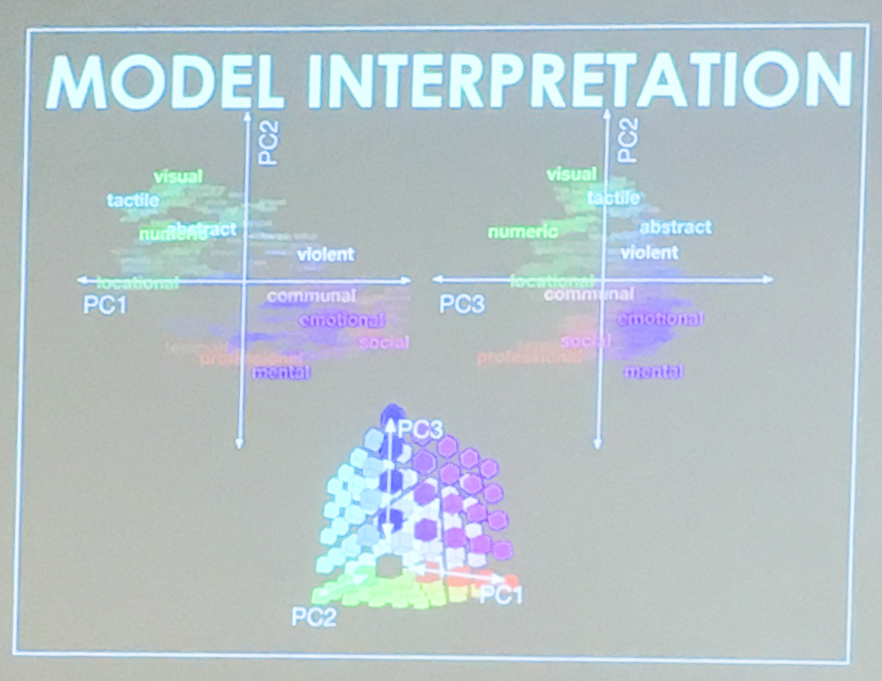
The Four Categories of Words Sorted in an X,Y-like Axis

These categories were then further simplified into four “areas” on what might resemble an x , y axis. On the top right was where violent words were located. The top left held social perceptual words. The lower left held words relating to “social.” The lower right held emotional words. Instead of x , y axis labels, there were PC labels. The words from the study were then colored based on where they appeared in the PC space.

By using this model, the Gallant could identify which patches of the brain were doing different things. Small patches of color showed which “things” the brain was “doing” or “relating.” The researchers found that the complex cortical maps showing semantic information among the subjects was consistent.

These responses were then used to create models that could predict BOLD responses from the semantic content in stories. The result of the study was that the parietal cortex, temporal cortex, and prefrontal cortex represent the semantics of narratives.

meg_shieh_100hedPost by Meg Shieh

Nature vs. Nurture: Predicting Our Futures

Sitting in The Connection at the Social Science Research Institute in Gross Hall was intimidating. I was surrounded by distinguished people: professors, visiting professors from distinguished universities, researchers, and postdocs, all of whom had gathered together to view a showing of the documentary, Predict My Future: The Science of Us.

moffit_temi_people

Dr. Terrie Moffitt, a Duke professor. Image courtesy of Moffitt and Caspi.

avshalom_caspi3_comp_sm

Dr. Avshalom Caspi, a Duke professor. Image Courtesy of Moffitt and Caspi.

Predict My Future documents the work of Terrie Moffitt and Avshalom Caspi, two Duke professors who study people in Dunedin, New Zealand. They have followed the lives of all the children born within a year in Dunedin for the last 40 years to measure genetics, personal habits, environment, jobs, physical attributes, and etc.  The Dunedin Longitudinal Study is the largest study of its kind and offers deep insights into how children become adults.

The episode, “The Early Years,” first posed the questions, “Why do some people become successful and others become outcasts? Why are we the way that we are?” By tracking all of these personal factors and some  behaviors, including risky sexual activities, criminal activities, and drinking and smoking habits, the Dunedin Longitudinal Study can answer these questions. The researchers can tell which children are likely to become “problem children,” “geniuses,” and so on, based on the child’s personality type.

30437237750_f6bf4f577f_o

Q&A Session After the Viewing of the Documentary. Image Courtesy of Duke SSRI and Taken By Shelbi Fanning.

The study first identified five different personality types in young children, and researchers discovered that the children’s’ personality types did not change in adulthood. The three personality types that are typically associated with doing well in life, having better health, having friends, and being more successful are: “well-adjusted,” “reserved,” and “confident.” The two personality types associated with having poorer quality of life in adulthood are “inhibited” and “undercontrolled.”

Then, the study identified other factors that lead to serious consequences later in life or simply predict futures. Children who experienced delays in walking and in talking were likely to have issues with brain development. Boys with these traits typically disliked school, did poorly in school, and were more likely to be involved in criminal activity.

30621009122_7a1db6d4e5_o

The full house watching the documentary. Image Courtesy of Duke SSRI and Taken by Shelbi Fanning.

The amount of sleep children received between the ages of five and eleven would determine obesity in adulthood. Adults who received the least amount of sleep as children tended to be obese by age 32.

Schizophrenia, researchers discovered, starts developing in young children, not just adults as had previously been thought. About half of the 11-year-olds in the study who said they had seen or heard things that weren’t there had developed schizophrenia two decades later.

Watching more TV was associated with a higher likelihood of smoking and having anxiety. Regardless of IQ or environment, children who watched more TV were more likely to leave school without qualifications.

The important lesson the documentary emphasized was that having a good childhood is important. Warm, sensitive, stimulating, family-feeling invoking environments are great protective factors to risk factors.

Overall, this was a brilliant, stimulating, easy-to-understand documentary.

meg_shieh_100hedPost by Meg Shieh

Violence: Risk vs Protection Factors

On Oct. 3, in place of a typical Interdisciplinary Discussion Course (IDC) for the Focus program, we were brought together in the White Lecture Hall on East to hear Dr. Jeremy G. Richman give a lecture on “Violence, Compassion, and the Brain”.

Dr. Jeremy G. Richman

Dr. Jeremy G. Richman

Since his daughter Avielle was killed in the Sandy Hook Elementary School shooting, Dr. Richman has been studying violence and the brain with the Avielle Foundation. Using his extensive research experience in neuroscience and neuropsychopharmacology, Dr. Richman has been working with a team on understanding the risk and protective factors for violence.

untitled-3

Dr. Richman lecturing in the White Lecture Hall on East Campus


The brain is very complex, and its processes and connections are still very much veiled. As a result, The Avielle Foundation currently conducts research on understanding violence by “bridging biochemical and behavioral sciences” using functional MRI brain scans, biochemistry, and genetics and epigenetics.

Based on the research he has analyzed, Dr. Richman identified four types of adverse childhood experiences (ACEs) that are risk factors for violence: psychological, physical, sexual, and household dysfunction/neglect.

Interestingly enough, research has shown that physically abused children are not necessarily more likely to be arrested for a violence-related crime. While it can happen, the situation is complicated because there is more than one factor involved. Adverse childhood experiences typically lead to violence towards oneself. When someone has at least four ACEs, their risk for alcoholism increases seven-fold. When males have more than five experiences within the ACE categories, their risk for drug use increases by 46 times. The more ACEs experienced, the greater exponentially the percentage of lifetime history of attempted suicides.

Studies have shown that firearm access in the home “is associated with an increased risk of firearm homicide and firearm suicide in the home.”

The debate of Nature vs. Nurture also comes into play. Humans have a monoamine oxidase A (MAOA) gene, also known as the Warrior Gene. MAOA is also associated with low dopamine levels. When males experience physical abuse and have low MAOA, the chance for them to have psychopathology increases. The interesting thing is that there is one main difference between a violent psychopath and a resilient leader: childhood experiences. A violent psychopath likely had an adverse childhood experience; the resilient leader had a nurturing childhood. Richman noted that studies involving neuroscience and psychology always have shortcomings because of the brain’s complexity. The brain’s processes and connections are still very much veiled.

Despite all these risk factors to violence, there are protective factors in place. One is less at risk when in a compassionate, kind, resilient, and connection-building environment with family and peers who embody these ideals. Healthy habits, good nutrition, and exercise help reduce stress, which in turn helps reduce the chances for violence.

Post by Meg Shiehmeg_shieh_100hed

Meet the New Blogger: Shanen Ganapathee

Hi y’all! My name is Shanen and I am from the deep, deep South… of the globe. I was born and raised in Mauritius, a small island off the coast of Madagascar, once home to the now-extinct Dodo bird.

Shanen Ganapathee

Shanen Ganapathee is a senior who wishes to be ‘a historian of the brain’

The reason I’m at Duke has to do with a desire to do what I love most — exploring art, science and their intersection. You will often find me writing prose; inspired by lessons in neuroanatomy and casting a DNA strand as the main character in my short story.

I’m excited about Africa, and the future of higher education and research on the continent. I believe in ideas, especially when they are big and bold. I’m a dreamer, an idealist but some might call me naive. I am deeply passionate about research but above all how it is made accessible to a wide audience.

I am currently a senior pursuing a Program II in Human Cognitive Evolution, a major I designed in my sophomore year with the help of my advisor, Dr. Leonard White, whom I had to luck to meet through the Neurohumanities Program in Paris.

This semester, I am working on a thesis project under the guidance of Dr. Greg Wray, inspired by an independent study I did under Dr. Steven Churchill, where we examined the difference in early human and Neandertal cognition and behavior. I am interested in using ancient DNA genomics to answer the age-old question: what makes us human? My claim is that the advent of artistic ventures truly shaped the beginning of behavioral modernity. In a sense, I want to be a historian of the brain.

My first exposure to the world of genomics was through the FOCUS program — Genome in our Lives — my freshman fall. Ever since, I have been fascinated by what the human genome can teach us. It is a window into our collective pasts as much as it informs us about our present and future. I am particularly intrigued by how the forces of evolution have shaped us to become the species we are.

I am excited about joining the Duke Research blog and sharing some great science with you all.

Aging Gracefully, and Cheaply, in a Small Space

The old joke is, “We’ve cured cancer several times — in mice!”

But the trouble with our favorite lab animal is that they aren’t nearly as close to humans as we had hoped.

Researchers who are working on human longevity obviously need a model organism — they can’t keep their funding going for 100 years to see how a person dies. And other primates aren’t ideal, either; they’re also pretty long-lived and expensive to house, besides.

microcebus mouse lemurs

Mouse lemurs at a lab outside Paris eagerly lap up their calories. Sometimes it’s great being in the control group. (CNRS photo)

So what if you had a primate that was relatively short-lived, say 13 years tops, and quite small, say 100 grams, a bit bigger than a mouse? Behold the Mouse Lemur, Microcebus, the smallest member of the primate family.

In a pair of presentations Friday during the Duke Lemur Center’s 50th Anniversary scientific symposium, gerontologists Fabien Pifferi of the French national lab CNRS, and Steven Austad, chair of biology at the University of Alabama-Birmingham (UAB), made their arguments for how well “le microcèbe” might work in studying aging in humans.

Pifferi works at one of two mouse lemur breeding colonies in France, which is housed in an elegant old chateau in Brunoy, a suburb southeast of Paris. There, a 450-member breeding colony of grey mouse lemurs produces about 100 pups a year, and the scientists have devised many clever, non-invasive ways to test their physical and mental abilities as they age.

“It seems like their normal aging is very similar to humans,” Pifferi said. But about 20 percent of the tiny lemurs follow a different trajectory, marked by the formation of brain plaques, atrophy of the brain and cognitive declines. It’s not exactly Alzheimer’s disease, he said, but it may be a useful scientific model of human aging.

Aging, UAB’s Austad began, is already the number one health challenge on the planet and will remain so for the foreseeable future. We need a good research model to understand not just how to achieve longevity, but how to live healthy longer, he said.

Filbert, a grey mouse lemur, was born at the Duke Lemur Center in June 2013, weighing less than two cubes of sugar. He should still be around in 2023 at least.

Filbert, a grey mouse lemur, was born at the Duke Lemur Center in June 2013, weighing less than two cubes of sugar.

Citing some early studies on using calorie restriction and rapamycin to increase longevity, Austad said mouse lemurs may be “a mid-way model between mice and humans.”

The CNRS colony at Brunoy tried to replicate a study on calorie restriction and longevity that had yielded mixed results in other animals. The mouse lemurs in the experimental condition thrived.

“I saw this colony last year,” Lemur Center Director Anne Yoder said. “The one remaining control animal was old and feeble and sort of pathetic. The four calorie-restricted animals were bouncing around, they were glossy.” Though suffering age-related blindness at that point, they were very much alive and frisky, Pifferi added.

“I think the mouse lemur is a great intermediate to do these sorts of studies,”  Austad said.

But, as you may imagine, some members of the lemur community who have dedicated their lives to preserving rare and critically endangered lemurs might struggle with the idea of  breeding up mouse lemurs to use as lab animals, even if the tests are non-invasive. Nobody asked hostile questions, but the discussion is sure to continue.

Karl Leif BatesPost by Karl Leif Bates

“Gastronauts” Decode Gut-Brain Communication

We like to think of our brains as the ultimate commanders-in-chief, dictating each and every heartbeat and muscle twitch within our bodies.

But our loopy insides may have a lot more say than we realize.

normal_gastric_mucosa_low_mag

Healthy mucosal cells in the human stomach, magnified. (credit: Nephron)

“Not only does the brain send information to the gut, but the gut sends information to the brain,” said Michael Gershon, professor of pathology and cell biology at Columbia University. “And much of that we don’t yet understand.”

Gershon was one of nearly 200 scientists gathered at Duke last Friday for Gastronauts, a symposium exploring how our twisty, slimy guts and our twisty, slimy brains communicate with each other. By decoding the cellular and molecular messaging behind this gut-brain chatter, these researchers hope to gain insight into a wide array of modern health challenges, from obesity to Alzheimer’s.

Scientists gathered in the Trent Semans Great Hall for the Gastronauts poster session

Nearly 200 scientists gathered in the Trent Semans Great Hall Sept. 9 for Gastronauts, sponsored by the Duke Institute for Brain Sciences.

Even if you sever all nerve connections between the brain and the gut, Gershon explained, your digestive tract will still carry on all that squeezing and acid-secreting necessary to digest food. The gut’s ability to ‘direct its own traffic’ led Gershon to dub the gut’s nervous system our “Second Brain.”

“The brain in the head deals with the finer things in life like religion, poetry, politics, while the brain in the gut deals with the messy, dirty, disgusting business of digestion,” Gershon said.

Our head brain and our gut brain talk to each other via long nerve fibers, such as a bundle of nerve cells called the vagus nerve that links the central nervous system to our abdominal organs, or via chemical signals, such as the neurotransmitter serotonin. Talks throughout the day delved into different aspects of these interactions – from how eating sugar can change our perception of taste to how the make-up of our gut microbiome might influence neural connectivity in the brain.

An illustration of human viscera

Our twisty loopy intestines can operate independently of our brains.

Duke professor Warren Grill presented his latest research on electrical stimulation of the vagus nerve. In projects led by graduate student Nikki Pelot and senior Eric Musselman, his group is building computer models to simulate the effects of electrical pulses on individual nerve cells within the vagus. These models might allow researchers to design devices to specifically block electrical signals going to the gut, a treatment that has been shown to help with obesity, Grill said.

And though we may think of the gut as the second brain, we should all remember that it came first, Duke professor Diego Bohórquez reminded the audience in the opening remarks.

“I like to say the gut is actually the first brain,” said Bohórquez. “If you go back to seafloor organisms, that was the first nervous system that was assembled.”


Kara J. Manke, PhD


Post by Kara Manke

Turning Duke Experiences into Science Fair Gold

Do we each have our own story about science fair? Mine is about that time my grandpa and I set fire to my parents’ garage while testing out the new corn stove we had built together. We were looking into cleaner fuels. It was a small fire, easily squelched, fortunately.

Katherine Yang presenting her poster

Katherine Yang presenting her poster

But in the rite of passage that is the science fair, two Duke-mentored high schoolers are not embarking on half-baked projects with non-scientific relatives like mine, but are instead blazing new trails in science with all of the high-end equipment and faculty mentoring that Duke has to offer.

Katherine Yang and Alisa Cui, of the North Carolina School of Science and Mathematics in Durham, are presenting their results in Phoenix this week in Intel’s International Science and Engineering Fair (ISEF), a prestigious annual science fair that convenes 1,700 of the best and brightest STEM students from around the world

Working in Qiu Wang’s group, Yang has discovered a potential new drug to treat cancer, focusing on a protein targeted called CARM1, which is known to cause breast and prostate cancers to grow uncontrollably.

Yang’s new molecule blocks CARM1. What’s more, in the process of narrowing her list of five candidates, she developed a new cell-based test that can inform the development of future screening tools for other CARM1 inhibitors.

Cui has worked in Jorg Grandl’s lab on the mechanism by which a family of proteins called Piezo ion channels allow cells to detect mechanical touch and eventually become desensitized to repeated stimulation and shut off. By recording the electrical activity of cells that express one type of Piezo, Cui determined that the channels do not use a particular type of shutdown mechanism that researchers had previously thought. Now, the group will move on to test another major mechanism.

NCSSM_Alisa Cui

Alisa Cui and her award-winning project.

On Friday, it was announced that Alisa had won a fourth place grand award in Cellular and Molecular Biology, which includes a $500 prize.

“I am very impressed by the impact Alisa made,” said Grandl, who is a member of the Duke Institute for Brain Sciences. “The data she collected helped starting a completely new line of research,” in understanding how these channels deal with repeated stimulations, such as vibrations.

Growing up, I was oblivious to the existence of international science fairs but my own experiences ignited a lifelong love for science. I can only hope that these young ladies felt something similar.

KellyRae_Chi_100Guest Post by Kelly Rae Chi

Curiosity Takes Center Stage at Visible Thinking 2016

Whether traipsing through the Duke Forest in search of a specific species of moss, using tiny scissors to dismember fruit fly larvae, or spicing up learning styles with celebrity memes and puppies, the quest for knowledge has led Duke students to some interesting pastimes.

Students

Inquisitive students shared their research stories with peers at Visible Thinking 2016

On April 20, those inquisitive students and their faculty mentors gathered to share their stories at Visible Thinking 2016, the annual poster session showcasing undergraduate research from across Duke’s campus.

More than 130 presentations extended to all three floors of the Fitzpatrick/CIEMAS Atrium and featured research subjects spanning from monkey flowers and color-changing chemicals to cardinal numbers and the death of Odysseus.

“As researchers, we are all working on problems that we find fascinating,” said Nina Sherwood, associate professor of the practice in the biology department and advisor to two of the student presenters. “There’s an appeal to seeing others get bitten by the same research bug and feeling that same excitement!”

Atrium

Over 130 posters lined all three levels of the Fitzpatrick/CIEMAS atrium during Visible Thinking on Wednesday.

Duke junior Ben Brissette’s passion to help people with mental and physical disabilities couldn’t be contained in just one project, so he did two.

The neuroscience major split his time between Sherwood’s biology lab, where he bred fruit flies and dissected their babies in search of nervous system abnormalities, and the library, where he surveyed recent literature on special education reform.

Brissette said the two approaches – one quantitative and reductionist, the other qualitative and complex – gave him a more nuanced perspective on the issue of disability.

And he had to learn to take each at its own pace.

“With a literature review, if you want to read for forty hours straight you can,” he said. “But if you are working with flies, you abide by their schedule.”

Brissette wasn’t the only student pulling double duty on Wednesday. Junior Logan Beyer bounced between two posters as well; one on her psychology research, examining differences the brain’s response to noise in typical children and children with autism spectrum disorder, and the other on her work with the Thompson Writing Program, designing a website to help students with learning disabilities tackle the writing process.

DSC_0753

Two students ponder a research question during a quiet moment at the event.

Junior Abi Amadin became curious about her research subject while entering data for a large survey on stress as part of her work study position in the Department of Community and Family Medicine. What caught her eye was a measure called the household Confusion, Hubbub, and Order Scale CHAOS. Chaos in the home is a factor that can negatively impact childhood development.

So Amadin asked if she could analyze some of the data for herself, and found some interesting results: in the families surveyed, household measured chaos was correlated not with income or the number of people in a household, as expected, but with the number of children in the household.

“It was interesting to see the process that you go through in research – first posing a question, and then figuring out how to analyze it.” said Amadin. “I definitely learned a lot.”

According to Sherwood, students aren’t the only ones who learn from the experience.

“Undergraduates researchers are great because they bring fresh eyes and a fresh outlook,” Sherwood said. “From them we get some questions that are naïve, and others that are quite profound, but both force us to think and talk about our work in a bigger context.”

Kara J. Manke, PhD

Post by Kara Manke

What Makes a Face? Art and Science Team Up to Find Out

From the man in the moon to the slots of an electrical outlet, people can spot faces just about everywhere.

As part of a larger Bass Connections project exploring how our brains make sense of faces, a Duke team of students and faculty is using state-of-the-art eye-tracking to examine how the presence of faces — from the purely representational to the highly abstract — influences our perception of art.

The Making Faces exhibit is on display in the Nasher Museum of Art’s Academic Focus Gallery through July 24th.

The artworks they examined are currently on display at the Nasher Museum of Art in an installation titled, “Making Faces: At the Intersection of Art and Neuroscience.”

“Faces really provide the most absorbing source of information for us as humans,” Duke junior Sophie Katz said during a gallery talk introducing the installation last week. “We are constantly attracted to faces and we see them everywhere. Artists have always had an obsession with faces, and recently scientists have also begun grappling with this obsession.”

Katz said our preoccupation with faces evolved because they provide us with key social cues, including information about another individual’s gender, identity, and emotional state. Studies using functional Magnetic Resonance Imaging (fMRI) even indicate that we have a special area of the brain, called the fusiform face area, that is specifically dedicated to processing facial information.

The team used eye-tracking in the lab and newly developed eye-tracking glasses in the Nasher Museum as volunteers viewed artworks featuring both abstract and representational images of faces. They created “heat maps” from these data to illustrate where viewers gazed most on a piece of art to explore how our facial bias might influence our perception of art.

This interactive website created by the team lets you observe these eye-tracking patterns firsthand.

When looking at faces straight-on, most people direct their attention on the eyes and the mouth, forming a triangular pattern. Katz said the team was surprised to find that this pattern held even when the faces became very abstract.

“Even in a really abstract representation of a face, people still scan it like they would a face. They are looking for the same social information regardless of how abstract the work is,” said Katz.


A demonstration of the eye-tracking technology used to track viewers gaze at the Nasher Museum of Art. Credit: Shariq Iqbal, John Pearson Lab, Duke University.

Sophomore Anuhita Basavaraju pointed out how a Lonnie Holley piece titled “My Tear Becomes the Child,” in which three overlapping faces and a seated figure emerge from a few contoured lines, demonstrates how artists are able to play with our facial perception.

“There really are very few lines being used, but at the same time it’s so intricate, and generates the interesting conversation of how many lines are there, and which face you see first,” said Basavaraju. “That’s what’s so interesting about faces. Because human evolution has made us so drawn towards faces, artists are able to create them out of really very few contours in a really intricate way.”

IMG_8354

Sophomore Anuhita Basavaraju discusses different interpretations of the face in Pablo Picasso’s “Head of a Woman.”

In addition to comparing ambiguous and representational faces, the team also examined how subtle changes to a face, like altering the color contrast or applying a mask, might influence our perception.

Sophomore Eduardo Salgado said that while features like eyes and a nose and mouth are the primary components that allow our brains to construct a face, masks may remove the subtler dimensions of facial expression that we rely on for social cues.

For instance, participants viewing a painting titled “Decompositioning” by artist Jeff Sonhouse, which features a masked man standing before an exploding piano, spent most of their time dwelling on the man’s covered face, despite the violent scene depicted on the rest of the canvas.

“When you cover a face, it’s hard to know what the person is thinking,” Salgado said. “You lack information, and that calls more attention to it. If he wasn’t masked, the focus on his face might have been less intense.”

In connection with the exhibition, Nasher MUSE, DIBS, and the Bass Connections team will host visiting illustrator Hanoch Piven this Thursday April 7th and Friday April 8th  for a lunchtime conversation and hands-on workshop about his work creating portraits with found objects.

Making Faces will be on display in the Nasher Museum of Art’s Academic Focus Gallery through July 24th.

Kara J. Manke, PhD

Post by Kara Manke

An Adventure Abroad in Brain-Machine Interfaces

11080630_10205422939006642_2749326952690554776_o copyMatthew McCann, Pratt ’16, spent his summer translating thoughts into movements.

A biomedical engineering and mathematics major, the Duke senior contributed to work in the field of prosthetics by creating a brain-machine interface that senses different brain waves of a subject and converts them into movements of a mechanical hand.

McCann, who had never traveled to Europe, let alone lived there for three months, took his foreign adventure one step further and pursued cutting-edge research in Rand Almajidy’s biomedical engineering lab in Germany. McCann was paired with the University of Freiburg for a Research Internship in Science and Engineering by the German Academic Exchange Service.

McCann combined two prominent biomedical techniques, tri-polar concentric electroencephalograms (tEEG) and near-infrared spectroscopy (NIRS), to pick up the brain activity of his subjects. EEGs are the typical devices one pictures when imagining recording brain activity: electrodes stuck all over a subject’s skull to pick up neuron firing when particular brain regions are active.

NIRS is a novel way of measuring brain activity. A common application of NIRS is in the pulse oximeter, or the plastic clip-like contraption doctors place on your finger to measure pulse and blood oxygenation. McCann used NIRS to measure the blood flow in different regions of the subject’s scalp. Different patterns of blood flow indicated dynamic brain activity.

Based on data obtained from these two techniques, McCann categorized brain activity into three specific intentions: thinking about moving the right hand, thinking about moving the left hand, and thinking about moving the feet. Each different intention to move was then connected with moving one finger of a mechanical hand. An example of the hand moving in response to different intentions is shown below (at 8x speed):

McCann’s major challenges in the project were processing complicated EEG signals and removing noise from these signals in order to correctly classify each of the movement intentions. He worked with vast amounts of training data from subjects who had practiced focusing acutely on each of the movements.

He ultimately isolated the specific frequency bands whose power was modulated most drastically during the three movement intentions he was targeting. These frequency bands served as the basis for his machine-learning algorithm, which matched known data the subjects had been trained to produce with unknown thoughts about movement.

After developing his algorithm, McCann tested it on unknown data, in which subjects thought about moving their right hand, their left hand, and their feet in some arbitrary sequence. McCann’s algorithm ultimately obtained impressive accuracy of up to 80% when categorizing unknown thoughts about movements.

Through his research, McCann demonstrated the feasibility of rapidly creating functional prosthetics from simple materials and only open-source software. His prosthetic hand proves promising to medical innovation, as it represents a non-invasive, functional brain-machine interface. Ultimately, his success sheds optimism on the future of prosthetics.

Learn more about McCann and his projects on his website.

professionalpictureby Olivia Zhu

Page 9 of 15

Powered by WordPress & Theme by Anders Norén