Following the people and events that make up the research community at Duke

Students exploring the Innovation Co-Lab

Category: Neuroscience Page 14 of 15

Probing our Internal Universe

By Prachiti Dalvi

Dr. Nicolelis was recently featured on the Daily Show with Jon Stewart to discuss his new book: Beyond Boundaries.

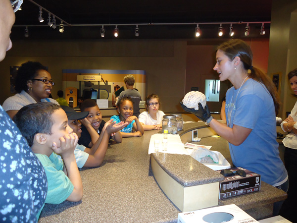
At the 2014 FIFA World Cup, Dr. Miguel Nicolelis hopes to see a quadriplegic child walk into the pitch and deliver the kickoff of the opening game. A pioneer in brain machine interface research and recent author of Beyond Boundaries, Nicolelis gave an evening talk on March 14 at the Nasher as a part of Brain Awareness Week.  Dr. Nicolelis grew up in São Paulo and came to Duke in 1993. Since then, he has focused his research efforts on facilitating two-way dialogue between brains and machines.

Recent advancements in biomedical engineering allow us to use filaments implanted in several parts of the brain to obtain brain function readings: something that was impossible several decades ago. In one of Dr. Nicolelis’s first experiments, monkeys learned to use a joystick to catch a moving object on a screen. After the monkey was able to accurately catch the object 90% of the time, a brain-machine interface was turned on linking the robotic arm to the brain signals. The joystick was eliminated from the setup. The only way to obtain the reward (Brazilian orange juice) was to imagine catching the object.

The brain-machine interface allows for the translation of mental movements into digital commands while recording muscle activity. Using data collected from this experiment, on March 28, 2003, the Nicolelis team was able to design and operate the first robotic arm.

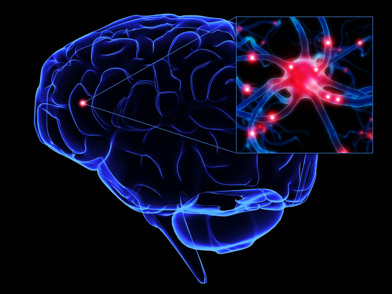
Until recently, neurons were considered the basic functional unit of the brain. More recently, scientists have focused their attention on populations of neurons as functional units instead.

Nicolelis and others are focusing on cell assembles as key functioning units of the brain, not simply neurons. “Populations of neurons across multiple brain structures are working together to make movement possible,” says Nicolelis. Thus, a holistic approach of looking at brain activation is necessary to understand and replicate movement in machines. Differences in the number of neurons activated have been observed when neurons are operating a robotic arm instead of a biological arm.

In a second set of studies, Nicolelis studied the effect of virtual simulations on the brain’s ability to assimilate other things as extensions of the human body. For example, if a professional tennis player is blindfolded and asked to point where his/her arm ends quickly after they have been playing tennis for an hour or so, they will point to the end of the racket as the end of the arm. In other words, the tennis player is assimilating the racket as an extension of the body. Similarly, if a monkey sees a knife approaching a rubber limb that is in the place where his arm should be, he will experience the anxiety, increase in heart rate, and even remove his real arm away from the perceived source of danger.

In an international collaboration with a team from Kyoto, Japan, researchers were able to send brain activity data of a monkey walking on a treadmill to a robot in Japan. The video of the robot walking was then transmitted back to the monkey. Even when the treadmill was stopped, and the monkey was rewarded for each step the robot took, the monkey began imagining that she was the one taking steps in order to be rewarded.

This brain-machine interface research has interesting implications in medicine, ranging from spinal lesions to Parkinson’s disease. When a spinal lesion forms, the brain continues to produce brainstorms to direct movement; however, the body does not have access to muscles. This is where the brain-machine interface comes into play: the brain can provide the directions that can be converted to digital commands, which can ultimately lead to functioning of the machine. To use the brain-machine interface to treat Parkinson’s, Dr. Nicolelis has been using a mouse model developed by Dr. Marc Caron in which 80% of the neurotransmitter dopamine is depleted. The rigid movements of Parkinson’s patients can be refined using brain-machine interface technology.

The brain-machine interface has the ability to alter medicine tremendously.

Dr. Nicolelis’s research implies that it is “possible to use brain activity beyond epithelial boundaries we have,” he says. Perhaps we will be able to do things using this technology, which we customarily cannot do because of the physical constraints of our body because there is no limit to what our minds are capable of doing. “There is a tremendous range of opportunities in this field.”

With the progress the Nicolelis lab is making, perhaps we will be able to see something truly unique at the 2014 FIFA World Cup in Brazil!


Animal emotions may mirror our own

Photo by Becky Phillips, WSU

By Becca Bayham

Did you know that rats can laugh?

All you have to do is tickle them. Oh, and get a supersonic noise detector so that you can hear their happy chirps. [Click here for video]

“It’s one of the most remarkable phenomena I’ve seen in my life,” renowned researcher Jaak Panksepp said during a lecture at Duke, March 15. Panksepp spoke as part of Brain Awareness Week, a series of events dedicated to increasing public awareness about brain research.

Panksepp is well-known for his work in the field of affective neuroscience, or the study of the neural mechanisms that underlie emotion. He argues that important inferences about human emotions can be made from studying emotion in animals. However, this idea has met resistance in the neuroscience community.

“Most scientists are skeptical that animal feelings can ever be studied,” Panksepp said. At this, he gestured toward the audience.

“Well, I will never know what any of you feel, nor will you ever know what I feel,” he said. “But do we close discussion on this important topic, or do we try to work past it?”

Panksepp considers the idea worth investigating, and he has conducted many experiments to test the relationship between human and animal emotions. For example, he found that rats exposed to cat hair exhibited signs of fear, even when they’d never seen a cat before.

“I think you can identify a category of human feelings that correspond with animal feelings. There are going to be differences – there have to be … [But] if we understand their basic feelings, we will begin to understand our own.”

Ewwww, Squishy! …and Kind of Cool

A museum volunteer shows an ACTUAL HUMAN BRAIN -- ewwwwww -- to some kids.

Guest post by Sandra Ackerman, Duke Institute for Brain Sciences.

Cries of “Eew!” and “Squishy!” and a fair amount of giggling marked the arrival of Brain Awareness Week at the North Carolina Museum of Life + Science last week as it offered visitors some great hands-on activities.

The museum’s second floor demonstration lab became the Brain Lab, where kids and adults learned about the three layers of protection that nature has provided to our brains. First the visitors examined half a dozen animal skulls and tried to identify them. There was a beaver, a boar, a deer…  What was that huge one, a toothed whale?  No way!

The next step was to check out a few specimens of preserved brains, each tightly encased in the tough membrane called the meninges.  The most courageous or curious visitors even got the opportunity, after donning latex gloves, to touch the surface of a human brain—an experience they will not soon forget.

Finally, for the purpose of active experimentation, each lab visitor was given a raw egg in its shell, representing the human brain within the meninges.  If the egg was shaken vigorously inside a plastic container (the “skull”), the shell often cracked, spilling its precious contents.  However, after adding water (“cerebrospinal fluid”) and a new egg to the container, a participant could shake the container or even slam it down on the counter, most often with the result that the egg just bobbed gently in the protective liquid.  In cases of extreme trauma, the eggshell sometimes cracked after all—thereby showing vividly why cyclists and skateboarders should always wear a fourth layer of protection, a helmet.

Skulls were compared for their relative brain cases.

Nearby, kids wearing vision-distorting “prism glasses” took turns throwing sponge balls at targets marked on the wall.  Before putting on the glasses, most participants took a few tries to hit the target, then quite a few more to home in on it while their vision was distorted by the prisms.  What really shocked them, though, was how hard it was throw accurately once they had taken the glasses off again.

Asked to give her overall opinion of the scientific experience, one tired participant said, “Hmm, kind of dumb.”

“You think it’s dumb?” her mother asked, surprised.

“Except—except the squishy part!”

A Brain Food You Never Heard Of

Guest post by Sandra Ackerman, Duke Institute for Brain Sciences

Did you know that certain foods are good for your brain — especially if you can persuade your mom to eat them before you’re born?

Christina Williams, Ph.D.

In the second public lecture for Brain Awareness Week, this one given at the Duke Center for Living, Christina Williams, professor of psychology and neuroscience, made a strong case for the long-term benefits of good prenatal nutrition. Of course, all-around good nutrition is important for the health of both an expectant mother and her baby, but in her talk on Tuesday evening Williams focused on choline, “the essential nutrient you’ve probably never heard of.”

Choline is one of those innumerable B vitamins that rarely make it onto a nutrition label. But it deserves to become famous, because choline is important not only for the building of membranes and the proper working of the liver (both very useful in the human body) but also as a basis for the neurotransmitter acetylcholine—the chemical signal that lets us move our muscles. Oh, and it serves another purpose as well: acetylcholine is the main neurotransmitter used in the function of memory.

It is in the realm of memory that prenatal choline really shines. In extensive studies with rats at all stages of their lifespan, Williams and her colleagues have found that adding choline to the usual prenatal diet gives a major boost to the developing brain. As a result, the offspring have brains that not only prove more resilient to shock or trauma but also demonstrate healthy, high-functioning memory well into old age. In the hippocampus, the brain structure most responsible for memory, the prenatally-choline-supplemented rats even show an advantage in the proliferation of new neurons, which enable them to keep learning long after the normal rat retirement age (about 24 months).

A newborn infant, still tapped into her mother's bloodstream, for the moment. (Photo by Gengiskanhg via Wikimedia Commons)

All this is not to say that pregnant women should start gulping down choline, says Williams—least of all as a “supplement” purchased from a health store, because these non-drug compounds don’t undergo strict standardization and scrutiny from the FDA.

You may, however, want to beef up the choline in your regular diet, and in fact beef liver is one of the best dishes for this purpose. Tofu and other soy-based foods, legumes, eggs, and fish will also fill the bill. So lift high your fork, egg cup, or peanut butter-covered knife, and let us keep in mind the power of choline.


Seeing You, Seeing Me …what are you thinking?

Guest Post by Sandra Ackerman, Duke Institute for Brain Sciences

“Don’t know much about history

Don’t know much biology. . .”

Lasana Harris, Ph.D.

Music from the West End Wine Bar in downtown Durham wafted up to the mezzanine where about 30 people gathered on Monday evening for a little light neuroscience.  In the first public lecture of this year’s Brain Awareness Week at Duke, Lasana Harris, assistant professor of psychology and neuroscience, introduced us to his research in a corner of the field where social psychology meets up with brain anatomy—and, despite the words of the popular song, history and biology also made it into the discussion.

Harris and his colleagues work on identifying the neural correlates of mental state inferences—in other words, the kinds of activity in your brain that allow you to think about what might be going on in someone else’s brain.

Functional MRI image of a person looking at images of others’ faces. (National Institutes of Health)

Through a combination of functional MRI scans and carefully delineated storytelling, the researchers have been able to map the two main areas of the brain most involved in inferring the mental state of another person.  These patterns of brain activity are instinctual, not learned — even very young children show evidence of the same patterns — and in normal life they take place with lightning speed, in tenths of a second.  Inferring the mental states of others is a brain exercise we perform dozens of times every day without ever being aware of it.  Truth to tell, most of us wouldn’t recognize a mental state inference if it were served up on a plate with a side of green beans.

And yet this pattern helps to shape so many of your interactions with the world — not just with other people but with pets, machines, or even corporations that skimp on customer service.  We’re so strongly inclined toward making these mental-state inferences that all too often we fall into the trap of treating some non-human entity as if it were a person.  Think of that the next time your computer freezes on you and you’re tempted to give it a swift smack in the monitor!

Brain Awareness Week Begins!

Guest post by Sandra Ackerman, Duke Institute for Brain Sciences.

FASTER than the blink of an eye . . .

MORE  POWERFUL than a champion bodybuilder .  .  .

ABLE to span whole galaxies in a single thought .  .  .

This marvel of multitasking is the human brain.

Brain Awareness Week Logo 2012

Brain Awareness Week Logo 2012

As you take a few seconds to skim this paragraph (perhaps while listening to music, or shifting your weight to keep your balance in a moving bus), various sites in your brain are firing off countless signals along pathways of nerve fiber in all directions.  These signaling pathways emerge, change, die back or grow stronger throughout the human lifespan, in a self-regulating, ever-evolving assemblage more complex than anything the human mind has ever created.  And we’re each carrying around one of these turbo-charged puppies in a bony box on top of our shoulders!

Now’s your chance to get to know your brain a little better, as the Duke Institute for Brain Sciences presents an array of activities for all ages during Brain Awareness Week, from Monday, March 12, through Saturday, March 17.

Each weeknight, in Durham locales ranging from the West End Wine Bar to the Nasher Museum to the Regulator Bookshop, distinguished faculty will give talks on different aspects of the brain, while during the day the Museum of Life and Science will give interactive demonstrations for school groups.  The week wraps up Saturday, March 17 with an all-hands-on-deck, multi-event Open House at the Levine Science Research Center.

Brain Awareness Week:  get ready to expand your mind!

Get Aware of Your Brain, Wouldya?

Are you even aware of your brain? Well you should be, and the good people at the Duke Institute for Brain Sciences are here to help you.

Their third annual Brain Awareness Week March 12 to 17 features a series of five great lectures in venues all over town. Social neuroscience in a wine bar? Why yes, in fact!

NIH color coded brain

Brain surgery and anatomy class would be so much easier if the brain were color-coded like this! (NIH image)

For more kid-friendly brain awareness, DIBS is also planning three days of fun lunch-time demonstrations at the Museum of Life and Science, and the ever-popular Saturday open house at LSRC on Saturday, March 17, which includes lab tours, brain-centric art projects, and lots of eager graduate students doing public outreach.

Please do your brain a favor. Show it a little love. Check out Brain Awareness Week.

(PS watch this space the following week for our student-bloggers’ coverage of some of the events!)

Brain Awareness Week Logo 2012

Brain Awareness Week Logo 2012

Brainnaroo!

By Pranali Dalvi

Human brain scan, in X-rays. Credit: Funderstanding.com

The complexity of the brain can be mind-boggling. After all, how can a jelly-like, three-pound mass of fatty tissue and protein give us the capacity for language, imagination and problem solving, while simultaneously coordinating movement and controlling vital processes such as breathing and digestion?

Duke’s neuroscience community came together on Thursday, Jan. 19 to marvel at the brain during the inaugural event of the Duke Consortium of Neuroscience Graduate Programs: Brainnaroo!

Held in the LSRC Hall of Science, the event included everything from a student poster session to a forum on the “Neuroscience of Self.”

The goal of the forum was to have one topic and approach it with many different ideas, incorporating different departments and techniques,” said Hrishikesh Rao, a graduate student in biomedical engineering and a member of the Brainnaroo Planning Committee.

The unifying theme of the forum was “the self,” and graduate students tackled this concept from a biomedical engineering perspective all the way to a behavioral medicine perspective. Graduate student Rolando Estrada delineated ‘the self’ in computer science. “Computers can do tasks that previously only biological entities could do. For instance, a robot can recognize itself in a mirror,” he said.

Kurtis Gruters, a graduate student specializing in Systems and Integrative Neuroscience, posed the question “Is ‘the self’ a sensory illusion?” in his neural circuitry analysis, and Kristen Batich explained a pathologist’s view of losing ‘the self’ in a spectrum of dementia disorders.

In the true spirit of science, awards followed the presentations and were given for best talk and best layman’s title. Best in Show went to Tina Tognoni for the project, “Sex Differences in Hippocampal Neuronal Responses to Changes in Environmental Stimuli, or “Do male rats pay attention to detail?”

Learning to Fear, By General Category

Guest post by graduate student Kia Walcott

If an animal is lucky enough to escape a predator, that animal will not only avoid the predator but it will have learned to avoid other animals that resemble it in the future. Survivors learn what can eat or hurt them and label them as harmful.

An example of the equipment used in this experiment

Duke neuroscientists Joseph Dunsmoor and Kevin LaBar and NIH neuroscientist Alex Martin wanted to see if we too can learn to fear and avoid related threats.

The researchers showed 26 healthy volunteers an assortment of images belonging to two categories: tools or animals. Some of the images associated with a particular category came along with an “annoying but not painful” electrical shock.  They saw 20 images during four training sessions. The images included typical objects such as a dog or hammer as well as more atypical objects such as an auger (a fancy type of drill) or a leaf insect.  The participants saw each image for six seconds, during which time they rated their shock expectancy. Researchers also measured the subjects’ sweat rate, which links to their emotional arousal as a result of fear.

The participants returned 24 hours after the last session for a surprise memory test which included the 80 total images (20 images over four training sessions) as well as 40 new images (20 tools and 20 animals), and were asked to rate whether each image was new or old and their level of confidence. The test was to see if participants were more likely to remember objects belonging to the shock category versus the ‘safe’ category.

By relating an object to a broader category, the participants quickly and effectively determined which group of objects was associated with the shock.  A difference in sweat rate was also seen in response to objects in the shock category versus those in the non-shock category. Participants were more likely to remember items from the shock category than items from the non-shock category.

These results suggest that fear can be learned and remembered by categories.  In the future, the researchers would like to use brain imaging to see how areas of the brain important for visual object recognition and conceptual knowledge interact with areas important for associating harmful and harmless things. Another important question is how learned fear and memory interact, especially in the case of traumatic experiences.

People who are unable to make connections between known threats and conceptually related things might fail to generalize fear properly, which could prove dangerous. On the other hand, people who make too many connections  to a known threat could suffer too, as in posttraumatic stress disorder.

Citation:  “Role of conceptual knowledge in learning and retention of conditioned fear,” Joseph Dunsmoor, Alex Martin, Kevin LaBar. Biological Psychology, Nov. 2011. doi: 10.1016/j.biopsycho.2011.11.002

http://www.sciencedirect.com/science/article/pii/S03010(2011)51111002730


Is He Conscious? Does He Want To Be?

Terri Schiavo of Florida, whose vegetative state and right to life became a national issue in 2005

By Jeannie Chung

The difference between a dead man and a man in a vegetative state used to be a thin line of whether
or not the body was still functioning. But what if the vegetative man is still conscious? That brings
the distinction into a whole new level.

Philosopher Walter Sinnott-Armstrong gave a talk titled “Is he conscious? Does he want to be?” at the Trent Center for Bioethics on Friday, Dec. 9. He discussed clinical studies which have shown that despite the unresponsive display, patients in vegetative state may be still conscious. With assistance from an fMRI or an EEG scan, doctors can tap into the patient’s brain activity and “read their thoughts.”

The scanning study’s control was patients who received severe brain trauma and were confirmed to be in a vegetative state. The studies focused on the specific brain activity when the patient was commanded to “think about tennis” and the brain activity that occurred when the patient was commanded to “imagine anything other than tennis.” The distinctive brain activities were then coupled with a series of yes or no questions. If their answer was yes, the patient was told to think about tennis and if their answer was no, the patient was told to think about navigating through a house. In one case study, the patient answered five out of seven questions right by showing brain activity associated with tennis to questions for which an affirmative was the correct answer. The other two questions showed no response, and the doctors assumed the patient had gone to sleep.

This confirmation of consciousness in some vegetative patients brings up an ethical issue. Those at the bedside can now ask questions, including “do you want to live?” The vegetative patient’s answer to such a question may inform the ethical issue that arises each time we worry about “pulling the plug” on a clearly “living” person.


Page 14 of 15

Powered by WordPress & Theme by Anders Norén