Following the people and events that make up the research community at Duke

Students exploring the Innovation Co-Lab

Author: Prachiti Dalvi

Collaboration, Mentorship Lead to Clinical Science Successes

By Prachiti Dalvi

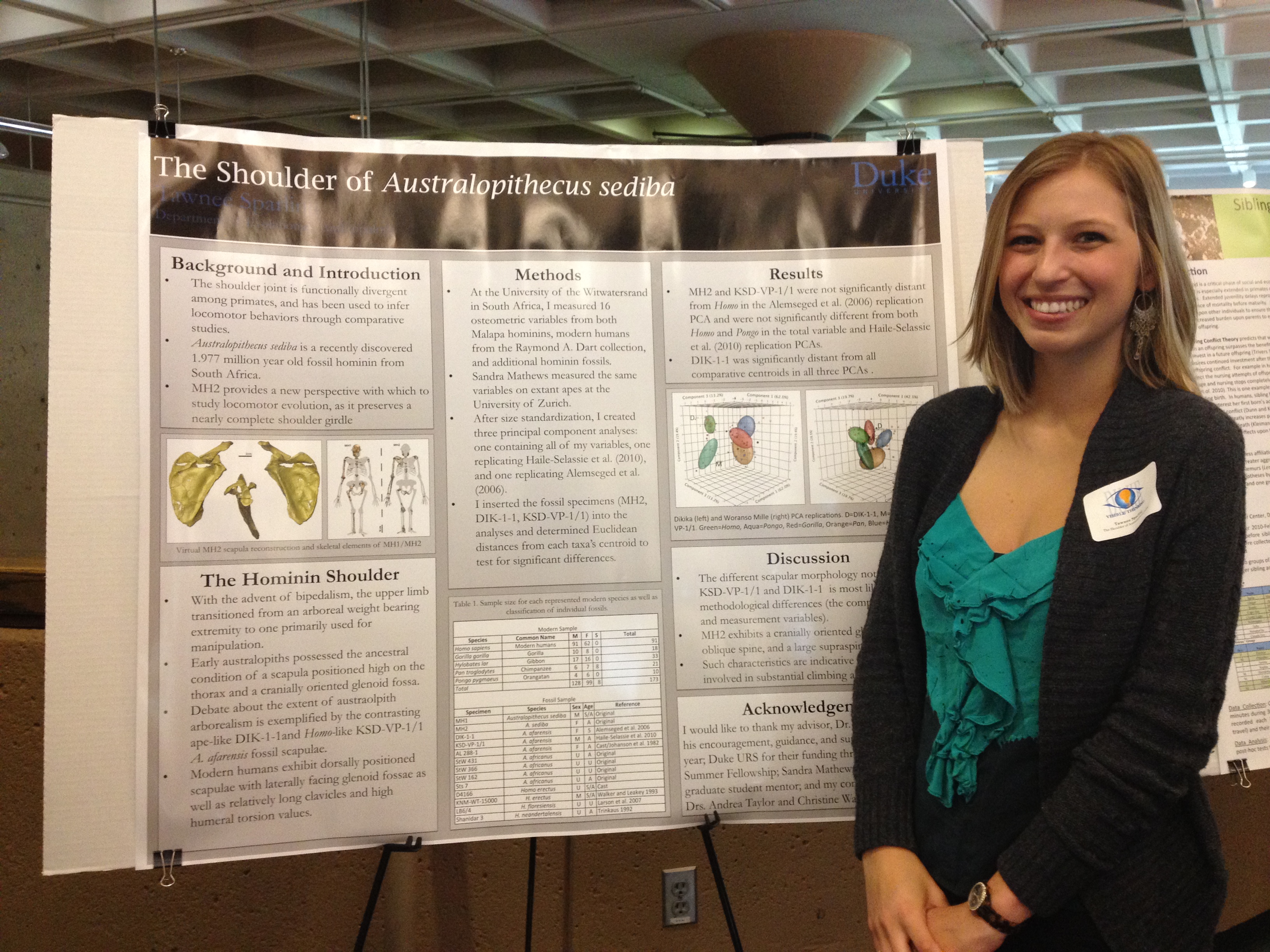
The School of Medicine held the third Clinical Science Day on Friday, October 18th during Duke Medical Alumni Weekend in the Great Hall of the Trent Semans Center. The half-day event brings together faculty, alumni, and students to discuss the cutting edge clinical research taking place in labs across Duke’s campus. The day started out with residents and fellows from a variety of clinical departments participating in a poster competition, followed by fifteen minute presentation by several key clinicians on campus.

Osteoarthritis causes a wearing away of the articular cartilage. (Image credit: http://images.rheumatology.org/viewphoto.php?albumId=77030&imageId=2897682)

Osteoarthritis causes a wearing away of the articular cartilage. (Image credit: http://images.rheumatology.org/viewphoto.php?albumId=77030&imageId=2897682)

Dr. Louis DeFrate from the Department of Orthopedic Surgery is interested in finding a new way to overcome osteoarthritis, a disease that has the potential to affect everyone as they age and is expensive to treat. Obesity increases the risk of osteoarthritis significantly by increasing strain on cartilage in between bones, or articular cartilage. DeFrate is focused on understanding how motion affects how the joints work. “There is very little data on how obesity affects cartilage deformation,” said DeFrate. This information is key to treating osteoarthritis effectively.

Dr. Kimberly Blackwell, who was on TIME’s 2013 list of the 100 most influential people in the world, discussed her research on HER2+ breast cancer tumors. These tumors, affecting 20% of breast cancer patients, are aggressive in coming back and reduce survival rate. The treatment Blackwell and her team have developed consists of an antibody specific to the tumor, loaded with an anticancer toxin. In general, cancer treatments focus on “killing the cancer more than you kill the patient,” says Blackwell. However, this new treatment is able to increase survival rates without many of the unpleasant side effects of other cancer treatments such as chemotherapy. Blackwell has been able to get two cancer-fighting drugs approved by the Federal Drug Administration.

Duke surgeons implanting the bioengineered vein. (Picture credit: http://www.newsobserver.com/2013/06/07/2944111/duke-surgeon-conducts-first-us.html)

Duke surgeons implanting the bioengineered vein. (Picture credit: http://www.newsobserver.com/2013/06/07/2944111/duke-surgeon-conducts-first-us.html)

Vascular surgeon Dr. Jeffrey Lawson discussed his success with implanting a bioengineered blood vessel into the arm of a patient with end-stage kidney disease. The technology designed by Lawson and colleagues involves cultivating donated human cells in a tubular apparatus. Any antibodies that may trigger an immune response are removed and then implanted into the patient. The first implantation took place in Poland in December. The first surgery in the United States took place in June at Duke. Lawson worked closely with Laura Niklason, MD, PhD, a former Duke faculty member who is now at Yale. Lawson and Niklason have worked together to found Humacyte, a spin-off company that makes these bioengineered vessels commercially available. Initially, researchers are interested in implanting these veins in kidney dialysis patients and seeing if they are beneficial. However, ultimately, researchers want to make readily available and durable grafts for heart bypass surgeries to treat blocked blood vessels in the limbs.

All researchers echoed the sentiment that the culture of collaboration and mentorship at Duke is unparalleled.

Making sense of smells

By Prachiti Dalvi

Dr. Richard Axel

Nobel Laureate Dr. Richard Axel, who visited Duke on March 14, 2013.

“A good scientist has to have a nose for which field to work in,” cancer researcher Bernard Weinstein once told his mentee Dr. Richard Axel.

It was a saying Axel took quite literally.

He and Linda Buck won the 2004 Nobel Prize in Physiology or Medicine for discovering the cell receptors that run our sensory system for olfaction, or the sense of smell.

Axel first stepped into the research world when he took a job as a glass washer to support himself during his undergraduate years at Columbia University. After a few broken test tubes and dirty beakers, he was fired as a glass washer and, instead, rehired to do research. He went on to publish three papers as an undergraduate, including one where he was senior author.

In 1979, he earned his medical degree from Johns Hopkins University. He then returned to Columbia, and in 1977 he, along with Michael Wigler and Saul Silverstein, discovered a way to insert foreign DNA into a host cell to produce particular proteins.

The finding grew into to the field of molecular cloning and earned Axel a spot in the National Academy of Sciences at 37.

Axel moved on to study how the brain represents the external world. This is a central issue in philosophy, psychology and neuroscience. But unlike, vision and touch, which have at least two dimensions in the external world, information about odors has no dimensionality at all.

So, how is smell represented in the various smell centers of the brain? Axel explained what scientists know about the representations during a March 14 lecture sponsored by the Ruth K. Broad Foundation and the Chancellor’s Lecture Series.

Screen shot 2013-03-17 at 12.39.56 AM

Pathway of odor perception (courtesy of nobelprize.org)

The process starts in the nose, where different kinds of smell-sensing cells line the nasal cavity. Each type of smell-sensing cell expresses only one of about 1400 odor-receptor genes. The cells expressing a given gene are randomly distributed through the nasal tissue and send projections back through the skull into the olfactory bulb.

This bulb is essentially first relay station for sensing smell in the brain. The incoming information gets consolidated in a part of the bulb called the glomerulus. There a given odor receptor activates nerve fibers called glomeruli. They send signals to higher brain regions, which then create a topographic map of incoming odors.

This topographic map is similar, or conserved, in all individuals of a species, Axel said.

His lab at Columbia is now focused on understanding how the sense of smell is established during development, how it may change over time and how certain smells can elicit specific thoughts and behaviors.

A Call For Action: Genetic Testing Before Prescriptions

By Prachiti Dalvi

Structure of Codeine

Codeine is an opioid pain medication; but if you are a poor metabolizer of a particular enzyme (CYP2D6), you will experience no pain relief from this drug. However, if your doctor could administer something called pharmacogenetic testing, she would know to simply give you morphine (an active metabolite of codeine) instead. For now, this kind of testing isn’t available.

Mary Relling, PharmD

Mary V. Relling, PharmD, the Chair of Pharmaceutical Sciences at St. Jude’s Children Hospital spoke about the need to implement pharmacogenetic testing on Thursday, January 10. A number of  tests have recently emerged that are ready for prime time. When we know that some drugs may have adverse effects for people with  particular genetic phenotypes, it is unethical to prescribe these drugs without knowing the patient’s genetic status.

However, Relling said there are a number of barriers to integrating pharmacogenetic tests into clinical care: fragmentation of our healthcare system, a focus on sick-care rather than disease prevention, a lack of evidence for clinical utility or cost-effectiveness, complex underlying lab results, and a lack of a centralized system for recording patient information.

The best way to break through these barriers is to conduct testing preemptively, Relling said. We can simply take drop of blood when the baby is born and run genetic tests. “Genetic tests are lifetime results. It makes sense to have it in the background, just as we know a patient’s age, weight, sex, etc.,” Relling said. The barriers discussed above can be avoided to a certain extent at St. Jude’s because they have adopted a team approach to patient care and a 100% electronic system for recording patient records.

The growing affordability of genotyping makes using preemptive pharmacogenetic testing more feasible, she said. The cost of sequencing one or two genes in the past will now produce results for 225 genes. Two years ago, the Clinical Pharmacogenetics Implementation Consortium (CPIC) studied how to migrate pharmacogenetic testing from the laboratory into routine patient care. They looked for gene-drug pairs associated with potential risks of life-threatening toxicity, serious adverse effects, or lack of effectiveness. Eleven of the genes CPIC determined met the threshold for high-risk were found to have profound effects on 33 drugs.

Relling said approximately 48% of patients receiving drugs at St. Jude’s received orders for at least one of those pharmacogenetically high-risk medications.

She said the question now is how to use genetic test results rather than whether a genetic test should be ordered. In the coming years, we will have to address how to maintain the fine balance of providing the clinician with enough information to treat the patient and overwhelming the patient with genetic testing results that are difficult to interpret.

This lecture was a part of the Genomics and Personalized Medicine Forum sponsored by the Duke Institute for Genome Sciences and Policy (IGSP).

A Passion for Research

By Prachiti Dalvi

Akash Shah, Trinity ’13

“Research enables me to think about a question that excites me and helps patients,” says Trinity senior Akash Shah.  A biology major, philosophy minor, and a candidate for the Genome Sciences and Policy Certificate, Akash became interested in genomics as a freshman in the Genome Focus. Originally from Fullerton, CA, Akash was drawn to Duke because of its its immense biomedical research enterprise. He also loved the fact that at Duke, the medical school, law school, and business school were on the same campus as the undergraduate campus.

Intrigued by the research his professor Dr. Hunt Willard was conducting, he asked to get involved. His work in Dr. Willard’s lab dealt with artificial human chromosomes. More specifically, he was working with others in the lab to identify which regions of the chromosome would be deleted when transformed into human cells.

Now, Akash works in the Nevins Lab, where he looks at candidate genes in the epidermal growth factor receptor (EGFR) pathway: an important pathway in many cancers. When growth factors bind to the external portion of the receptor, the receptor becomes activated. Side effects of receptor activation include tumor growth and metastasis. When scientists target genes associated with this pathway, they can increase tumor cells’ sensitivity to pathway inhibitors and better prevent tumor cell reproduction.

http://www.youtube.com/watch?v=zE4BkAw_lL4

The advent of computational genomics has allowed for major advances in the field. Fifteen to twenty years ago, cloning genes was considered a PhD project, and now, it is something an undergraduate can do.

Akash’s favorite aspect of research at Duke is its collaborative nature. Faculty members work with another and across departments. His research is not limited to labs at Duke. In fact, he as also worked with professors at UCLA and Harvard. The culture of research varies from one university to the next; thus, Shah encourages undergraduates to do research at different institutions. “It gives you a chance to succeed in different cultures.”

When he is not in the lab, Akash enjoys playing cricket and exploring local restaurants with friends. During his time at Duke, he has been involved with numerous organizations, and has become an integral part of the Genome Research and Education Society (GRES). During his sophomore year, he founded a program in which undergraduates shadowed other undergraduates doing genomics research. In order to make research more accessible to undergraduates, Akash has helped organize career talks, including MD/PhD information sessions. After graduating from Duke in the spring, Akash hopes to begin medical school, and eventually pursue a career in academic medicine so he can continue conducting research. He has worked extensively in cancer genomics research and hopes to explore cancer stem cells in the future.

Visible Thinking

By Prachiti Dalvi & Pranali Dalvi

(See our video from this event! It also appears below.)

In every field from humanities, to behavioral sciences, to biological sciences, Duke undergraduates are tackling current research questions, developing new technologies, and proposing new theories on Duke’s campus and around the world!

Approximately 200 students showcased their research findings from summer internships, independent research projects, and field work projects at Visible Thinking, an undergraduate research symposium sponsored by the Duke Undergraduate Research Support Office and the Duke Undergraduate Research Society (DURS). The event was held in the Bryan Center on Wednesday, April 18.

Joshua Weiss (T'14)

Joshua Weiss found his scientific niche in the laboratory of Daniel Wechsler, MD, PhD, while doing a summer research project funded by the Dean’s Summer Research Fellowship the summer after his freshman year. Hoping to eventually attend medical school, Josh was intrigued by a chromosomal change leading to the development of leukemia in children. It’s a translocation that causes increased activity in two genes called Hox a7 and Hox a9. Hox genes play an enormous role in development and are believed to ultimately cause the leukemia. The Wechsler Lab is interested in understanding the molecular basis behind the genetic change with the hope that it could lead to targeted therapies. Josh is looking at a recent discovery of some molecules that inhibit this chain of events to see whether they’d work with chemotherapy.

Senior Stephanie Patterson found a project that combined her interests of public health, pharmacology, and psychology in the lab of Dr. Nicole-Schramm-Saptya, PhD. A chemistry-major with a concentration in pharmacology, Stephanie was interested in understanding how ethanol exposure during adolescence would affect acute impulsivity and chronic impulsivity in rats. Adolescents have immature brain development, especially in regions of the brain associated with impulsivity and decision-making. Exposure to drugs of abuse has been demonstrated to increase impulsivity. Rats were exposed to ethanol at two developmental time points (adolescence and adulthood) and the delay discounting paradigm was used to measure impulsivity.

Charmaine Mutucumarana (T'13)

Charmaine Mutucumarana, a Trinity junior double-majoring in Biology and French, became involved with research in the laboratory of Dr. Marilyn Telen, MD through the Howard Hughes Research Fellows Program. For the past two years, she has been studying the adhesive properties of blood cells. In sickle cell disease, red blood cells become more adhesive and stick to vascular endothelium. Previously, researchers have found elevated levels of laminin, a plasma factor, in sickle cell patients than in non-sickle cell patients.  Charmaine tested whether soluble purified laminin and plasma laminin enhanced the ability of sickle cell red blood cells to adhere to endothelial cells. She hopes to continue working in the Telen lab to see whether laminin levels in plasma correlate with cell adhesion. These findings could lead to a better understanding of the role of laminin in sickle cell disease.

Melanie Sperling (T'14)

Melanie Sperling is a sophomore majoring in Psychology with a minor in Philosophy and a certificate in Children in Contemporary Society. During her freshman year, she received summer funding from the Hart Leadership Program to study the barriers and opportunities for engaging parents in their children’s education in the Boston area. Melanie worked with the BELL Foundation, which provides after-school and summer opportunities for underprivileged children, to understand why some parents are invested in their children’s educations while others are not. After emailing and setting up phone interviews with more than 500 parents, Melanie explored home-to-school and school-to-home communication and how frequently parents engaged in “learning at home,” or assisting students with homework and other curricular activities.

Sperling found that many parents did not perceive the importance of being involved in their child’s education, while others were busy due to work, other  children, and personal relationships. For instance, some parents returned home late after work when their children were already sleeping. Additionally, parents often felt they lacked the technical knowledge to help their kids with homework. There was a general consensus among the parents that school-to-home communication is lacking. Thus, Melanie suggested that the BELL Foundation can improve communication by sending children home with a checklist every day of homework assignments. She also suggested that parents start weekly meetings with other parents to discuss personal struggles and get advice from one another and also increasing the frequency of parent-teacher conferences. By working with the BELL Foundation to implement these leaders, Melanie hopes to launch parents as leaders in their children’s educations.

Tawnee Sparling, a senior in biological anthropology and anatomy, studied the

Tawnee Sparling (T'12)

shoulder of Autralopithecus sediba, a species of hominids dating to 2 million years ago that was discovered from the partial skeletons of a juvenile male (MH1), an adult female (MH2), one other adult, and an 18-month infant. Taking Steven Churchill’s osteology class during her sophomore year piqued Tawnee’s interest in anthropological research. Her passion for research took her to Johannesburg, South Africa where she compared bone collections from other finds to understand the evolution of the human shoulder with funding from the Dean’s Summer Research Fellowship.

Tawnee explained that there is controversy over the evolution of the hominin shoulder. With the advent of bipedalism, the upper limb became used for manipulation rather than climbing trees. Early australopiths had the ancestral condition in which the shoulder blade was positioned high on the thorax, while modern humans exhibit a lower shoulder blade. However, it’s unclear how much australopiths still climbed trees because anthropologists have found both ape-like and Homo-like fossil shoulder blades. To figure out this controversy, Tawnee  compared measurements from modern humans as well as hominin fossils at the University of Witwatersrand. Her measurements of MH2’s shoulder blades revealed that australopiths were probably involved in substantial climbing. After graduation, Tawnee plans to work in an orthopedics biomechanics lab for a year before attending medical school.
[youtube http://www.youtube.com/watch?v=biK-gPR8C1U]

Probing our Internal Universe

By Prachiti Dalvi

Dr. Nicolelis was recently featured on the Daily Show with Jon Stewart to discuss his new book: Beyond Boundaries.

At the 2014 FIFA World Cup, Dr. Miguel Nicolelis hopes to see a quadriplegic child walk into the pitch and deliver the kickoff of the opening game. A pioneer in brain machine interface research and recent author of Beyond Boundaries, Nicolelis gave an evening talk on March 14 at the Nasher as a part of Brain Awareness Week.  Dr. Nicolelis grew up in São Paulo and came to Duke in 1993. Since then, he has focused his research efforts on facilitating two-way dialogue between brains and machines.

Recent advancements in biomedical engineering allow us to use filaments implanted in several parts of the brain to obtain brain function readings: something that was impossible several decades ago. In one of Dr. Nicolelis’s first experiments, monkeys learned to use a joystick to catch a moving object on a screen. After the monkey was able to accurately catch the object 90% of the time, a brain-machine interface was turned on linking the robotic arm to the brain signals. The joystick was eliminated from the setup. The only way to obtain the reward (Brazilian orange juice) was to imagine catching the object.

The brain-machine interface allows for the translation of mental movements into digital commands while recording muscle activity. Using data collected from this experiment, on March 28, 2003, the Nicolelis team was able to design and operate the first robotic arm.

Until recently, neurons were considered the basic functional unit of the brain. More recently, scientists have focused their attention on populations of neurons as functional units instead.

Nicolelis and others are focusing on cell assembles as key functioning units of the brain, not simply neurons. “Populations of neurons across multiple brain structures are working together to make movement possible,” says Nicolelis. Thus, a holistic approach of looking at brain activation is necessary to understand and replicate movement in machines. Differences in the number of neurons activated have been observed when neurons are operating a robotic arm instead of a biological arm.

In a second set of studies, Nicolelis studied the effect of virtual simulations on the brain’s ability to assimilate other things as extensions of the human body. For example, if a professional tennis player is blindfolded and asked to point where his/her arm ends quickly after they have been playing tennis for an hour or so, they will point to the end of the racket as the end of the arm. In other words, the tennis player is assimilating the racket as an extension of the body. Similarly, if a monkey sees a knife approaching a rubber limb that is in the place where his arm should be, he will experience the anxiety, increase in heart rate, and even remove his real arm away from the perceived source of danger.

In an international collaboration with a team from Kyoto, Japan, researchers were able to send brain activity data of a monkey walking on a treadmill to a robot in Japan. The video of the robot walking was then transmitted back to the monkey. Even when the treadmill was stopped, and the monkey was rewarded for each step the robot took, the monkey began imagining that she was the one taking steps in order to be rewarded.

This brain-machine interface research has interesting implications in medicine, ranging from spinal lesions to Parkinson’s disease. When a spinal lesion forms, the brain continues to produce brainstorms to direct movement; however, the body does not have access to muscles. This is where the brain-machine interface comes into play: the brain can provide the directions that can be converted to digital commands, which can ultimately lead to functioning of the machine. To use the brain-machine interface to treat Parkinson’s, Dr. Nicolelis has been using a mouse model developed by Dr. Marc Caron in which 80% of the neurotransmitter dopamine is depleted. The rigid movements of Parkinson’s patients can be refined using brain-machine interface technology.

The brain-machine interface has the ability to alter medicine tremendously.

Dr. Nicolelis’s research implies that it is “possible to use brain activity beyond epithelial boundaries we have,” he says. Perhaps we will be able to do things using this technology, which we customarily cannot do because of the physical constraints of our body because there is no limit to what our minds are capable of doing. “There is a tremendous range of opportunities in this field.”

With the progress the Nicolelis lab is making, perhaps we will be able to see something truly unique at the 2014 FIFA World Cup in Brazil!


Catching the Space Bug

Prachiti Dalvi

Robert Satcher, MD, PhD –the first orthopedic oncologist to orbit the Earth –discussed his interest in telemedicine and telesurgery during a school of medicine seminar last month.

Growing up not to far from Duke in Denmark, South Carolina, Dr. Satcher developed a profound interest in science and chose to pursue chemical engineering at MIT. After graduating at the top of his class, he entered the MD/PhD program just across the river at Harvard Medical School and returned to MIT to complete his PhD in chemical engineering.

Then, he followed the more conventional route of interning in general surgery and spending his time as a resident at UCLA. Deciding to further specialize, Dr. Satcher proceeded with an orthopedic oncology fellowship at the University of Florida. For a short time period, Dr. Satcher was an assistant professor at Northwestern before he caught the space bug. Satcher successfully completed a rigorous application and interview process and was elected to begin space training at NASA.

Although his interests span chemical engineering and orthopedic oncology, he is particularly interested in bone mineralization, nanomaterials, and bone metastasis in cancer. At the MD Anderson Cancer Center he is exploring telesurgery and telemedicine. In November 2009, Dr. Satcher went into space as a mission specialist on Atlantis, spending more than 200 hours in space and engaging in more than twelve hours of spacewalk.

“Medical knowledge comes into play when people are going through adaptation in aerospace,” Satcher said. While in space, Satcher performed maintenance and conducted research on how the human body reacts in space. His own research interests resonated through when he was able to study how bone density and skeletal muscles are affected by zero gravity. Dr. Satcher likened walking in space while inspecting the station’s outside equipment to surgery: attention to precision is vital. To complete this task, he was able to use his surgical skills to navigate a robotic arm to scan the shuttle for damage.

Although space exploration comes with some dangers and difficulties, Satcher believes space exploration is important because there is a lot we still do not know. According to Dr. Robert Satcher, the common thread of curiosity for the unknown ties space exploration and medicine.

The Paul Farmer Way of Life

By Prachiti Dalvi

One of Duke’s most distinguished alumni and one of global health’s biggest names, Paul Farmer, MD, PhD, discussed his latest book “Haiti After the Earthquake at Duke on Saturday afternoon.

(See Complete Video of his Talk)

So, what should Duke students take from this individual who just like us lived in Wannamaker, wrote for the Chronicle, and once roamed this Gothic wonderland? “Start as early as you can.”

Upon graduating from Duke in 1982 with a BA in medical anthropology, Farmer spent a year in Haiti: a decision that would help him become the kind of physician he always imagined himself as and would help him uncover his lifelong passion. Farmer then returned to Harvard University (which he humorously referred to as “the Duke of the North”) to obtain his MD and PhD in medical anthropology.

Now, Dr. Farmer is Chair of the Department of Global Health and Social Medicine at Harvard Medical School and the Kolokotrones University Professor, one of the highest honors that can be granted to a faculty member. He has been awarded the Conrad N. Hilton Humanitarian Prize and the MacArthur Foundation Genius Award for his work.

“We must counter failures of imagination.”

Just five years after leaving Duke, Farmer co-founded Partners in Health (PIH), an international health organization committed to improving health care in developing countries. This nonprofit organization focuses on building sustainable health care systems rather than simply treating patients. However, Farmer believes that their biggest challenge lies in breaking the cycle of poverty and disease. And, in essence, helping the Haitian community imagine a world where these hurdles can be overcome.

PIH aids developing countries establish sustainable health systems by recruiting community members to be involved in all aspects of designing and implementing a health system. The hospitals that PIH helps establish in Haiti are operated by Haitian doctors, nurses, and medical students.

This nonprofit organization is grounded in the belief that health and education are vital for development. Partners in Health works with partner organizations and national ministries of health to operate projects in twelve different countries all over the globe.

“You might lose the battle, but you won’t lose the war.”

When a magnitude 7 earthquake struck near Port-au-Prince, Haiti’s capital, on January 10, 2010, the effects were everlasting. The toll the earthquake took on Haiti was augmented by the lack of resources and the unexpectedness of the disaster. In fact, the earthquake depleted resources to the extent that it was difficult for Haitian firms to meet minimal process requirements. Resources had to be brought in from outside sources.

One of the most striking issues was facing the largest epidemic of cholera Haiti had ever witnessed. Academic institutions such as Duke and Harvard played a huge role in providing medical relief and “we can be proud of the role academic medicine played” during this critical time period. Farmer claims “Universities have a role to play” in helping bring healthcare equality to developing countries.

Although the earthquake brought with it a great deal of destruction, it also brought with it the opportunity to invest resources in building a sturdier foundation for the health care system. For example, rather than accentuating either treatment or prevention we can now integrate treatment and prevention.

“It’s wonderful to be around, hopeful, optimistic, young people like you.”

Dr. Farmer emphasized the role teaching plays in innovation. According to Farmer, innovation comes from change. Change comes from critical feedback loops. And, critical feedback loops come from discussion. Education is critical in generating conversation and new ideas. As one ages, pessimism begins to sink through and we need fresh minds to bring light to the optimistic aspects of life.

Yet Farmer’s work in Haiti remains fresh and optimistic. “Although Haiti does not have a teaching hospital, doesn’t it deserve a good teaching hospital?” asks Farmer. Currently, PIH is constructing Mirebalais Teaching Hospital. When completed, the hospital will house 320 beds, serve approximately 500 patients daily and will train the next generation of Haiti’s doctors, nurses, and medical professionals.


Us vs. Them

By Prachiti Dalvi

Bacteria occupied Earth long before the evolution of humans. Inhabiting a wide array of environments and exhibiting unparalleled genetic diversity, bacteria share a unique relationship with humans.  Some of these microscopic organisms are essential to life while others threaten our very existence.

Sir Richard Roberts, a 1993 Nobel Laureate in Physiology or Medicine and a Fellow of the Royal Society, spoke about the importance of bacteria to the world of science on Thursday in an event co-sponsored by the Duke Institute for Genome Sciences and Policy (IGSP) and Quintiles, a clinical research organization.

Roberts articulated the importance of understanding the structure of macromolecules in order to understand their precise function: a principle he has used to build his own career. The molecular biologist was awarded a Nobel for his discovery of introns in eukaryotic DNA as well as for explaining the mechanism of gene-splicing.

According to Roberts, although bacteria are generally looked at in a negative light, most bacteria are harmless. He expressed concern about the indiscriminate use of antibiotics. For example, the strength of the farm lobby has prevented the strict regulation of antibiotics administered to animals in the food industry. Here, antibiotics are being used to make animals weightier rather than to fight an infection.

Misuse of antibiotics can help bacteria generate resistance, making fighting these microorganisms in the future more difficult. In fact, when we develop an antibiotic we are simply “selecting for the next drug-resistant bacteria, and we are polluting ourselves out of existence.”

“We tend to lose track of just how interesting bacteria are,” says Roberts. “There was a time when an awful lot of NIH funding was set aside exclusively for bacterial research.” However, now, NIH funding is focused primarily on eukaryotic organisms despite the indispensible role bacteria play.

Hello!

A very warm welcome to you from one of the Research Blog’s newest bloggers!

My name is Prachiti Dalvi, and I am a sophomore in Trinity from Lakeland, Florida. For now, I am studying biology and chemistry; however, I have yet to officially declare my major.

I am absolutely STOKED to be a blogger for Duke’s Research Blog! I started blogging this past summer when I worked in a cell biology lab in the Medical Center. I fell in love with the idea of archiving and reflecting on my experiences. (And, I also thought it was really cool to check my site stats everyday to see how many hits my blog was getting!)

In my free time, I love playing the piano, watching Duke basketball, hanging out with friends on the plaza, reading the New York Times, dancing, travelling with my family and spending time with my twin sister, who happens to be my roommate and is (surprise, surprise…) a blogger for this site too!

Luckily, I stumbled on research when I was a freshman in high school and have been going strong since. I am wholly fascinated by the world of research because it allows scientists to flit along the fine boundary between the known and the unknown.

The cohesive efforts of many individuals, the diligence exhibited when experiments fail and an undying curiosity for the unfamiliar are embodied in a discovery – a discovery that has the potential to explain the simplest of phenomena or to change thousands of lives. I am a true believer in the ability of innovative thinking to find solutions to even the most intricate problems.

As a student at a world-renowned research institution, I look forward to sharing Duke’s research sphere with you through my eyes and hope that you enjoy reading my posts as much as I enjoy writing them!

Powered by WordPress & Theme by Anders Norén