Following the people and events that make up the research community at Duke

Students exploring the Innovation Co-Lab

Category: Genetics/Genomics Page 3 of 11

A Computer Scientist Investigating the Source Code of Life

We are all born with defining physical characteristics. Whether it be piercing blue eyes or jet black hair, these traits distinguish us throughout our entire lives. However, there is something that all of our attributes have in common, a shared origin: genes.

Beyond dictating our individual features, genes instruct cells to create proteins that are essential for a variety of processes, from controlling muscle function to managing digestive systems. Despite their importance in the workings of our body, genes can also code for detrimental diseases, such as Huntington’s disease or Duchenne muscular dystrophy.

Raluca Gordân, Ph.D.

These types of diseases are exactly what Raluca Gordân, Ph.D. is battling through her research. She and her group are trying to figure out how to decode the non-coding genome, the DNA apart from protein-coding genes. They are deepening their understanding of the role non-coding areas of the genome play in the expression of the coding genes and the production of proteins.

Gordân, an associate professor in biostatistics and bioinformatics at Duke, said a majority of disease-causing genetic mutations derive from the genome outside of genes.

“That is a huge search space,” she says, chuckling. “Genes only make up about 2% of the genome. If we don’t understand what those non-coding regions are doing, it’s hard to make predictions about what the mutation in those regions would be doing and how to connect that to the development of a disease.”

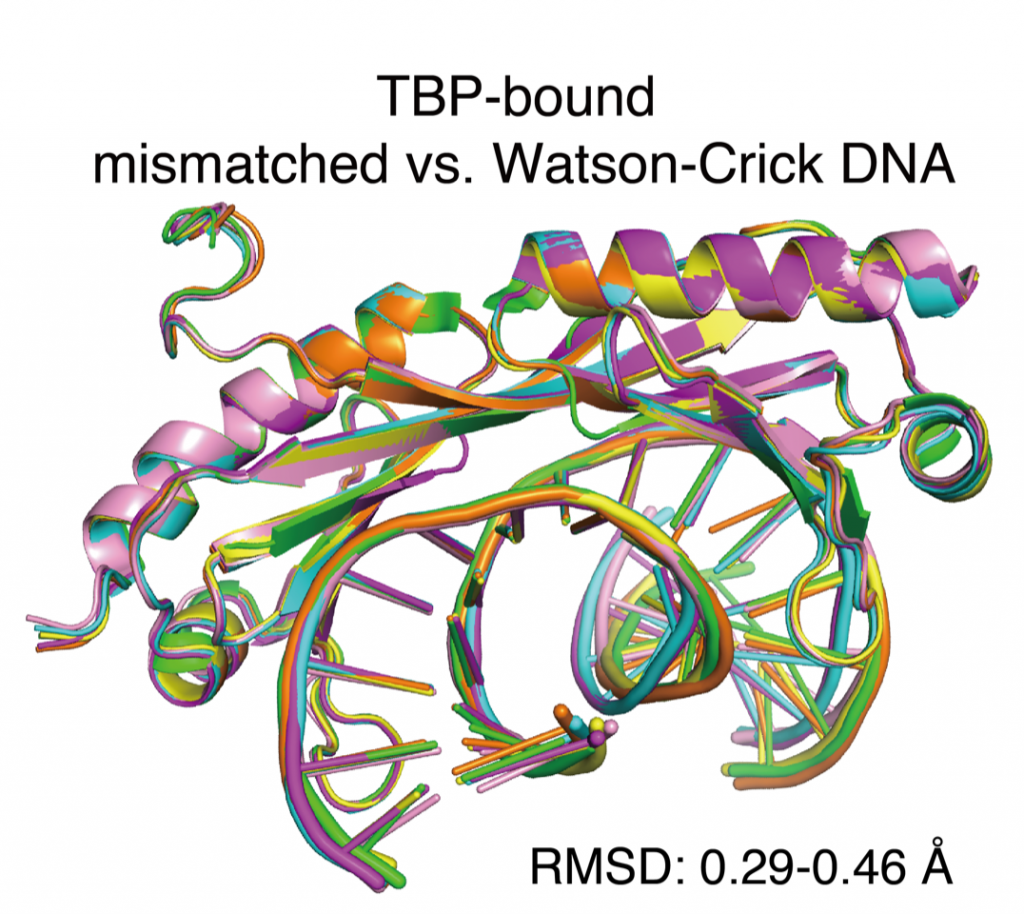
Gordân recently published a paper, entitled “DNA mismatches reveal conformational penalties in protein–DNA recognition,” which focuses on transcription factors and their exceptional ability to bind to mispaired DNA, misspellings that occur as DNA is copied. During regular replication, nucleotide bases (the building blocks of our DNA) are paired correctly, where adenine pairs with thymine and cytosine goes with guanine. However, when an error occurs during replication, mispairs start to appear, as adenine may pair with guanine instead.

“Normally, those are mistakes that get repaired by specific mismatch repair pathways but that repair might not happen if one of these transcription factors sits on the replication error and doesn’t allow the repair mechanism to see it,” Gordân explains. “Normally, one would expect the transcription factors not to bind to those errors. But we found that they can bind way better than their actual genomic targets.”

Modeling of the binding between mismatched DNA and transcription factors.

To expand on her computational discovery, Gordân is now following up with a study of transcription factor binding to mismatches in living cells, observing whether they adopt their usual role of regulating gene expression or contribute to the development of mutations.

Gordân’s research is a product of her passion and desire to make change. It also can be attributed to a series of realizations she made during college and inspirational mentors who guided her along the way.

While pursuing her undergraduate degree, Gordân was a purely computer science major, concentrating on cryptography. However, as she was nearing the end of her four years of college, she soon found herself yearning for the opportunity to do more. She began looking into machine learning applications and enrolled in a course based around genetic algorithms which she credits for launching her career path.

At that point, she attained what she describes as her “first taste of genetics” and her interest in bioinformatics was irrevocably piqued. Thereafter, Gordân applied for a PhD at Duke, where she worked with advisor Alex Hartemink investigating transcription factor proteins in regulatory genomics. At Duke, her work was primarily computational.  But with her postdoctoral advisor Martha Bulyk of Harvard Medical School, Gordan was exposed to the more experimental aspects of biology.

Today, she recognizes these experiences as integral to her ongoing research, which requires her to frequently iterate between observational approaches and computational work.

Gordân is acclimating to the newly quarantined world. While she strives to continue her research, in the pandemic, it has changed her routine.

“I think what was affected a lot since the pandemic started is the fact that we don’t meet in person,” she says. “A lot of the quick progress was being made when we were in the same physical space and were able to get feedback immediately, with students learning about each other’s results in the lab, in real time. That was replaced with Zoom meetings, where students get to see the other students’ results mainly at lab meetings, weeks or months later. Those continuous discussions that were going on in the lab all the time. We’re missing that.”

Gordân offered some thoughtful parting advice to aspiring computational biologists, like me.

“I was trained as a computer scientist, so I wasn’t really sure about experimental work. But after actually doing the experimental work, I realized how much value there is in doing both,” she said. “You have to pick what you’re strongest at, either the computational or experimental part, but you should not be afraid of the other side.”

Guest Post by Akshra Paimagam, Class of 2021, NC School of Science and Math

Claire Engstrom, a Student Researcher Working to Treat Duchenne’s Muscular Dystrophy by Optimizing CRISPr-cas9

Meet Claire Engstrom, a Senior from Pasadena California. Claire is a Biology major who works in the Gersbach Lab at Duke. 

Claire first got involved with on-campus research through her pre-orientation program, PSearch that introduces incoming first-years to undergraduate research. Following her experience in PSearch, Claire got her first work-study research position in the Tung Lab where she worked closely with Jenny Tung, an Associate Professor in the Departments of Evolutionary Anthropology and Biology at Duke and a Faculty Associate of the Duke University Population Research Institute. 

In the Tung Lab, Claire’s research focused on how DNA methylation is passed through generations. Essentially looking at the inheritance of DNA whose methylation was impacted by environmental factors and how that affects future generations. 

Duke has research opportunities available in all disciplines as well as across departments. Approximately 53% of undergraduates graduate with research experience. Not only can students participate in groundbreaking research, but they can receive funding from the university as well to support the work they are doing.

Within the Biology department, there is a fellowship called B-SURF, the Biological Sciences Undergraduate Research Fellowship, an 8-week summer research program for rising sophomores. Claire applied for and was accepted to the fellowship and placed in one of Duke’s biomedical science laboratories. She also received a $4,000 stipend for her summer research.

Claire was placed in Charles Gersbach’s Lab focused on researching Genome Editing for Gene and Cell Therapy. Dr, Gersbach is a Rooney Family Associate Professor of Biomedical Engineering and has conducted groundbreaking work in genome editing.

Members of the Gersbach Lab in Fall 2019

Gersbach is doing research in several different domains of biomedical engineering. Claire’s project focuses on using CRISPR-Cas9, a technology that allows scientists to change an organism’s DNA using clustered regularly interspaced short palindromic repeats and CRISPR-associated protein 9. faster, cheaper, more accurate, and more efficient than other existing genome editing methods. 

Prior to joining his lab, Claire had already heard a lot about Gersbach in her course Biology 201 as well as through reading his papers. The project she would spend the next two and a half years working on focused on using and optimizing CRISPR-Cas9 to treat Duchenne’s Muscular Dystrophy and lessen the severity of the symptoms. 

Duchenne’s Muscular Dystrophy is a muscle wasting disease that affects one in every five thousand male births.

“People are diagnosed when they are around five and then they lose the ability to walk and their heart can’t pump blood because of the lack of muscles.” Claire explained.  

“CRISPR-based genome editing restores dystrophin expression in mouse models of Duchenne muscular dystrophy. Cross-sections of muscle tissue where the dystrophin protein has been labeled green, including normal, healthy tissue (left), tissue from a mouse model of Duchenne muscular dystrophy (middle), and tissue from the same mouse model that has been treated with the CRISPR gene editing system (right). Nelson et al., Science (2016)”

Thus, those affected often die in early adulthood despite current advances in cardiovascular and respiratory treatments. Duchenne’s Muscular Dystrophy generally occurs as a result of a frameshift mutation of the dystrophin gene. As a result, one’s muscles can no longer connect to anything making it nearly impossible to contract and function properly. In the Gersbach lab they are trying to treat the mutation by using CRISPR-Cas9 to remove an exon or coding region of the gene in order to shift the reading frame back into its normal place. 

This shift produces a less severe phenotype that lessens the effects of Duchenne’s Muscular Dystrophy. The result will significantly improve the quality of life and life spans for affected patients. 

Claire will be continuing her work in the Gersbach lab full time in Spring 2021 as she graduated early, with distinction in the Fall. Her thesis on the work she did in the Gersbach lab was recently approved and her results will be published in a larger paper in the future. After this year she plans to take a gap year an then return to California to hopefully attend grad school and pursue a Ph.D. in Biology.

By Anna Gotskind

Dealing With Lead for Life

Though lead has been widely eliminated from use in products due to proven health risks, the lifelong consequences of childhood lead exposure for children born in the era of lead use in gasoline are still unknown.

Aaron Reuben, fifth-year Ph.D. candidate in clinical psychology at Duke, spoke about the long-term implications of childhood lead exposure Friday, September 18th through the Nicholas School’s Environmental Health and Toxicology Seminar series. He conducts research as a member of the Moffitt and Caspi Lab, studying genes, environment, health, and behavior.

Aaron Reuben

Reuben started with a brief history of lead exposure. After the United States’ initial use of lead in gasoline in 1923, the practice became widespread with the U.S. Public Health Services approval for expansion. Five decades later, in the mid-1970s, the Environmental Protection Agency issued the first restrictions on lead use in gasoline products. Simultaneously, surveillance of population-level blood-lead levels indicated cause for concern. Though lead was phased of out of gas completely by 1995, the peak led exposures in the 70s were on average three to four times higher than current levels that demand clinical attention. Despite lead regulations, the impacts of exposure did not miraculously cease as well.

Lead use in gasoline quickly increased after its initial introduction.

The research Reuben covered in his talk centers on the Dunedin Study. This study of 1,037 people born between April 1972 and March 1973 in Dunedin, New Zealand is an ongoing longitudinal research project comprised of over 30 years of data. The cohort of participants provide a unique chance for research in which social and economic factors do not have to be detangled from findings as they represent the full range of socioeconomic statuses in their city.

Reuben’s first question was about the impact of lead exposure on psychiatric and personality differences in adulthood. Study members were asked about symptoms such as substance dependence, depression, fears and phobias, or mania. These reports were transformed into a continuous measure of general psychopathology, which indicated that children with high lead levels experienced more psychiatric problems across adulthood. Though the developmental differences were modest, the associations between lead and psychopathological issues are of a similar magnitude to other known risk factors like childhood maltreatment and family history of mental illness. Yet, unlike the latter two risk factors, Reuben said, “Lead exposure is not preordained – it’s modifiable.”

The research team also measured participant personality using the Big Five Inventory and found that individuals with high-blood level levels as children exhibited more difficult personality styles as adults. The biggest difference between groups with high and low childhood blood-lead level was the trait of conscientiousness, which has impacts on goal obtainment within one’s education and occupation, as well as overall satisfaction with relationships.

Findings from the Big Five Inventory of Dunedin participants.

The next question of the presentation centered on differences in adulthood cognitive ability. At midlife, defined as age 38 for this question, children with higher blood-lead levels had lower cognitive ability, experiencing a deficit of two IQ points per five microgram per deciliter increase of blood-lead level. Once again, though these findings were relatively modest, the loss of IQ points was accompanied by downward social mobility compared to participants’ parents. Further, when evaluations that took place at age 45 were included in the data, researchers saw even larger declines in IQ points between exposure-level groups, which Reuben predicts may even represent a trend of acceleration. He believes that as the study continues with the participants, they will find rapid decline around age 65, with higher levels of dementia symptoms among participants compared to same-aged peers.

The last question evaluated the structural integrity of the brain at midlife. The team found that children with higher lead exposure had lower gray-matter integrity, lower white-matter integrity, and older estimated brain age at age 45. Estimated brain age was predicted by an algorithm based on MRI scans, as brains look physically different as they age and gray- and white-matter integrity refers to the conditions of physical structures in the brain. These findings suggest that childhood led exposure may result in an overall lowered brain integrity at midlife, as well as accelerated brain aging.

Reuben’s take-away findings from his presentation.

Reuben’s work is important for understanding how childhood exposure to this neurotoxin has the ability to influence continued development, behavior, emotion, and life outcomes decades later. It is crucial to evaluate long-term ramifications of childhood lead exposure – a phenomena experience by hundreds of millions of people across the globe during the era of lead in gasoline who are likely unknowingly dealing with impacts now.

Post by Cydney Livingston

Duke Scientists Studying the Shape of COVID Things to Come

The novel coronavirus pandemic has now resulted in more than 3 million confirmed cases globally and is pushing scientists to share ideas quickly and figure out the best ways to collaborate and contribute to solutions.

SARS-CoV-2 surface proteins illustrated by We Are Covert, via Wikimedia Commons

Recently, Duke researchers across the School of Medicine came together for an online symposium consisting of several short presentations to summarize the latest of what is known about the novel coronavirus, SARS-CoV-2.

This daylong event was organized by faculty in the Department of Molecular Genetics and Microbiology and researchers from different fields to share what they know about the virus and immunity to guide vaccine design. This conference highlighted the myriad new research pathways that Duke researchers are launching to better understand this pandemic virus.

One neat area of research is understanding viral processes within cells to identify steps at which antivirals may block the virus. Stacy Horner’s Laboratory studies how RNA viruses replicate inside human cells. By figuring out how viruses and cells interact at the molecular level, Horner can inform development of antivirals and strategies to block viral replication. Antivirals stop infections by preventing the virus from generating more of copies of itself and spreading to other cells. This controls damage to our cells and allows the immune system to catch up and clear the infection.

At the symposium, Horner explained how the SARS-CoV viral genome consists of 29,891 ribonucleotides, which are the building blocks of the RNA strand. The viral genome contains 14 areas where the RNA code can be transcribed into shorter RNA sequences for viral protein production. Though each RNA transcript generally contains the code for a single protein, this virus is intriguing in that it uses RNA tricks to code for up to 27 proteins. Horner highlighted two interesting ways that SARS-CoV packs in additional proteins to produce all the necessary components for its replication and assembly into new viral progeny.

The first way is through slippery sequences on the RNA genome of the virus. A ribosome is a machine inside the cell that runs along a string of RNA to translate its code into proteins that have various functions. Each set of 3 ribonucleotides forms one amino acid, a building block of proteins. In turn, a string of amino acids assembles into a distinct structure that gives rise to a functional protein.

One way that SARS-CoV-2 packs in additional proteins is with regions of its RNA genome that make the ribosome machinery slip back by one ribonucleotide. Once the ribosome gets offset it reads a new grouping of 3 ribonucleotides and creates a different amino acid for the same RNA sequence. In this way, SARS-CoV-2 makes multiple proteins from the same piece of RNA and maximizes space on its genome for additional viral proteins.

An example of an RNA ‘hairpin’ structure, which might fool a ribosome to jump across the sequence rather than reading around the little cul de sac. (Ben Moore, via Wikimedia Commons)

Secondly, the RNA genome of SARS-CoV-2 has regions where the single strand of RNA twists over itself and connects with another segment of RNA farther along the code to form a new protein. These folds create structures that look like diverse trees made of repetitive hairpin-like shapes. If the ribosome runs into a fold, it can hop from one spot in the RNA to another disjoint piece and attach a new string of amino acids instead of the ones directly ahead of it on the linear RNA sequence. This is another way the SARS-CoV-2 packs in extra proteins with the same piece of RNA.

Horner said a step-by-step understanding of what the virus needs to survive at each step of its replication cycle will allow us to design molecules that are able to block these crucial steps.

Indeed, shapes of molecules can determine their function inside the cell. Three Duke teams are pursuing detailed investigation of SARS-CoV-2 protein structures that might guide development of complementarily shaped molecules that can serve as drugs by interfering with viral processes inside cells.

Some Duke faculty who participated in the virtual viral conference. (L-R from, top) Stacy Horner, Nick Heaton, Micah Luftig, Sallie Permar, Ed Miao and Georgia Tomaras. (image: Tulika Singh)

For example the laboratory of Hashim Al-Hashimi, develops computational models to predict the diversity of structures produced by these tree-like RNA folds to identify possible targets for new therapeutics. Currently, the Laboratories of Nicholas Heaton and Claire Smith are teaming up to identify novel restriction factors inside cells that can stop SARS-CoV-2.

However, it is not just the structures of viral components expressed inside the cells that matter, but also those on the outside of a virus particle. In Latin, corona means a crown or garland, and coronaviruses have been named for their distinctive crown-like spikes that envelop each virus particle. The viral protein that forms this corona is aptly named the “Spike” protein.

This Spike protein on the viral surface connects with a human cell surface protein (Angiotensin-converting enzyme 2, abbreviated as ACE2) to allow the virus to enter our cells and cause an infection. Heaton proposed that molecules designed to block this contact, by blocking either the human cell surface protein or the viral Spike protein, should also be tested as possible therapies.

One promising type of molecule to block this interaction is an antibody. Antibodies are “Y” shaped molecules that are developed as part of the immune response in the body by the second week of coronavirus infection. These molecules can detect viral proteins, bind with them, and prevent viruses from entering cells. Unlike several other components on our immune defense, antibodies are shaped to specifically latch on to one type of virus. Teams of scientists at Duke led by Dr. Sallie Permar, Dr. Georgia Tomaras, and Dr. Genevieve Fouda are working to characterize this antibody response to SARS-CoV-2 infection and identify the types of antibodies that confer protection.

Infectious disease specialist Dr. Chris Woods is leading an effort to test whether plasma with antibodies from people who have recovered can prevent severe coronavirus disease in acutely infected patients.

Indeed, there are several intriguing research questions to resolve in the months ahead. Duke scientists are forging new plans for research and actively launching new projects to unravel the mysteries of SARS-CoV-2. With Duke laboratory scientists rolling up their sleeves and gowning up to conduct research on the novel coronavirus, there will be soon be many more vaccine and therapeutic interventions to test.

Guest post by Tulika Singh, MPH, PhD Candidate in the Department of Molecular Genetics and Microbiology (T: @Singh_Tulika)

Duke’s Fundamental Research Can Turn Viruses Into Marvels

The COVID-19 epidemic has impacted the Duke research enterprise in profound ways. Nearly all laboratory-based research has been temporarily halted, except for research directly connected to the fight against COVID-19. It will take much time to return to normal, and that process of renewal will be gradual and will be implemented carefully.

Trying to put this situation into a broader perspective, I thought of the 1939 essay by Abraham Flexner published in Harper’s magazine, entitled “The Usefulness of Useless Knowledge.” Flexner was the founding Director of the Institute for Advanced Study at Princeton, and in that essay, he ruminated on much of the type of knowledge acquired at research universities —  knowledge motivated by no objective other than the basic human desire to understand. As Flexner said, the pursuit of this type of knowledge sometimes leads to surprises that transform the way we see that which was previously taken for granted, or for which we had previously given up hope. Such knowledge is sometimes very useful, in highly unintended ways.

Gregory Gray, MD MPH

The 1918 influenza pandemic led to 500 million confirmed cases, and 50 million deaths. In the Century since, consider how far we have come in our understanding of epidemics, and how that knowledge has impacted our ability to respond. People like Greg Gray, a professor of medicine and member of the Duke Global Health Institute (DGHI), have been quietly studying viruses for many years, including how viruses at domestic animal farms and food markets can leap from animals to humans. Many believe the COVID-19 virus started from a bat and was transferred to a human. Dr. Gray has been a global leader in studying this mechanism of a potential viral pandemic, doing much of his work in Asia, and that experience makes him uniquely positioned to provide understanding of our current predicament.

From the health-policy perspective, Mark McClellan, Director of the Duke Margolis Center for Health Policy, has been a leading voice in understanding viruses and the best policy responses to an epidemic. As a former FDA director, he has experience bringing policy to life, and his voice carries weight in the halls of Washington. Drawing on faculty from across Duke and its extensive applied policy research capacity, the Margolis Center has been at the forefront in guiding policymakers in responding to COVID-19.

Through knowledge accrued by academic leaders like Drs. Gray and McClellan, one notes with awe the difference in how the world has responded to a viral threat today, relative to 100 years ago. While there has been significant turmoil in many people’s lives today, as well as significant hardship, the number of global deaths caused by COVID-19 has been reduced substantially relative to 1918.

One of the seemingly unusual aspects of COVID-19 is that a substantial fraction of the population infected by the virus has no symptoms. However, those asymptomatic individuals shed the virus and infect others. While most people have no or mild symptoms, other people have very adverse effects to COVID-19, some dying quickly.

This heterogeneous response to COVID-19 is a characteristic of viruses studied by Chris Woods, a professor medicine in infectious diseases. Dr. Woods, and his colleagues in the Schools of Medicine and Engineering, have investigated this phenomenon for years, long before the current crisis, focusing their studies on the genomic response of the human host to a virus. This knowledge of viruses has made Dr. Woods and his colleagues leading voices in understanding COVID-19, and guiding the clinical response.

A team led by Greg Sempowski, a professor of pathology in the Human Vaccine Institute is working to isolate protective antibodies from SARS-CoV-2-infected individuals to see if they may be used as drugs to prevent or treat COVID-19. They’re seeking antibodies that can neutralize or kill the virus, which are called neutralizing antibodies.

Barton Haynes,MD
Barton Haynes, MD

Many believe that only a vaccine for COVID-19 can truly return life to normal. Human Vaccine Institute Director Barton Haynes, and his colleagues are at the forefront of developing that vaccine to provide human resistance to COVID-19. Dr. Haynes has been focusing on vaccine research for numerous years, and now that work is at the forefront in the fight against COVID-19.

Engineering and materials science have also advanced significantly since 1918. Ken Gall, a professor of mechanical engineering and materials science has led Duke’s novel application of 3D printing to develop methods for creatively designing personal protective equipment (PPE). These PPE are being used in the Duke hospital, and throughout the world to protect healthcare providers in the fight against COVID-19.

Much of the work discussed above, in addition to being motivated by the desire to understand and adapt to viruses, is motivated from the perspective that viruses must be fought to extend human life.

In contrast, several years ago Jennifer Doudna and Emmanuelle Charpentier, academics at Berkeley and the Max Planck Institute, respectively, asked a seemingly useless question. They wanted to understand how bacteria defended themselves against a virus. What may have made this work seem even more useless is that the specific class of viruses (called phage) that infect bacteria do not cause human disease. Useless stuff! The kind of work that can only take place at a university. That basic research led to the discovery of clustered regularly interspaced short palindromic repeats (CRISPR), a bacterial defense system against viruses, as a tool for manipulating genome sequences. Unexpectedly, CRISPR manifested an almost unbelievable ability to edit the genome, with the potential to cure previously incurable genetic diseases.

Charles Gersbach, a professor of Biomedical Engineering, and his colleagues at Duke are at the forefront of CRISPR research for gene and cell therapy. In fact, he is working with Duke surgery professor and gene therapy expert Aravind Asokan to engineer another class of viruses, recently approved by the FDA for other gene therapies, to deliver CRISPR to diseased tissues. Far from a killer, the modified virus is essential to getting CRISPR to the right tissues to perform gene editing in a manner that was previously thought impossible. There is hope that CRISPR technology can lead to cures for sickle cell and other genetic blood disorders. It is also being used to fight cancer and muscular dystrophy, among many other diseases and it is being used at Duke by Dr. Gersbach in the fight against COVID-19. 

David Ashley, Ph.D.
David Ashley, Ph.D.

In another seemingly bizarre use of a virus, a modified form of the polio virus is being used at Duke to fight glioblastoma, a brain tumor. That work is being pursued within the Preston Robert Tisch Brain Tumor Center, for which David Ashley is the Director. The use of modified polio virus excites the innate human immune system to fight glioblastoma, and extends life in ways that were previously unimaginable. But there are still many basic-science questions that must be overcome. The remarkable extension of life with polio-based immunotherapy occurs for only 20% of glioblastoma patients. Why? Recall from the work of Dr. Woods discussed above, and from our own observation of COVID-19, not all people respond to viruses in the same way. Could this explain the mixed effectiveness of immunotherapy for glioblastoma? It is not known at this time, although Dr. Ashley feels it is likely to be a key factor. Much research is required, to better understand the diversity in the host response to viruses, and to further improve immunotherapy.

The COVID-19 pandemic is a challenge that is disrupting all aspects of life. Through fundamental research being done at Duke, our understanding of such a pandemic has advanced markedly, speeding and improving our capacity to respond. By innovative partnerships between Duke engineers and clinicians, novel methods are being developed to protect frontline medical professionals. Further, via innovative technologies like CRISPR and immunotherapy — that could only seem like science fiction in 1918 (and as recently as 2010!) — viruses are being used to save lives for previously intractable diseases.

Viruses can be killers, but they are also scientific marvels. This is the promise of fundamental research; this is the impact of Duke research.

“We shall not cease from exploration
And the end of all our exploring
Will be to arrive where we started
And know the place for the first time.”

T.S. Eliot, Four Quartets

Post by Lawrence Carin, Vice President for Research

The evolution of a tumor

The results of evolution are often awe-inspiring — from the long neck of the giraffe to the majestic colors of a peacock — but evolution does not always create structures of function and beauty.

In the case of cancer, the growth of a population of malignant cells from a single cell reflects a process of evolution too, but with much more harrowing results.

Johannes Reiter uses mathematical models to understand the evolution of cancer

Researchers like Johannes Reiter, PhD, of Stanford University’s Translational Cancer Evolution Laboratory, are examining the path of cancer from a single sell to many metastatic tumors. By using this perspective and simple mathematical models, Reiter interrogates the current practices in cancer treatment. He spoke at Duke’s mathematical biology seminar on Jan. 17.

 The evolutionary process of cancer begins with a single cell. At each division, a cell acquires a few mutations to its genetic code, most of which are inconsequential. However, if the mutations occur in certain genes called driver genes, the cell lineage can follow a different path of rapid growth. If these mutations can survive, cells continue to divide at a rate faster than normal, and the result is a tumor.

As cells divide, they acquire mutations that can drive abnormal growth and form tumors. Tumors and their metastases can consist of diverse cell populations, complicating treatment plans out patient outcomes. Image courtesy of Reiter Lab

With each additional division, the cell continues to acquire mutations. The result is that a single tumor can consist of a variety of unique cell populations; this diversity is called intratumoral heterogeneity (ITH). As tumors metastasize, or spread to other locations throughout the body, the possibility for diversity grows.

Intratumoral heterogeneity can exist within primary tumors, within metastases, or between metastases. Vogelstein et al., Science, 2013

Reiter describes three flavors of ITH. Intra-primary heterogeneity describes the diversity of cell types within the initial tumor. Intrametastatic heterogeneity describes the diversity of cell types within a single metastasis. Finally, inter-metastatic heterogeneity describes diversity between metastases from the same primary tumor.

For Reiter, inter-metastatic heterogeneity presents a particularly compelling problem. If treatment plans are made based on biopsy of the primary tumor but the metastases differ from each other and from the primary tumor, the efficacy of treatment will be greatly limited.

With this in mind, Reiter developed a mathematical model to predict whether a cell sample collected by biopsy of just the primary tumor would provide adequate information for treatment.

Using genetic sequence data from patients who had at least two untreated metastases and a primary tumor, Reiter found that metastases and primary tumors overwhelmingly share a single driver gene. Reiter said this confirmed that a biopsy of the primary tumor should be sufficient to plan targeted therapies, because the risk of missing driver genes that are functional in the metastases proved to be negligible.

 In his next endeavors as a new member of the Canary Center for Cancer Early Detection, Reiter plans to use his knack for mathematical modeling to tackle problems of identifying cancer while still in its most treatable stage.  

Post by undergraduate blogger Sarah Haurin

Post by Sarah Haurin

First-Year Students Designing Real-World Solutions

In the first week of fall semester, four first-year engineering students, Sean Burrell, Teya Evans, Adam Kramer, and Eloise Sinwell, had a brainstorming session to determine how to create a set of physical therapy stairs designed for children with disabilities. Their goal was to construct something that provided motivation through reward, had variable step height, and could physically support the students. 

Evans explained, “The one they were using before did not have handrails and the kids were feeling really unstable.”

,
Teya Evans is pictured stepping on the staircase her team designed and built. With each step, the lightbox displays different colors.

The team was extremely successful and the staircase they designed met all of the goals set out by their client, physical therapists. It provided motivation through the multi-colored lightbox, included an additional smaller step that could be pulled out to adjust step height, had a handrail to physically support the students and could even be taken apart for easy transportation.

This is a part of the Engineering 101 course all Pratt students are required to take. Teams are paired with a real client and work together throughout the semester to design and create a deliverable solution to the problem they are presented with. At the end of the semester, they present their products at a poster presentation that I attended. It was pretty incredible to see what first-year undergraduates were able to create in just a few months.

The next poster I visited focused on designing a device to stabilize hand tremors. The team’s client, Kate, has Ataxia, a neurological disorder that causes her to have uncontrollable tremors in her arms and hands. She wanted a device that would enable her to use her iPad independently, because she currently needs a caregiver to stabilize her arm to use it. This team, Mohanapriya Cumaran, Richard Sheng, Jolie Mason, and Tess Foote, needed to design something that would allow Kate to access the entire screen while stabilizing tremors, being comfortable, easy to set up and durable.

The team was able to accomplish its task by developing a device that allowed Kate to stabilize her tremors by gripping a 3D printed handlebar. The handlebar was then attached to two rods that rested on springs allowing for vertical motion and a drawer slide allowing for horizontal motion.

“We had her [Kate] touch apps in all areas of the iPad and she could do it.” Foote said. “Future plans are to make it comfier.”

The team plans to improve the product by adding a foam grip to the handlebar, attaching a ball and socket joint for index finger support, and adding a waterproof layer to the wooden pieces in their design. 

The last project I visited created a “Fly Flipping Device.” The team, C. Fischer, E. Song, L. Tarman, and S. Gorbaly, were paired with the Mohamed Noor Lab in the Duke Biology Department as their client. 

Tarman explained, “We were asked to design a device that would expedite the process of transferring fruit flies from one vial to another.”

The Noor lab frequently uses fruit flies to study genetics and currently fly flipping has to be done by hand, which can take a lot of time. The goal was to increase the efficiency of lab experiments by creating a device that would last for more than a year, avoid damaging the vials or flies, was portable and fit within a desk space. 

The team came up with over 50 ideas on how to accomplish this task that they narrowed down to one that they would build. The product they created comprised of two arms made of PVC pipe resting on a wooden base. Attached to the arms were “sleeves” 3D printed to hold the vials containing flies. In order to efficiently flip the flies, one of the arms moves about the axis allowing for multiple vials to be flipped that the time it would normally take to flip one vial. The team was very successful and their creation will contribute to important genetic research.

The Fly Flipping Device

It was mind-blowing to see what first-year students were able to create in their first few months at Duke and I think it is a great concept to begin student education in engineering through a hands-on design process that allows them to develop a solution to a problem and take it from idea to implementation. I am excited about what else other EGR 101 students will design in the future.

By Anna Gotskind


Working Through Frustrations to Understand Nature Better

This is the fifth of six posts written by students at the North Carolina School of Science and Math as part of an elective about science communication with Dean Amy Sheck.

Research is a journey full of uncertainty in which scientists have to construct their own path, even if they’re unsure of what the end of the journey actually is. Despite this unpredictability, researchers continue their journey because they believe their work will one day drive their fields forward. At least, that’s why Kate Meyer Ph.D. says she has investigated something called m6A for several years.

Kathryn Meyer, Ph.D.

“Virtually every study that I have ever been part of had some frustrations involved because everything can fall apart in just one night,” Meyer said. “Despite all the frustrations you might have, you are still in the research because you know that at the end of the day, you will get new knowledge that is worthy to your field, or perhaps to the world.”

N6-methyladenosine (or m6A) is a modification to one of the four main bases of RNA – adenosine. Because RNA plays a significant role as a bridge between genetic information and functional gene products, modifications in RNA can alter how much of a certain product will be produced, which then controls how our cells and eventually our whole body functions.

The idea of this tiny but powerful modification was first proposed in the 1970s. But scientists struggled to find where m6A was located in the cell before research Meyer made a major contribution to as a trainee was published in 2012. Combining a newly developed antibody that could recognize m6A and gene sequencing techniques that became more accessible to the researchers, Meyer’s work led to the first method that can detect and sequence all of the m6A regions in a cell.

m6A’s interaction with a neuron, as depicted on Dr. Meyer’s laboratory site.

Meyer’s work was transformative research. Her method allowed laboratories around the world to investigate what regulates m6A and what its consequences are. Meyer said this first study which ignited m6A field is one of her most prideful moments as a researcher. 

Significant progress has been made since 2012, but there are still lots of questions that need to be answered. Currently, Meyer’s research team is investigating the relationships between m6A and various neurological issues. She believes that regulation of m6A controls the expression, or activity level, of various genes in the brain. As such, m6A may play an important role in neurodegenerative diseases and memory.

Author Jun Hee Shin, left, and Kate Meyer in her lab.

As an assistant professor of both biochemistry and neurobiology at Duke, Meyer is definitely one of the most important figures in the m6A field. Despite her many accomplishments, she said she had experienced and overcome many frustrations and failures on her way to the results.

Guest Post by Jun Shin, NCSSM 2020

The Anthropology of “Porkopolis”

Alex Blanchette, cultural anthropologist and lecturer in anthropology and environmental studies at Tufts University, is a scholar of pork production.

As America’s pork industry is continually pushed to ever greater production, so are the human beings who labor to breed, care for, and slaughter these animals.

Blanchette, who gave a talk hosted by the Ethnography Workshop at Duke on November 4th, said there is an intimate relationship between pig and person. The quality of the factory farm worker’s life is tied to that of the porcine species.

Alex Blanchette of Tufts University

Blanchette’s current work will be published in the 2020 ethnographic book – Porkopolis: American Animality, Standardized Life, and the “Factory” Farm. The book is focused on the consequences of human labor and identity that are bound to the pig – an animal which has become more industrialized over time due to corporations’ goal of a mass produced, standardized pig predictable in nature, uniform in existence, and easy to slaughter.

A common practice in factory farming is the ‘runting’ of litters, genetically making piglets smaller to increase the number each sow produces. But this practice has propelled a fundamental shift in the need for human workers to act as neonatal nurses, what Blanchette calls “external prosthetics,” to care for the newborns. Blanchette described one extraordinary worker responsible for taking care of piglet litters, saving the weak and deformed after birth. She has taken measures so drastic as to give a piglet mouth-to-mouth, incubate them in her pockets, and quickly form body-casts out of duct-tape for the small creatures. This worker has had the chance to study over 400,000 piglets in her seven-year career, encountering conditions of the pig body that no scientist has seen in real life.

Blanchette explained the active engagement required in any portion of the factory production. For example, people working with pregnant sows have to be extremely conscious of the way that the pigs are perceiving them to keep the sensory state of the mother pigs balanced. This means avoiding touching them unless work requires it, not wearing perfumes on the job, and taking overall care and precision in every motion throughout the workday. The danger is the risk of causing mass miscarriages and spontaneous abortions within a barn of sows because of their genetically engineered weakness and inability to handle stresses.

Piglets nursing in a device known as a farrowing crate.

Blanchette said one worker could be seen standing in the exact same place over the course of 1,000 compiled picture frames. He developed this habit to prevent large hogs in open pens from knocking him down and biting his legs while he was working. This is something that Blanchette said he couldn’t manage for more than a few minutes even though he too has worked within the pork industry before.

Workers on slaughter and “disassembly” lines are responsible for making the same exact cut or slice 9,500 times a day.

And finally, the conformation of human labor to the precisions of the factory pig often does not stop at the end of the work shift. In rural factory farming areas, corporations try to re-engineer the human communities in which their workers live to further regulate the human body outside of work because of potential impacts on the pigs. For example, workers’ socialization has been monitored by companies in some cases due to the threat of communicable disease reaching the hogs through human kinship.

No worker knows the pig from birth to death, but for the individual portion of the pig’s life for which they are responsible, they are bound intimately and intricately to the hog, Blanchette said. These people are also disproportionately people of color and immigrant workers who are underpaid for how strenuous, demanding, and encapsulating this labor is. Workers in factory farms often have little protections, and Blanchette’s work gives new life to the consequences of industrial capitalism in America as the pig has become a product of vertical integration in rural communities.

We have long been moving at the speed limits of human physiology in the pork industry,  Blanchette said. In 2011, one company’s annual effort to improve their corporation was to build a new human clinic on the jobsite to treat cuts and injuries acquired on the slaughter lines. This clinic was also responsible for assessing new hires in order to match the strongest part of their body to a place on the line where they would be most productive.

The interior of a typical confined animal feeding operation (CAFO).

Factory farms are actively searching for new money to be found in the pig and to have a closed-loop system which uses every aspect of its life and death for profit. This has caused a deep integration of the “capital swine” into everyday human life for the laborers and communities sustained by these economic ventures.

The Trump administration recently removed standards for pork slaughter line speeds and ultimately reduced overall regulations. People like Blanchette are already considering something you too might be wondering, What happens next? Where does pork and the human labor behind it go from here?

Post by Cydney Livingston

Malaria Hides In People Without Symptoms

It seems like the never-ending battle against Malaria just keeps getting tougher. In regions where Malaria is hyper-prevalent, anti-mosquito measures can only work so well due to the reservoir that has built up of infected humans who do not even know they carry the infection.

In high-transmission areas, asymptomatic malaria is more prevalent than symptomatic malaria. Twenty-four percent of the people in sub-Saharan Africa are estimated to harbor an asymptomatic infection, including 38 to 50 percent of the school-aged children in western Kenya. Out of the 219 million malaria cases in 2017 worldwide, over 90%  were in sub-Saharan Africa.  

Using a special vacuum-like tool, Kelsey Sumner, a former Duke undergraduate now completing her Ph.D. at UNC-Chapel Hill, collected mosquitoes in households located in rural western Kenya. These weekly mosquito collections were a part of her pre-dissertation study on asymptomatic, or invisible, malaria. She visited Duke in September to catch us up on her work in Data Dialogue event sponsored by the mathematics department.

Sumner and colleague Verona Liao, in front of a sticky trap for mosquitoes

People with asymptomatic malaria carry the infection but have no idea they do because they do not have any indicators. This is incredibly dangerous because without symptoms, they will not get treated and can then infect countless others with the disease. As a result, people with an asymptomatic infection or infections have become a reservoir for malaria — a place for it to hide. Reservoirs are a group that is contributing to transmission at a higher rate or proportion than others.

Sumner’s study focused on examining the effect of asymptomatic malaria on malaria transmission as well as whether asymptomatic malaria infections would protect a person against future symptomatic infections from the same or different malaria infections. They were particularly looking into Plasmodium falciparum malaria. In Kenya, more than 70% of the population lives in an area with a high transmission of this potentially lethal parasite.

“P. falciparum malaria is very diverse in the region,” she said. “It’s constantly mutating, which is why it’s so hard to treat. But because of that, we’re able to actually measure how many infections people have at once.” 

The researchers discovered that many study participants were infected with multiple, genetically-distinct malaria infections. Some carried up to fourteen strains of the parasite.

Participants in the study began by filling out an enrollment questionnaire followed by monthly questionnaires and dried blood spot collections. The project has collected over nearly 3,000 dried blood spots from participants. These blood spots were then sent to a lab where DNA was extracted and tested for P. falciparum malaria using qPCR

“We used the fact that we have this really diverse falciparum species in the area and sequenced the DNA from falciparum to actually determine how many infections people have,” Sumner said. “And then, if there’s a shared infection between humans and mosquitoes.”

Sumner and her team also visited symptomatic participants who would fill out a behavioral questionnaire and undergo a rapid diagnostic test. Infected participants were able to receive treatment. 

While people in the region have tried to prevent infection through means like sleeping under insecticide-treated nets, malaria has persisted. 

One of the Kenyan staff members hanging a CDC light trap for mosquitoes

Sumner is continuing to analyze the collected DNA to better understand asymptomatic malaria, malarial reservoirs and how to best intervene to help stop this epidemic. 

“We’re basically looking at how the number of shared infections differ between those that have asymptomatic malaria versus those that have symptomatic malaria.”

She and her team hypothesize that there are more asymptomatic infections that would result in and explain the rapid transmission of malaria in the region.

Post by Anna Gotskind

Page 3 of 11

Powered by WordPress & Theme by Anders Norén