Following the people and events that make up the research community at Duke

Students exploring the Innovation Co-Lab

Category: Genetics/Genomics Page 2 of 10

Quorum Sensing: The Social Network of Bacteria

Dr. Bonnie L. Bassler, the Chair of Molecular Biology at Princeton University, is an advocate for diversity in science.

Bonnie Bassler of Princeton University

Throughout her presentation of the Ingrid Daubechies Lecture on Jan. 31 during Research Week, she emphasized that the diversity of her scientific team allowed every lab member to contribute to different steps of the process of studying quorum sensing, a form of microbial communication. (Watch her talk.)

Bacteria are everywhere. They’re sitting at the tables you sit at, burrowing in the clothes you wear and unfortunately, also crawling around on your skin. For a long time, they’ve had a pretty bad reputation, and for good reason! They have caused plagues that have wiped out masses across the globe, driven up breath mint sales by thriving in your mouth and mutated into every biologist’s Boogeyman when you try to kill them with antibiotics – super bugs!

However, the bacterial redemption arc is also quite compelling. “Good guy” bacteria in the gut help us break down food, produce essential vitamins and also sometimes fight off their evil siblings.

So, good or bad, the impact of bacteria in our daily lives is undeniable. But how does a microscopic little being have the capacity to influence the macroscopic world so greatly?

It doesn’t. At least, not by itself.

Lactococcus lactis is one of the starter bacteria in a cheese culture!

A bacterium never works alone because its strength lies in its numbers! Groupwork and communication (as any Pratt star going through recruiting season will swear to their interviewers) are what make bacteria so powerful. A bacterium by its lonesome will act differently than a bacterium surrounded by its daughters, sisters and cousins (bacterial family tree dynamics can get a little unusual).

Knowing that bacteria optimize their behaviors to work efficiently in a group answers the question of how they are so powerful, but it raises another.

How do bacteria know when they have company?

In a world where social media helps us stay connected, it is easy to take rapid status updates for granted. But for tech-deprived microbial colony, how does one member gauge the population of their surroundings? This question is one that Bassler’s lab answered: with a special chemical compound called autoinducers.

Autoinducers are little chemical signaling molecules that each bacterium sends out into its immediate environment. These molecules allow for quorum sensing, or cell-to-cell communication, to take place among bacteria.

Basic quorum sensing: How bacteria know who’s around them. (Nidhi Srivaths)

Every bacterium senses changes in the concentration of these autoinducers in their surroundings. Sensing a sudden increase in autoinducer concentration will change a bacterium’s gene expression, protein synthesis, and consequently, behavior. It will adapt to group behavior, while a bacterium that senses a drop in autoinducers will adapt to individual behavior.

Bacteria not only sense how many others are around them but also who their neighbors are. Autoinducers are universal to both Gram positive and negative bacteria and are unique to the type of bacteria that produce them.

This provides the bacterium with qualitative information on the population of its surroundings. Are they friends or foes?

Quorum sensing also includes nametags so bacteria can tell friend from foe. (Nidhi Srivaths)

In marine vibrio (a genus of Gram-negative bacteria), Bassler’s lab found that quorum sensing could perform intra-species, intra-genus and inter-species identification. This additional information helps the microbes adjust their behavior – from being friendly and supportive towards their relatives to being aggressive and competitive with their enemies.

Bassler provided a real-world perspective on quorum sensing. One species of the vibrio genus, Vibrio cholerae, is responsible for causing Cholera a deadly food and water-borne disease that has plagued low- and middle-income countries for centuries.

When the cholera bacteria enter the host, they are highly virulent and create a sticky biofilm around themselves that helps them clump into aggregates. Their cell density increases with bacterial division until the bacteria sense a certain concentration of autoinducers. Then their gene expression is modified to reduce virulence and biofilm production and the bacterial gene expression patterns shift to escape mechanisms. The bacteria soon break out in large numbers in search of a new host. (Their human host has voluminous, watery diarrhea in response and that becomes the vector for infecting new hosts.)

While the sequence of events that occurred in a cholera infection was known, the discovery of quorum sensing in V. cholerae opens doors for possible treatments, Bassler said. As bacterial communication sets the cycle of infection and division in motion, interfering with the autoinducers produced or disrupting bacteria’s ability to sense them sets the stage for innovative therapies for several infectious diseases.

Quorum sensing is another step towards understanding the world of the tiny microorganisms that influence our world and Bassler and her team are another example of the incredible research that can come from diverse teams in science.

Post by Nidhi Srivaths, Class of 2024

Medicine Under a Microscope

Duke Research Week 2022 featured a range of speakers from across all disciplines. The Lefkowitz Distinguished Lecture on January 31st highlighted some of our favorite things here at Duke Research Blog: ingenuity and perspective. 

Dr. Huda Yahya Zoghbi’s career spans decades; her Wikipedia page sports an “Awards and Honors” section that takes up my entire computer screen. She is a geneticist, neuroscientist, pediatric neurologist, pharmaceutical executive, and literature lover. Her presentation kicked off 2022 Research Week with a discussion of her work on Rett Syndrome. (View the session)

Rett Syndrome is a rare genetic disorder. The gene that researchers identified as the driver of the syndrome is MeCP2, which is especially active in brain cells. Certain mutations of this one gene can be responsible for a loss of speech, development issues, and persistent fidgeting. 

This image has an empty alt attribute; its file name is _Sk5Y-Q39YYuBd_Ntz_77pXwX8uwtEhTnl4yokbul2AdWHu0xpP_mTKi8mQQHlG7GjmP32Nc4xb1oq-InXReJVfbBai5DF8Enf6LvkuPNZlPou9rc5RCjjjc6B_fRj1oCaVMGWCS
The MeCP2 protein. Image: Wikipedia Commons

Children with Rett Syndrome faced chronic misdiagnosis, and even with proper care were limited by a lack of research.

Duke’s Dr. Robert Lefkowitz introduced Zoghbi at the beginning of the seminar and explained how she came to become the leading expert on this relatively unknown disorder. After completing medical school in Beirut in the midst of the ravaging Lebanese Civil War, she came to Texas Children’s Hospital, where she was able to observe and diagnose her first case of the syndrome, a process spurred by a simple interest in a newly-published journal article.

Holistic knowledge of Rett Syndrome is completely dependent on genetic research. A mutation on the MeCP2 gene causes errors in transcription, the reading out of DNA in your cells which leads to the production of proteins.

The mutated gene’s MeCP2 protein is then lacking the ability to do its job, which is helping other genes be expressed, or actively transcribed.

It’s a vicious cycle; like when you go to sleep late one night, so you sleep in the next day, then go to sleep late the next night, then sleep in the next day, and so forth.

In order to simulate and measure the effect of different kind of mutations on the MeCP2 gene, Zoghbi and her team studied genetically modified mice. While Rett Syndrome is caused by a lack of MeCP2 function, an overactive MeCP2 gene causes MeCP2 duplication syndrome. Varying degrees of gene efficiency then produce varying degrees of severity in the syndrome’s traits, with fatality at either end of the curve.  

Varying degrees of phenotype severity.

Zoghbi’s talk focused mainly on the mechanics of the disorder on a genetic level, familiar territory to both Nobel Laureate Lefkowitz and Duke Medicine Dean Mary Klotman, who shared some discussion with Zoghbi.

This medicine on a microscale is applicable to treating genetic disorders, not just identifying them. Zoghbi has been able to experimentally correct MeCP2 duplication disorder in mice by modifying receptors in a way that reverses the effects of the disorder.

The symptoms of Rett Syndrome are physical; they present themselves as distinct phenotypes of a subtle difference in genotype that’s too small to see. The field of genetics in medicine is responsible for making that connection.

Post by Olivia Ares, Class 2025

This image has an empty alt attribute; its file name is ARES_Olivia.jpg

Introducing Muser – A Better Way to Find Student Research

An effortlessly simple research platform where Duke students and Duke research projects can connect? Yes, please!

If you are anything like me, Duke University’s incredible research opportunities were extremely enticing when considering this school. One of the top 10 research institutions in the United States, Duke University’s research community spends over 1 billion dollars annually to fund its projects, which includes notable research facilities like the Duke Center for Human Genetics, the Duke Cancer Institute, the Duke Center for AIDS Research, and the Duke Human Vaccine Institute.

However, the amount of opportunity in this area can be overwhelming to approach, and as a student you often have no clue where to start.

Summer undergraduate research in cancer biology at the Duke University School of Medicine.

That’s where Muser comes in.

Duke introduces: Muser.com

Muser is a website created by Sheila Patek, a Duke biology professor who used grant money from the National Science Foundation to create a more equitable and straightforward way to connect undergraduates with professors with research opportunities. The resource allows researchers to post ongoing research positions with a direct application through the website.

Muser can sort research projects by compensation, hours, year, and project category, simplifying Duke’s incredibly complex research community by a lot.

“Muser posts research projects in 4 rounds throughout the year, a Fall round (August), a late Fall round for Spring projects (October/November), a Spring round for Summer projects (February/March), and a Spring round for Fall projects (March/April),” according to its website. Muser makes it easy to accommodate research positions into the part of your semester that works with your busy schedule.

I connected with some Duke students who have found success with the growing research platform, and though their interests were diverse, the success was all-encompassing.

“My experience with my Muser Project for the summer of 2021 was great overall,” said Elaijah Lapay, class of 2025. “It was essentially a history research assistantship helping a professor in the history department conduct research on elderly and eldercare in North Carolina. I was able to go to the NC State archives as well as archives across eastern North Carolina to really dive into the question of treatment of the elderly during the 20th century.”

Lapay’s research is so fruitful that the professor, James Chappel, the Gilhuly Family Associate Professor of History, is continuing to pursue this project for the rest of the school year. “I truly felt one-of-a-kind… I definitely feel like I’ve learned a lot and it’s sparked a passion in me for geriatrics and eldercare.”

A look inside Dr. Laurie Sanders’s lab here at Duke University.

“I got the chance to work in the Sanders lab under principal investigator Dr. Laurie Sanders and post-doctorate Dr.Claudia Gonzalez-Hunt!” said Shreya Goel, class of 2025. This lab was the first to link a genetic mutation to mitochondrial DNA damage which was ultimately discovered to be a marker for sporadic Parkinson’s disease.

“I get to work with human cells to induce and track mitochondrial and nuclear DNA mutations to determine their effect on the progression of the cell cycle,” Goel said. Her research position is making a difference and it allows her to gain tangible experience in a field she is passionate about.

The success stories are copious, and the opportunity that this platform has brought to prodigious students like these is without question.

At a billion-dollar research school, understanding where to begin can be intimidating. Muser alleviates these worries by connecting researchers and students through an accessible platform.

Have more questions? Visit Muser’s FAQ page to get more information and get into contact with one of Muser’s staff.

Post by Skylar Hughes
Class of 2025

Blake Fauskee and the ‘Little Typos’ of Fern DNA

Blake Fauskee, third-year Biology PhD student, initially pitched his graduate project to advisor Kathleen Pryer (Ph.D.) as an undergrad.

Fauskee, who researches RNA editing sites in ferns, told me about the project that he’s been working on for the last several years. His research could push back against the idea that DNA is the end-all, be-all molecule for encoding life as we know it.

Blake Fauskee, third-year Biology PhD student

Fauskee broke down RNA editing for me. “RNA editing is this extra step in the whole central dogma, the whole gene expression process, that happens in plant organellar DNA,” he said. This process takes place in plant mitochondrial and chloroplast genomes.

Fauskee uses a lot of metaphors to describe his work, which I find both helpful and admirable. Science can often be dense and lack feasible connections to processes that most of us are familiar with. “Basically, in [plant] DNA, there are little typos almost. The wrong nucleotide is encoded at certain spots. When those genes are going to be expressed, they get turned into RNA and then other proteins from the nucleus come in and find the little typos so that in the end you get the correct protein.”

This image shows a simplified diagram of how RNA editing works.

Fauskee calls RNA editing an “interesting and strange process” that neither animals nor humans have. His work attempts to study the evolution of this process, the patterns of RNA editing, and why it came to be. He uses DNA and RNA sequence data and the help of computational tools to do his work. He explains that when sequencing DNA, you can think of the fragmented base pairs “as little puzzle pieces.”

“So, I take all those little puzzle pieces and try to put back together the chloroplast genome, which is about 150,000 base pairs. It’s like a thousand-piece puzzle.”

Next, he figures out where the fern’s gene sequences are on the DNA strands, making use of genomic databases that contain known genomes. He then aligns RNA sequences to the genes he has mapped. Fauskee looks for the “typos” or “little differences” between the DNA and RNA: “That’s how we find the RNA editing sites.” Finally, he evaluates how the proteins would be changed by the typos in the DNA if the RNA was not edited after being transcribed.

“So, a lot of these fern genes will have a STOP codon right in the middle, which is really, really bad if you don’t fix because you are only going to get half a protein,” Fauskee said. STOP codons signal to the protein-building ribosomes that the protein is finished once it reads this portion of the RNA. Fauskee explained that these types of errors are the ones would expect organisms to lose, but it turns out they are the ones that are conserved in ferns. “Is there an extra function there? Is it helpful? Is it adaptive?” Fauskee asked.

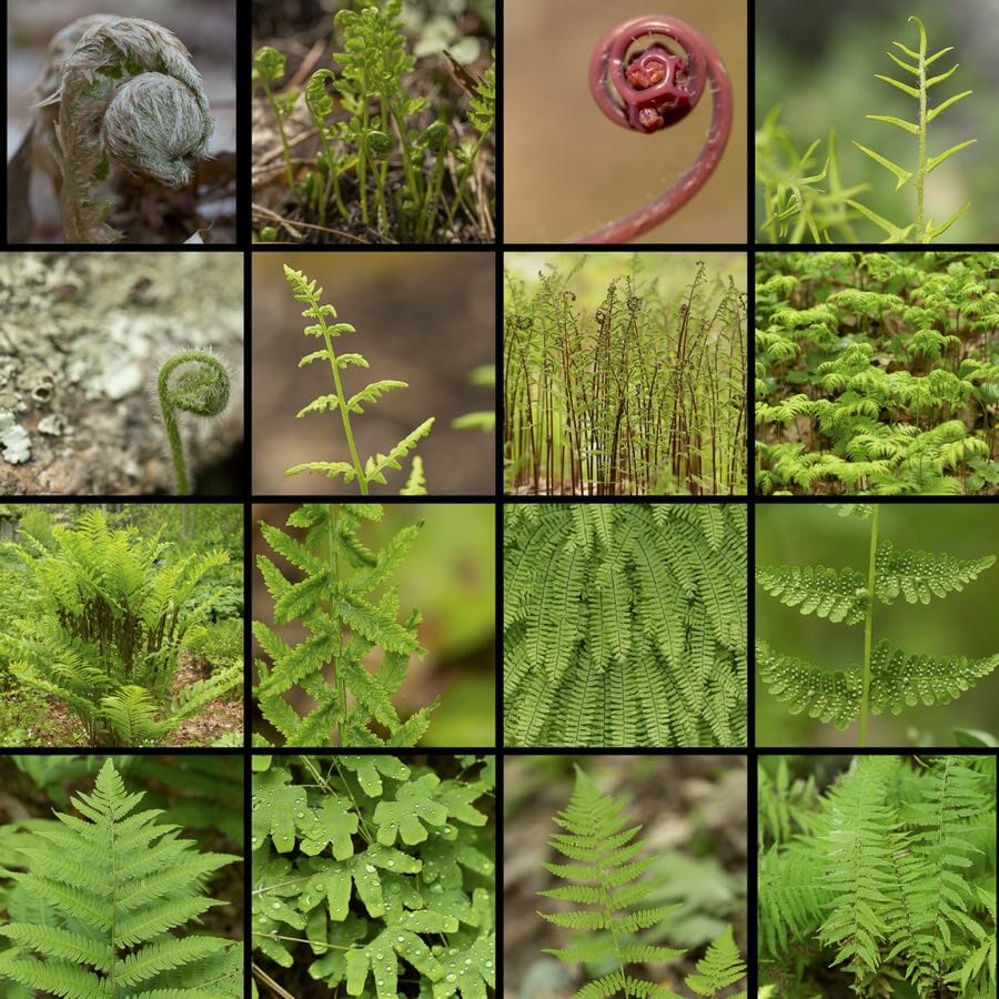
An image of different ferns.

Comparative analyses between fern species are important. By looking at whether there are common editing sites and common amino acid changes, Fauskee says, “we’re trying to understand if certain editing sites may be advantageous and what kinds of fluctuation we see between certain types of changes.”

Fauskee underscored the importance of his work. “RNA editing is a really interesting process that kind of undermines what I learned in molecular biology…They always tell you DNA is the bedrock, it’s the be-all, end-all. But what happens when the DNA is wrong? What’s the other added layer on this?”

Simply put, Fauskee, says that because of RNA editing, “We have to rethink central dogma a little bit.” In some plants, 10% of all their gene products contribute to RNA editing, Fauskee tells me. “That’s a big chunk and that’s got to be important,” Fauskee said, “Why would evolution keep such a burden going?”

Biology’s central dogma is the idea that DNA is transcribed into RNA and then translated into proteins. RNA editing adds an extra step before translation and protein production.

There may also be implications for how RNA editing sites affect the way that genetic relationships are mapped through phylogenetics. If differences between the DNA of different species at RNA editing sites, this could be misleading. Though the DNA indicates a change in base pair, RNA editing could lead to the same output in protein despite the seeming change. “If you took [RNA editing] into account,” Fauskee says, “does it give you a different answer?”

Fauskee studies ferns because of the amount of editing sites found in these plants. While flowering plants have lost editing sites over time, ferns have not. “For RNA editing, you can look at all angiosperms (flowering plants) and for the whole chloroplast genome, they might have 30-50 RNA editing sites. When you get into ferns, that number jumps up to 300-500 and I am trying to understand why.”

Botanical science first captured Fauskee’s interest while he completed his undergraduate degree in his home state at the University of Minnesota Duluth (UMD). As a sophomore at UMD, Fauskee was taken under the wing of Amanda Grusz (Ph.D.). Grusz received her PhD in biology from Duke and worked under Pryer during her own time at the university. “I’m like my advisor’s academic grandson, which is kind of funny.” Clara Howell, who is part of Fauskee’s PhD cohort and who I spoke with last Fall, is also an academic grandchild in her own lab.

Being an “academic grandson” has worked out well for Fauskee. His key advice to me for any person considering a PhD, “Make sure your advisor is not someone you just admire as a scientist, but as a person.” On a day-to-day basis, Fauskee says that advisor Katheen Pryer “is pretty hands off” but is also “one of the most supportive people ever. I’m pretty much the driver of my own ship. If I am falling off the road, she’ll push me back on the road, but she’ll give me freedom to swerve around on that road.”

Fauskee also emphasized a piece of wisdom that Pryer passed down to him. “If whatever you’ve got going on is working and everyone else is doing something different, who cares?” he said.

Though Fauskee says that “lab work can be frustrating,” getting his long analyses to run after wrangling lots of data is very rewarding. Fauskee, who does not have a background in coding or computer languages, likes to “tell people that [his] floor of biology combined is one competent coder.” When he’s not stealing bits of his biology neighbor’s code, Fauskee loves to attend Duke Basketball games and is a fan of the television show Survivor.

Post by Cydney Livingston, Class of 2022

Nobel Laureate Dr. Jennifer Doudna and Groundbreaking Applications of CRISPR

In 2011, Dr. Jennifer Doudna began studying an enzyme called Cas9. Little did she know, in 2020 she would go on to win the Nobel Prize in Chemistry along with Emmanuelle Charpentier for discovering the powerful gene-editing tool, CRISPR-Cas9. Today, Doudna is a decorated researcher, the Li Ka Shing Chancellors Chair, a Professor in the Department of Chemistry and Molecular as well as Cell Biology at the University of California Berkeley, and the founder of the Innovative Genomics Institute.

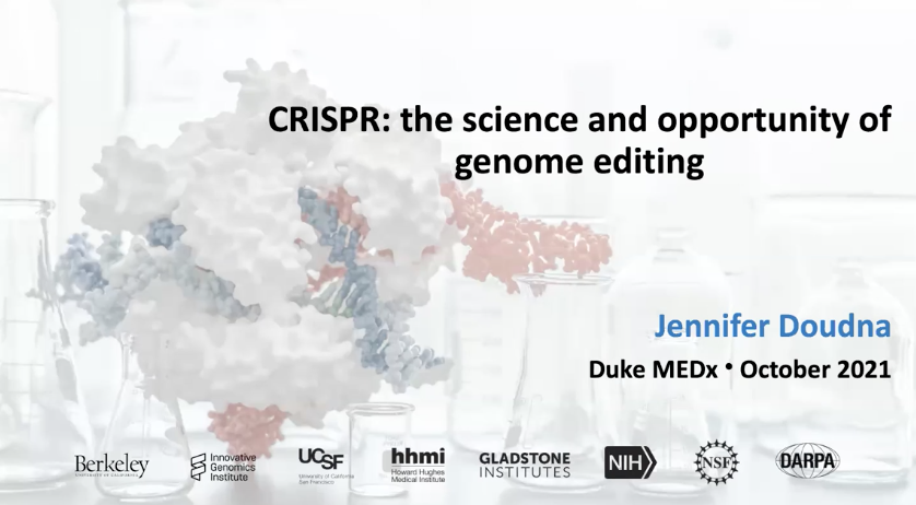
Doudna was also this year’s speaker for the MEDx Distinguished Lecture in October where she delivered presented on “CRISPR: Rewriting DNA and the Future of Humanity.”

“CRISPR is a system that originated in bacteria as an adaptive immune system” Doudna explained.

Dr. Jennifer Doudna holding the Nobel Prize in Chemistry

When bacterial cells are infected by viruses those viruses inject their genetic material into the cell. This discovery, a couple decades ago, was the first indication that there may be ways to apply bacteria’s ability to acquire genetic information from viruses.

CRISPR itself was discovered in 1987 and stands for “Clustered Regularly Interspaced Short Palindromic Repeats.” Doudna was initially studying RNA when she discovered Cas-9, a bacterial RNA-guided endonuclease and one of the enzymes produced by the CRISPR system. In 2012, Doudna and her colleagues found that Cas9 used base pairing to locate and splice target DNAs when combined with a guide RNA.

Essentially, they designed guide RNA to target specific cells. If those cells had a CRISPR system encoded in their genome, the cell is able to make an RNA copy of the CRISPR locus. Those RNA molecules are then processed into units that each include a sequence derived from a virus and then assemble with proteins. This RNA protein then looks for DNA sequences that match the sequence in the RNA guide. Once a match occurs, Cas9 is able to bind to and cut the DNA, leading to the destruction of the viral genome. The cutting of DNA then triggers DNA repair allowing gene editing to occur.

“This system has been harnessed as a technology for genome editing because of the ability of these proteins, these CRISPR Cas-p proteins, to be programmed by RNA molecules to cut any desired DNA sequence,” Doudna said.

Jennifer Doudna holding a Model of CRISPR-cas9

While continuing to conduct research, Doudna has also been focused on applying CRISPR in agriculture and medicine. For agriculture, researchers are looking to make changes to the genomes of plants in order to improve drought resistance and crop protection. 

CRISPR-cas9 is also being applied in many clinical settings. In fact, when the COVID-19 pandemic hit, Doudna along with several colleagues organized a five-lab consortium including the labs of Dan Fletcher, Patrick Hsu, Melanie Ott, and David Savage. The focus was on developing the Cas13 system to detect COVID-19. Cas13 is a class of proteins, that are RNA guided, RNA targeting, CRISPR enzymes. This research was initially done by one of Doudna’s former graduate students, Alexandra East-Seletsky. They discovered that if the reporter RNA is is paired with enzymes that have a quenched fluorophore pair on the ends, when the target is activated, the reporter is cleaved and a fluorescent signal is released. 

One study out of the Melanie Ott group demonstrated that Cas13 can be used to detect viral RNA. They are hoping to apply this as a point-of-care diagnostic by using a detector as well as a microfluidic chip which would allow for the conduction of these chemical reactions in much smaller volumes that can then be read out by a laser. Currently, the detection limit is similar to what one can get with a PCR reaction however it is significantly easier to run.

Graphical Abstract of Cas13 Research by the Melanie Ott lab

“And this is again, not fantasy, we’ve actually had just fabricated devices that will be sitting on a benchtop, and are able to use fabricated chips that will allow us to run the Cas13 chemistry with either nasal swab samples or saliva samples for detection of the virus,” Doudna added.

Another exciting development is the use of genome editing in somatic cells. This involves making changes in the cells of an individual as opposed to the germline. One example is sickle cell disease which is caused by a single base pair defect in a gene. Soon, clinicians will be able to target and correct this defect at the source of the mutation alleviating people from this devastating illness. Currently, there are multiple ongoing clinical trials including one at the Innovative Genomics Institute run by Doudna. In fact, one patient, Victoria Gray, has already been treated for her sickle cell disease using CRISPR.

Victoria Gray being treated for Sickle Cell Anemia
Meredith Rizzo/NPR

“The results of these trials are incredibly exciting and encouraging to all of us in the field, with the knowledge that this technology is being deployed to have a positive impact on patient’s lives,” Doudna said.

 Another important advancement was made last summer involving the use of CRISPR-based therapy to treat ATR, a rare genetic disease that primarily affects the liver. This is also the first time CRISPR molecules will be delivered in vivo.

In just 10 years CRISPR-cas9 has gone from an exciting discovery to being applied in several medical and agricultural settings. 

“This powerful technology enables scientists to change DNA with precision only dreamed of a few years ago,” said MEDx director Geoffrey Ginsburg, a Professor of Medicine at Duke. “Labs worldwide have redirected the course of research programs to incorporate this new tool, creating a CRISPR revolution with huge implications across biology and medicine.”

Examples of further CRISPR-Cas9 research can also be found in the Charles Gersbach lab here at Duke. 

By Anna Gotskind, Class of 2022

Trust-Building, Re-Visited History, and Time Pertinent to Achieve Health Equity for Black Americans

Along with being a beautiful person and leading a productive life, Henrietta Lacks is the mother of modern medicine. Her scientific child was born without Henrietta’s consent through the clinical breakthroughs and medical miracles achieved with the help of her cervical cells – HeLa cells – stolen without her knowledge when she sought healthcare. Ironically, the same treatments developed from the cells of this Black woman are inaccessible for many Black Americans contemporarily. Though Ms. Lacks passed away from cervical cancer at the premature age of 31, her unique cells have become immortal. Her story lives on as a pertinent reminder of the importance of building trust between medicine and the Black community. In honor of her birthday, expert panelists met to both celebrate Ms. Lacks and discuss the path forward in trust-building, equity, and reckoning with our history to change the narrative of healthcare for Black Americans.

The panel honored Henrietta Lacks through discussion of the path forward for biomedical research and Black communities. The panel was hosted in August in remembrance of Ms. Lacks’ birthday on August 1st.

The panel, which took place on Tuesday, August 31, began as a conversation between Nadine Barrett (Ph.D.), Robert A. Winn (M.D.) and Vanessa B. Sheppard (Ph.D.). Among their many other titles and positions, Barrett is Director, Center for Equity in Research, Dukev CTSI and Associate Director of Equity, Community and Stakeholder Strategy, Duke Cancer Institute, Dr. Winn is the Director of the Virginia Commonwealth University (VCU) Massey Cancer Center, and Sheppard is the Associate Director of Community Outreach Engagement and Health Disparities at VCU Massey Cancer Center. The trio were joined by Reuben Warren (D.D.S., M.P.H., Dr. P.H., M.DIV.), Director of Tuskegee University’s Bioethics Center, along with a handful of other contributors including Veronica Robinson – Henrietta Lacks’ great-granddaughter and a registered nurse who represents the Lacks family on the NIH panel that reviews applications to conduct research using the HeLa genome.

A screenshot of panelists who took part in Tuesday’s conversation.

Winn began by referencing the U.S. 1932 public health service study that took place in Tuskegee, Alabama. The experiment exploited Black men in Tuskegee when an effective form of treatment for syphilis was discovered 15 years into the study but withheld from participants “to track the disease’s full progression.” In 1972, 40 years after the study began, it was the associated press, not the scientific community that finally led to the experiment’s demise and the issue of an apology from the U.S. President.

As Warren pointed out, the issue with the study was less about the treatment and more about the dishonesty, the falsifying information, and lies. “Stop calling them poor, stop calling them all sharecroppers,” Warren said of the Black men who participated in the study, “They were far more than that.” “[The study] was an issue of trust, not an issue of ignorance,” he continued. Unfortunately, when talking about this story, Winn said that Black Americans “don’t always talk about the power of us standing up and saying not again.

Bioethics violations have been a continuous part of the biomedical research enterprise in the U.S., and race and racism have been part of scientific inquiry, which continues to be of great concern, Warren said. Often, rather than putting preventative protections in place, bioethics regulations have come as a reaction to extreme violations of justice. Thus, Warren laid out a central theme of the panel that “You build trust by making yourself trustworthy and that takes time.” Rather than initiating transactional research with Black communities when the scientific and medical community needs something, Warren offered that they should start when they want to help with something.

Dr. Rueben Warren presenting examples of bioethics violations in the history of biomedical research, with most examples stemming from the United States

As Sheppard said, “[Black people] have earned a mistrust” for medical communities. This is largely hinged on Barrett’s argument that the American systems from health to education to criminal justice “are working as they were designed” – to ensure that the very inequalities that exist today came to be. Using the analogy of a marathon, Barrett said while white men in the U.S. started the race 450 years ago, Black men and women only began running this race hundreds of years later. “Those who start the race are going to…ensure that they thrive,” Barrett said. This has led to Black people dying disproportionately from often treatable diseases, Sheppard said, continuing to add that these sorts of disparities were front and center for the world to see during the COVID-19 pandemic.

In the creation of our structural inequalities, the system created “two bookends: Black and white.” But there has to be a narrative that keeps this story alive. “In order to create the change, we have got to do the work to change the narrative,” said Barrett.

Nadine Barrett (Ph.D.), Director of Health Equity and Disparities at Duke Cancer Institute

Robinson pointed to the importance of history, paralleling Warren’s comments that in focusing on health equities we are fully focusing on the future in a way that ignores the past and does not deal with “what really brought us into health disparities” in the first place. Robinson said that we “can no longer sweep [conversations on the historical injustices of medical racism] under the rug.” She continued to say that the reason why Tuesday’s conversation and the ongoing dialogue that is sure to follow is so powerful is because “we are no longer victims in our own legacies” by taking over conversations at the table rather than being the topics of discussion at the table.

Mistrust in the Black community for systems of medicine and healthcare are based on hundreds of years of action. Hesitancy – from Covid-19 vaccinations to participation in clinical trials for cancer research – amongst Black Americans “aren’t us saying no,” said Robinson, “We’re saying something happened.” Sharon Ribera Sanchez, Founder-Director of Saving Pennies 4 A Cure, is a cancer survivor and advocate for people of color to engage in clinical trials because of the difference they can make in medical developments that draw on more diverse and robust data.

But there is a bigger conversation than just having more Black folks take place in research and clinical trials, Winn said. “How are you going to look at my biology without looking at my history?” he asked, referencing the genetic implications of environmental conditions and stressors from socially constructed race that impact DNA.

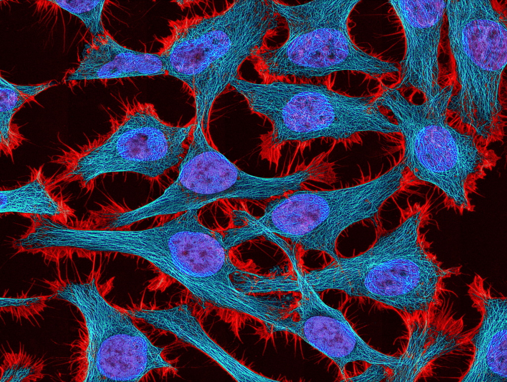
An image of HeLa cells

The dialogue, which was opened and closed with a prayer, also spoke to the importance of establishing regular, ongoing, transparent relationships between the Black faith community and the medical community. This should happen, not just in times of crisis, because “mass hysteria is prime for miscommunication,” Ralph Hodge, pastor of the Second Baptist Church in South Richmond, Virginia, said.

“Today was a big way of us looking back at the past, looking at where we are at now, and moving forward to the solutions,” said Barrett. This comes by letting communities know that we care, said Winn, along with “doing things with our communities, not through them.”

A key factor in deconstructing this issue and achieving health equity is time. Time to reflect on the past in order to avoid reliving it; time to generate innovative solutions to the problems at hand; and time to invest in Black communities – to learn from them, support them, and earn their trust not because they can offer science something, but because science has something to offer them.

Post by Cydney Livingston

In Drawers of Old Bones, New Clues to the Genomes of Lost Giants

DNA extracted from a 1,475-year-old jawbone reveals genetic blueprint for one of the largest lemurs ever.

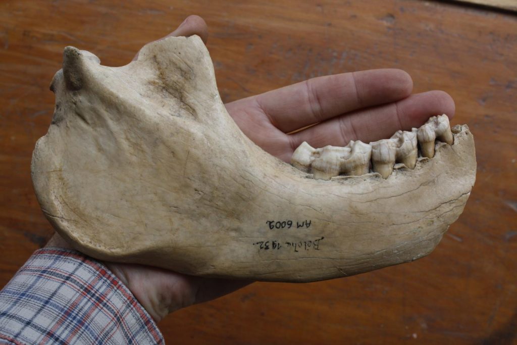
By teasing trace amounts of DNA from this partially fossilized jawbone, nearly 1,500 years after the creature’s death, scientists have managed to reconstruct the first giant lemur genome. Credit: University of Antananarivo and George Perry, Penn State

If you’ve been to the Duke Lemur Center, perhaps you’ve seen these cute mouse- to cat-sized primates leaping through the trees. Now imagine a lemur as big as a gorilla, lumbering its way through the forest as it munches on leaves.

It may sound like a scene from a science fiction thriller, but from skeletal remains we know that at least 17 supersized lemurs once roamed the African island of Madagascar. All of them were two to 20 times heftier than the average lemur living today, some weighing up to 350 pounds.

Then, sometime after humans arrived on the island, these creatures started disappearing.

The reasons for their extinction remain a mystery, but by 500 years ago all of them had vanished.

Coaxing molecular clues to their lives from the bones and teeth they left behind has proved a struggle, because after all this time their DNA is so degraded.

But now, thanks to advances in our ability to read ancient DNA, a giant lemur that may have fallen into a cave or sinkhole near the island’s southern coast nearly 1,500 years ago has had much of its DNA pieced together again. Researchers believe it was a slow-moving 200-pound vegetarian with a pig-like snout, long arms, and powerful grasping feet for hanging upside down from branches.

A single jawbone, stored at Madagascar’s University of Antananarivo, was all the researchers had. But that contained enough traces of DNA for a team led by George Perry and Stephanie Marciniak at Penn State to reconstruct the nuclear genome for one of the largest giant lemurs, Megaladapis edwardsi, a koala lemur from Madagascar.

Ancient DNA can tell stories about species that have long since vanished, such as how they lived and what they were related to. But sequencing DNA from partially fossilized remains is no small feat, because DNA breaks down over time. And because the DNA is no longer intact, researchers have to take these fragments and figure out their correct order, like the pieces of a mystery jigsaw puzzle with no image on the box.

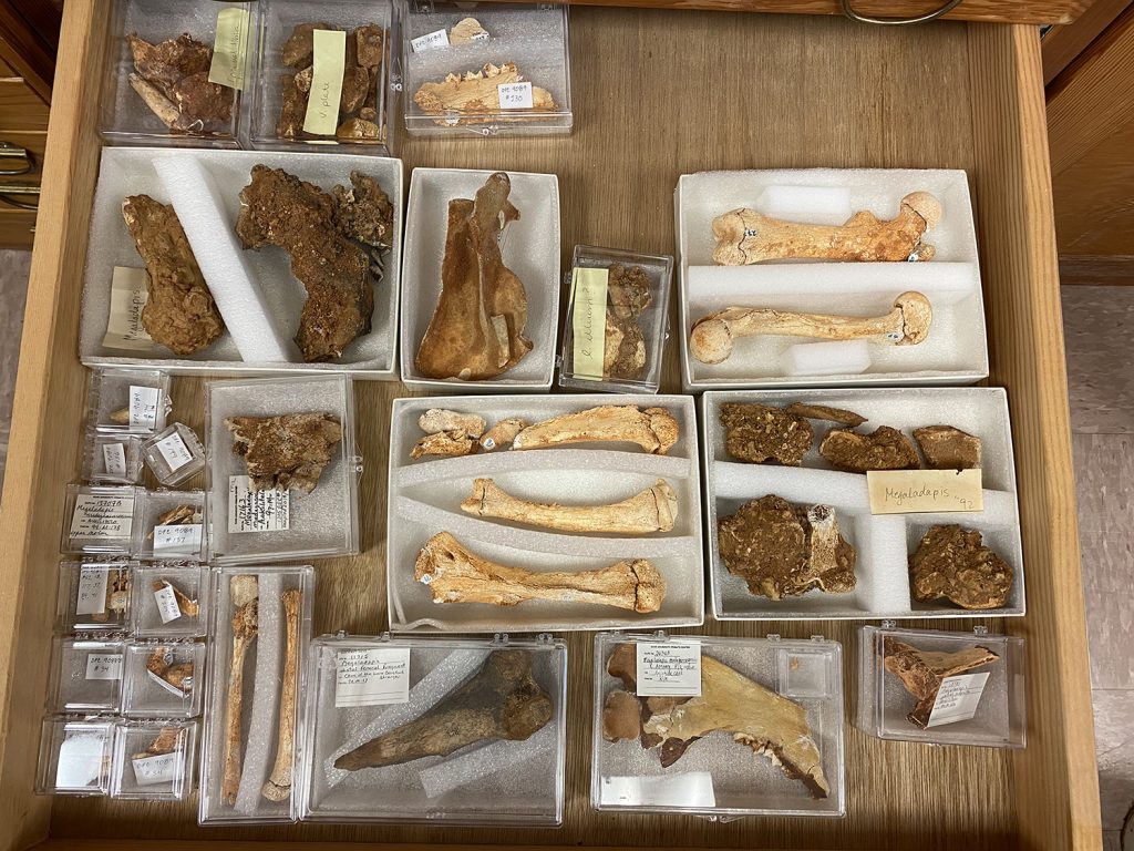
Bones like these are all that’s left of Madagascar’s giant lemurs, the largest of which weighed in at 350 pounds — 20 times heftier than lemurs living today. Credit: Matt Borths, Curator of the Division of Fossil Primates at the Duke Lemur Center

Hard-won history lessons

The first genetic study of M. edwardsi, published in 2005 by Duke’s Anne Yoder, was based on DNA stored not in the nucleus — which houses most of our genes — but in another cellular compartment called the mitochondria that has its own genetic material. Mitochondria are plentiful in animal cells, which makes it easier to find their DNA.

At the time, ancient DNA researchers considered themselves lucky to get just a few hundred letters of an extinct animal’s genetic code. In the latest study they managed to tease out and reconstruct some one million of them.

“I never even dreamed that the day would come that we could produce whole genomes,” said Yoder, who has been studying ancient DNA in extinct lemurs for over 20 years and is a co-author of the current paper.

For the latest study, the researchers tried to extract DNA from hundreds of giant lemur specimens, but only one yielded enough useful material to reconstitute the whole genome.

Once the creature’s genome was sequenced, the team was able to compare it to the genomes of 47 other living vertebrate species, including five modern lemurs, to identify its closest living relatives. Its genetic similarities with other herbivores suggest it was well adapted for grazing on leaves.

Despite their nickname, koala lemurs weren’t even remotely related to koalas. Their DNA confirms that they belonged to the same evolutionary lineage as lemurs living today.

To Yoder it’s another piece of evidence that the ancestors of today’s lemurs colonized Madagascar in a single wave.

Since the first ancient DNA studies were published, in the 1980s, scientists have unveiled complete nuclear genomes for other long-lost species, including the woolly mammoth, the passenger pigeon, and even extinct human relatives such as Neanderthals.

Most of these species lived in cooler, drier climates where ancient DNA is better preserved. But this study extends the possibilities of ancient DNA research for our distant primate relatives that lived in the tropics, where exposure to heat, sunlight and humidity can cause DNA to break down faster.

“Tropical conditions are death to DNA,” Yoder said. “It’s so exciting to get a deeper glimpse into what these animals were doing and have that validated and verified.”

See them for yourself

Assembled in drawers and cabinets cases in the Duke Lemur Center’s Division of Fossil Primates on Broad St. are the remains of at least eight species of giant lemurs that you can no longer find in the wild. If you live in Durham, you may drive by them every day and have no idea. It’s the world’s largest collection.

In one case are partially fossilized bits of jaws, skulls and leg bones from Madagascar’s extinct koala lemurs. Nearby are the remains of the monkey-like Archaeolemur edwardsi, which was once widespread across the island. There’s even a complete skeleton of a sloth lemur that would have weighed in at nearly 80 pounds, Palaeopropithecus kelyus, hanging upside down from a branch.

Most of these specimens were collected over 25 years between 1983 and 2008, when Duke Lemur Center teams went to Madagascar to collect fossils from caves and ancient swamps across the island.

“What is really exciting about getting better and better genetic data from the subfossils, is we may discover more genetically distinct species than only the fossil record can reveal,” said Duke paleontologist Matt Borths, who curates the collection. “That in turn may help us better understand how many species were lost in the recent past.”

They plan to return in 2022. “Hopefully there is more Megaladapis to discover,” Borths said.

A fossil site in Madagascar. Courtesy of Matt Borths, Duke Lemur Center Division of Fossil Primates

CITATION: “Evolutionary and Phylogenetic Insights From a Nuclear Genome Sequence of the Extinct, Giant, ‘Subfossil’ Koala Lemur Megaladapis Edwardsi,” Stephanie Marciniak, Mehreen R. Mughal, Laurie R. Godfrey, Richard J. Bankoff, Heritiana Randrianatoandro, Brooke E. Crowley, Christina M. Bergey, Kathleen M. Muldoon, Jeannot Randrianasy, Brigitte M. Raharivololona, Stephan C. Schuster, Ripan S. Malhi, Anne D. Yoder, Edward E. Louis Jr, Logan Kistler, and George H. Perry. PNAS, June 29, 2021. DOI: 10.1073/pnas.2022117118.

Duke Researcher Busts Metabolism Myths in New Book

Herman Pontzer explains where our calories really go, and what studying humanity’s past can teach us about staying healthy today.

Photo by Elena Georgiou, My City /EEA

Duke professor Herman Pontzer has spent his career counting calories. Not because he’s watching his waistline, exactly. But because, as he sees it, “in the economics of life, calories are the currency.” Every minute, everything the body does — growing, moving, fighting infection, even just existing — “all of it takes energy,” Pontzer says.

In his new book, “Burn,” the evolutionary anthropologist recounts the 10-plus years he and his colleagues have spent measuring the metabolisms of people ranging from ultra-athletes to office workers, as well as those of our closest animal relatives, and some of the surprising insights the research has revealed along the way.

Much of his work takes him to Tanzania, where members of the Hadza tribe still get their food the way our ancestors did — by hunting and gathering. By setting out on foot each day to hunt zebra and antelope or forage for berries and tubers, without guns or electricity or domesticated animals to lighten the load, the Hadza get more physical activity each day than most Westerners get in a week.

So they must burn more calories, right? Wrong.

Herman Pontzer
Herman Pontzer, associate professor of evolutionary anthropology at Duke

Pontzer and his colleagues have found that, despite their high activity levels, the Hadza don’t burn more energy per day than sedentary people in the U.S. and Europe.

These and other recent findings are changing the way we understand the links between energy expenditure, exercise and diet. For example, we’ve all been told that if we want to burn more calories and fight fat, we need to work out to boost our metabolism. But Pontzer says it’s not so simple.

“Our metabolic engines were not crafted by millions of years of evolution to guarantee a beach-ready bikini body,” Pontzer says. But rather, our metabolism has been primed “to pack on more fat than any other ape.” What’s more, our metabolism responds to changes in exercise and diet in ways that thwart our efforts to shed pounds.

What this means, Pontzer says, is you can walk 16,000 steps each day like the Hadza and you won’t lose weight. Sure, if you run a marathon tomorrow you’ll burn more energy than you did today. But over time, metabolism responds to changes in activity to keep the total energy you spend in check.

Pontzer’s book is more than a romp through the Krebs cycle. For anyone suffering pandemic-induced pangs of frustrated wanderlust, it’s also filled with adventure. He takes readers on an hours-long trek to watch a Hadza man track a wounded giraffe across the savannah, to the rainforests of Uganda to study climbing chimpanzees, and to the foothills of the Caucasus Mountains to unearth the 1.8 million-year-old remains of some of the first people who trekked out of Africa.

His humor shines through along the way. Even when awoken by a chorus of 300-pound lions just a few hundred yards from his tent, he stops to ponder whether his own stench gives him away, and what he might do if they come for his “soft American carcass, the  warm triple crème brie of human flesh.”

Pontzer spoke via email with Duke Today about his book:

Q: What’s the lesson the Hadza and other hunter-gatherers teach us about managing weight and staying healthy?

A: The Hadza stay incredibly fit and healthy throughout their lives, even into their older ages (60’s, 70’s, even 80’s). They don’t develop heart disease, diabetes, obesity, or the other diseases that we in the industrialized world are most likely to suffer from. They also have an incredibly active lifestyle, getting more physical activity in a typical day than most Americans get in a week.

My work with the Hadza showed that, surprisingly, even though they are so physically active, Hadza men and women burn the same number of calories each day as men and women in the U.S. and other industrialized countries. Instead of increasing the calories burned per day, the Hadza physical activity was changing the way they spend their calories — more on activity, less on other, unseen tasks in the body.

The takeaway for us here in the industrialized world is that we need to stay active to stay healthy, but we can’t count on exercise to increase our daily calorie burn. Our bodies adjust, keeping energy expenditure in a narrow range regardless of lifestyle. And that means that we need to focus on diet and the calories we consume in order to manage our weight. At the end of the day, our weight is a matter of calories eaten versus calories burned — and it’s really hard to change the calories we burn!

Q: You’re saying that exercise doesn’t matter? What’s the point, if we can’t eat that donut?

A: All those adjustments our bodies make responding to exercise are really important for our health! When we burn more calories on exercise, our bodies spend less energy on inflammation, stress reactivity (like cortisol), and other things that make us sick.

Q: What’s the biggest misunderstanding about human metabolism?

A: We’re told — through fitness magazines, diet fads, online calorie counters — that the energy we burn each day is under our control: if we exercise more, we’ll burn more calories and burn off fat. It’s not that simple! Your body is a clever, dynamic product of evolution, shifting and adapting to changes in our lifestyle.

Q: In your book you say we’re driven to magical thinking when it comes to calories. What do you mean by that?

A: Because our body is so clever and dynamic, and because humans are just bad at keeping track of what we eat, it’s awfully hard to keep track of the calories we consume and burn each day. That, along with the proliferation of fad diets and get-thin-quick schemes, has led to this idea that “calories don’t matter.” That’s magical thinking. Every ounce of your body — including every calorie of fat you carry — is food you consumed and didn’t burn off. If we want to lose weight, we must eat fewer calories than we burn. It really comes down to that.

Q: Some people say that if the cavemen didn’t eat it, we shouldn’t either. What does research show about what foods are “natural” for humans to eat?

A: There’s no singular, natural human diet. Hunter-gatherers like the Hadza eat a diverse mix of plant and animal foods that varies day to day, month to month, and year to year. There’s even more dietary diversity when we look across populations. Humans are built to thrive on a wide variety of diets — just about everything is on the menu.

That said, the ultra-processed foods we’re inundated with in our modern industrialized world really are unnatural. There are no Twinkies to forage in the wild. Those foods are literally engineered to be overconsumed, with a mix of flavors that overwhelm our brain’s ability to regulate our appetites. Now, it is still possible to lose weight on a Twinkie diet (I’m not recommending it!), if you’re very strict about the calories eaten per day. But we need to be really careful about how we incorporate ultra-processed foods into our daily diets, because they are calorie bombs that drive us to overconsume.

Q: If we could time travel, what would our hunter-gatherer ancestors make of our industrialized diet today?

A: We don’t even need to imagine — We are those hunter-gatherers! Biologically, genetically, we are the same species that we were a hundred thousand years ago, when hunting and gathering were the only game in town. When we’re confronted with modern ultra-processed foods, we struggle. They are engineered to be delicious, and we tend to overconsume.

Q: Has the COVID-19 pandemic brought any of these lessons home for you? What can we do to keep active and watch what we eat, even while working from home?

The pandemic has been a tragedy on so many levels — the loss of life, those suffering with long-term effects, the social and economic impacts. The impact on diet and exercise have been bad as well, for many of us. Stress eating is a real phenomenon, and the stress and emotional toll of the pandemic — along with having easy access to the snacks in our kitchen — have led many to gain weight. Physical activity seems to have declined for many. There aren’t easy answers, but we should try to make a point to get active every day. And we can help ourselves make better decisions about food by keeping ultra-processed foods out of our houses. You can’t plow through a bag of chips if you don’t have chips in your cupboard.

Q: You’ve measured the energy costs of activities ranging from taking a breath to doing an Ironman. What is one of the more extreme or surprising calorie-burning activities that you’ve measured, or would like to measure, in humans or some other animal?

A: With colleagues from Japan, I measured the energy cost of a heartbeat – a tricky bit of metabolic measurement! Turns out each beat of your heart burns about 1/300th of a kilocalorie! Amazing how efficient our bodies can be.

Q: What is something people have questions about that we just don’t know the answer to yet? What would it take to find out?

A: Right now we’re excited about measuring the adjustments our bodies make when we increase our exercise: how exactly does burning more energy on physical activity impact our immune system, our stress response, our reproductive system? It will take a long-term study of exercise to see how these systems change over time.

Robin Smith - University Communications
Robin Smith – University Communications

A Computer Scientist Investigating the Source Code of Life

We are all born with defining physical characteristics. Whether it be piercing blue eyes or jet black hair, these traits distinguish us throughout our entire lives. However, there is something that all of our attributes have in common, a shared origin: genes.

Beyond dictating our individual features, genes instruct cells to create proteins that are essential for a variety of processes, from controlling muscle function to managing digestive systems. Despite their importance in the workings of our body, genes can also code for detrimental diseases, such as Huntington’s disease or Duchenne muscular dystrophy.

Raluca Gordân, Ph.D.

These types of diseases are exactly what Raluca Gordân, Ph.D. is battling through her research. She and her group are trying to figure out how to decode the non-coding genome, the DNA apart from protein-coding genes. They are deepening their understanding of the role non-coding areas of the genome play in the expression of the coding genes and the production of proteins.

Gordân, an associate professor in biostatistics and bioinformatics at Duke, said a majority of disease-causing genetic mutations derive from the genome outside of genes.

“That is a huge search space,” she says, chuckling. “Genes only make up about 2% of the genome. If we don’t understand what those non-coding regions are doing, it’s hard to make predictions about what the mutation in those regions would be doing and how to connect that to the development of a disease.”

Gordân recently published a paper, entitled “DNA mismatches reveal conformational penalties in protein–DNA recognition,” which focuses on transcription factors and their exceptional ability to bind to mispaired DNA, misspellings that occur as DNA is copied. During regular replication, nucleotide bases (the building blocks of our DNA) are paired correctly, where adenine pairs with thymine and cytosine goes with guanine. However, when an error occurs during replication, mispairs start to appear, as adenine may pair with guanine instead.

“Normally, those are mistakes that get repaired by specific mismatch repair pathways but that repair might not happen if one of these transcription factors sits on the replication error and doesn’t allow the repair mechanism to see it,” Gordân explains. “Normally, one would expect the transcription factors not to bind to those errors. But we found that they can bind way better than their actual genomic targets.”

Modeling of the binding between mismatched DNA and transcription factors.

To expand on her computational discovery, Gordân is now following up with a study of transcription factor binding to mismatches in living cells, observing whether they adopt their usual role of regulating gene expression or contribute to the development of mutations.

Gordân’s research is a product of her passion and desire to make change. It also can be attributed to a series of realizations she made during college and inspirational mentors who guided her along the way.

While pursuing her undergraduate degree, Gordân was a purely computer science major, concentrating on cryptography. However, as she was nearing the end of her four years of college, she soon found herself yearning for the opportunity to do more. She began looking into machine learning applications and enrolled in a course based around genetic algorithms which she credits for launching her career path.

At that point, she attained what she describes as her “first taste of genetics” and her interest in bioinformatics was irrevocably piqued. Thereafter, Gordân applied for a PhD at Duke, where she worked with advisor Alex Hartemink investigating transcription factor proteins in regulatory genomics. At Duke, her work was primarily computational.  But with her postdoctoral advisor Martha Bulyk of Harvard Medical School, Gordan was exposed to the more experimental aspects of biology.

Today, she recognizes these experiences as integral to her ongoing research, which requires her to frequently iterate between observational approaches and computational work.

Gordân is acclimating to the newly quarantined world. While she strives to continue her research, in the pandemic, it has changed her routine.

“I think what was affected a lot since the pandemic started is the fact that we don’t meet in person,” she says. “A lot of the quick progress was being made when we were in the same physical space and were able to get feedback immediately, with students learning about each other’s results in the lab, in real time. That was replaced with Zoom meetings, where students get to see the other students’ results mainly at lab meetings, weeks or months later. Those continuous discussions that were going on in the lab all the time. We’re missing that.”

Gordân offered some thoughtful parting advice to aspiring computational biologists, like me.

“I was trained as a computer scientist, so I wasn’t really sure about experimental work. But after actually doing the experimental work, I realized how much value there is in doing both,” she said. “You have to pick what you’re strongest at, either the computational or experimental part, but you should not be afraid of the other side.”

Guest Post by Akshra Paimagam, Class of 2021, NC School of Science and Math

Claire Engstrom, a Student Researcher Working to Treat Duchenne’s Muscular Dystrophy by Optimizing CRISPr-cas9

Meet Claire Engstrom, a Senior from Pasadena California. Claire is a Biology major who works in the Gersbach Lab at Duke. 

Claire first got involved with on-campus research through her pre-orientation program, PSearch that introduces incoming first-years to undergraduate research. Following her experience in PSearch, Claire got her first work-study research position in the Tung Lab where she worked closely with Jenny Tung, an Associate Professor in the Departments of Evolutionary Anthropology and Biology at Duke and a Faculty Associate of the Duke University Population Research Institute. 

In the Tung Lab, Claire’s research focused on how DNA methylation is passed through generations. Essentially looking at the inheritance of DNA whose methylation was impacted by environmental factors and how that affects future generations. 

Duke has research opportunities available in all disciplines as well as across departments. Approximately 53% of undergraduates graduate with research experience. Not only can students participate in groundbreaking research, but they can receive funding from the university as well to support the work they are doing.

Within the Biology department, there is a fellowship called B-SURF, the Biological Sciences Undergraduate Research Fellowship, an 8-week summer research program for rising sophomores. Claire applied for and was accepted to the fellowship and placed in one of Duke’s biomedical science laboratories. She also received a $4,000 stipend for her summer research.

Claire was placed in Charles Gersbach’s Lab focused on researching Genome Editing for Gene and Cell Therapy. Dr, Gersbach is a Rooney Family Associate Professor of Biomedical Engineering and has conducted groundbreaking work in genome editing.

Members of the Gersbach Lab in Fall 2019

Gersbach is doing research in several different domains of biomedical engineering. Claire’s project focuses on using CRISPR-Cas9, a technology that allows scientists to change an organism’s DNA using clustered regularly interspaced short palindromic repeats and CRISPR-associated protein 9. faster, cheaper, more accurate, and more efficient than other existing genome editing methods. 

Prior to joining his lab, Claire had already heard a lot about Gersbach in her course Biology 201 as well as through reading his papers. The project she would spend the next two and a half years working on focused on using and optimizing CRISPR-Cas9 to treat Duchenne’s Muscular Dystrophy and lessen the severity of the symptoms. 

Duchenne’s Muscular Dystrophy is a muscle wasting disease that affects one in every five thousand male births.

“People are diagnosed when they are around five and then they lose the ability to walk and their heart can’t pump blood because of the lack of muscles.” Claire explained.  

“CRISPR-based genome editing restores dystrophin expression in mouse models of Duchenne muscular dystrophy. Cross-sections of muscle tissue where the dystrophin protein has been labeled green, including normal, healthy tissue (left), tissue from a mouse model of Duchenne muscular dystrophy (middle), and tissue from the same mouse model that has been treated with the CRISPR gene editing system (right). Nelson et al., Science (2016)”

Thus, those affected often die in early adulthood despite current advances in cardiovascular and respiratory treatments. Duchenne’s Muscular Dystrophy generally occurs as a result of a frameshift mutation of the dystrophin gene. As a result, one’s muscles can no longer connect to anything making it nearly impossible to contract and function properly. In the Gersbach lab they are trying to treat the mutation by using CRISPR-Cas9 to remove an exon or coding region of the gene in order to shift the reading frame back into its normal place. 

This shift produces a less severe phenotype that lessens the effects of Duchenne’s Muscular Dystrophy. The result will significantly improve the quality of life and life spans for affected patients. 

Claire will be continuing her work in the Gersbach lab full time in Spring 2021 as she graduated early, with distinction in the Fall. Her thesis on the work she did in the Gersbach lab was recently approved and her results will be published in a larger paper in the future. After this year she plans to take a gap year an then return to California to hopefully attend grad school and pursue a Ph.D. in Biology.

By Anna Gotskind

Page 2 of 10

Powered by WordPress & Theme by Anders Norén