“My name is Meg Stalter I’m 5’7 I’m living in LA and a fun fact about me is something bad happened to my cousin.”
As made evident by her Twitter profile, my favorite comedian, Megan (“Meg”) Stalter, knows how to make an introduction. Stalter is best known (as far as I know) for her role in the HBO comedy “Hacks,” in which she plays Kayla (whoever that is).
I do not have a Twitter account and I have never seen the show. While we are talking about me, I will explain that I do not really watch TV, with the one exception of West Wing.
Since we are still talking about me, you should know that I fibbed. There are two exceptions. The other one is Grantchester, a Masterpiece Mystery about a hot priest who solves crime (but that was sort of a given, no?).
I share Stalter’s bio for a few reasons. For starters, it makes me smile, and sharing a smile is a tried-and-true way to score a friend (cha-ching!).
Meg Stalter once again proved her knack for making a first impression at her Emmy’s debut
On top of that, it is a good example of someone who knows how to make a first impression. I expect to have made a great impression by the time I finish this, but to ensure things got started on the right foot, hedging my bets if you will, I thought it best to leave the preamble to someone at the top of the trade.
Stalter’s bio also proves a simple point; it is not merely what you say that counts, but how you say it.
I am something of a sub-par reader. I love to read, it is just not my biggest strength (doesn’t mean it can’t be (growth mindset)! Just facing today’s facts). I don’t think I read enough as a child, so now I am slow and I usually fall asleep.
But I get by. I power through my class readings, I keep a book on my bedside table, and I get my news through the radio (that and two free tickets to the Hoppin’ John’s Fiddler’s Convention–it pays to be tuned into WUNC on Saturday nights at 10. Cha-ching!).
This relationship with reading influences my writing style. When I write, I try to keep my readers awake. Not with what I write — I have full faith in the topic at hand’s capacity to speak for itself — but with the way I write it.
My past experience writing for a published paper was in high school, where I spent four years as co-editor of the “Hustle and Bustle” page. I authored a satirical advice column in which troubled high schoolers (me) could send their personal woes to someone who would publish them for the whole school to read (also me). I like writing as a secondary form of chatting.
My senior year, I retitled my column “Dear Addy,” after the well-known advice column “Dear Abby.”
And so it is with this laudable writing background that I report to you on the groundbreaking discoveries from one of the top research universities in the U.S.
Why write for a research blog? Research is interesting. Research makes the world go round. Just ask a freshman. They all came here for the “research opportunities,” as did all the other freshmen at all the other universities.
Before I sign off, I will let you know where you might catch me in my free time. This is a key element of the standard student bio, and I am prone to severe FOMO, so let me get right to it.
I am a sophomore from Hickory, North Carolina hoping to major in Public Policy and minor in Math. In my free time you might catch me listening to NPR, jogging, potting, singing to myself, making a smoothie, telling people about my smoothie, spamming my contacts for an ice cream date, or for the not-so-lucky, trying my best at Appalachian-style fiddle.
When I write about myself, it always reads like a poorly crafted match.com zinger. Boring, awkward, and something along the lines of:
I’m Alex. Aquarius. Love dogs, classic rock, old NCIS episodes. $1 Goodwill paperback thrillers, marked with “Happiest 53rd Richard! All my love, Janet” and “8/17/2005, Saw this and thought of you!” And I like to ask myself why Steven King’s Carrie conjures up thoughts of said person? Who’s Richard? How’s Janet?
I also love coffee. And tea. Peppermint, of course. Irish breakfast, sure. Chamomile, why not. But I think I really just like collecting mugs — hearty ceramics, dainty porcelain, hand-painted, non-dishwashable, chipped, stained monstrosities. It might be a problem though (as I don’t have much shelf space).
Favorite genre of film? It’s got to be anything in the Meg Ryan romcom cinematic universe. Or the Brat Pack coming-of-age cannon. Breakfast Club, St. Elmo’s Fire, About Last Night, Pretty in Pink. Really just the Judd Nelson je ne sais quoi.
My dog and I celebrating her 11th birthday this summer!
I think my 2nd grade superlative was “Wormiest Bookworm,” whatever that means. That might’ve been the year I read every Nancy Drew book in the library and founded the neighborhood’s first and only detective business. I do wish I could say I’ve Jules Verne’d the world in 80 days — circumnavigating all five nebulous oceans, frozen Arctic plains, Swiss peaks, and continental slopes; Phileas Fogging my way through the Mediterranean, aperitivo in hand. But I’m a bit unworldly in the geographic sense. I’ve only been out of the country once to boat up next to Niagara Falls, wearing a thin, plastic poncho and an I <3 Canada tee (though I’ve possibly made it a second time to Canada after getting lost on the circumference of a lake in Vermont).
I’ve only ever lived in Charleston, SC, never straying too far from its labyrinth of intercostals and waterways, its Theseus-like shrimpers, gliding into port. At Duke, I spend half my time majoring in molecular/cellular biology and the other lamenting my landlockedness, missing Charleston’s temperate sea breeze.
Beach in the middle of winter
Growing up there was all briny inlet and Waffle House, midnight bacon, butter pats, cordgrass, molting blue crab, churches on every street corner and in every denomination, weak coffee and greasy hash brown breakfast, September hurricanes, salt, cicadas, farm stands packed with peaches, a once-in-a-hundred year 6-inch snowfall that closed school for two weeks.
On Saturdays, I sharktooth-hunted with my sisters in pluff mud plots now developed (strangers tend to find the smell of the marsh pungent, but I think it’s character building). Fished for red drum. Searched for pearls in half-mooned oyster mouths. Kayaked down creeks.
Charleston’s a literary city, or so I’ve always heard. I think Edgar Allen Poe’s ghost haunts a cobble-stoned alley downtown or something like that. And if not an alley then a quaint B&B, its porch bearing creaky rocking chairs and purple coneflower. I went to an arts-specialized middle and high school for creative writing, wrote some bad poetry in my formative years and a couple of questionable short films, then went to college and somehow fell into the field of cell bio and now I spend a decent chunk of my free time researching genetic heart disease in a campus lab. Feeding cardiomyocytes via gentle pipette like they’re sea monkeys.
I like to picture the act of writing and that of science as similar — fraternal twins or first cousins — and I don’t think it a coincidence that early philosophers were our first physicians, mathematicians, physicists, chemists, etc. Both fields challenge us to pose questions about our world, about its inhabitants, its oddities, its nuances. We just go about answering them differently.
For this reason, I’m incredibly excited to join Duke’s Research Blog, to write about science and innovation, to poeticize protein structures or to search for lyricism in neuronal action potentials the way a deep sea troller searches for the elusive giant squid. I just think there’s something so wonderful about learning new things, cradling little curiosities that often lead nowhere, and doing so through an accessible, enjoyable medium.
Saxicolous lichens (lichens that grow on stones) from the Namib Desert, and finger lichen, Dactylina arctica (bottom left insert), common in the Arctic, on display in Dr. Jolanta Miadlikowska’s office. The orange color on some of the lichen comes from metabolites, or secondary chemicals produced by different lichen species. The finger lichen is hollow.
Lichens are everywhere—grayish-green patches on tree bark on the Duke campus, rough orange crusts on desert rocks, even in the Antarctic tundra. They are “pioneer species,” often the first living things to return to barren, desolate places after an extreme disturbance like a lava flow. They can withstand extreme conditions and survive where nearly nothing else can. But what exactly are lichens, and why does Duke have 160,000 of them in little envelopes? I reached out to Dr. Jolanta Miadlikowska and Dr. Scott LaGreca, two lichen researchers at Duke, to learn more.
Dr. Jolanta Miadlikowska looking at lichen specimens under a dissecting microscope. The pale, stringy lichen on the brown bag is whiteworm lichen (Thamnolia vermicularis), used to make “snow tea” in parts of China.
According to Miadlikowska, a senior researcher, lab manager, and lichenologist in the Lutzoni Lab (and one of the Instructors B for the Bio201 Gateway course) at Duke, lichens are “obligate symbiotic associations,” meaning they are composed of two or more organisms that need each other. All lichens represent a symbiotic relationship between a fungus (the “mycobiont”) and either an alga or a cyanobacterium or both (the “photobiont”). They aren’t just cohabiting; they rely on each other for survival. The mycobiont builds the thallus, which gives lichen its structure. The photobiont, on the other hand, isn’t visible—but it is important: it provides “food” for the lichen and can sometimes affect the lichen’s color. The name of a lichen species refers to its fungal partner, whereas the photobiont has its own name.
Lichen viewed through a dissecting microscope. The black speckles visible on some of the orange lichen lobes are a “lichenicolous” fungus that can grow on top of lichen. There are also “endolichenic fungi… very complex fungal communities that live inside lichen,” Miadlikowska says. “We don’t see them, but they are there. And they are very interesting.”
Unlike plants, fungi can’t perform photosynthesis, so they have to find other ways to feed themselves. Many fungi, like mushrooms and bread mold, are saprotrophs, meaning they get nutrients from organic matter in their environment. (The word “saprotroph” comes from Greek and literally means “rotten nourishment.”) But the fungi in lichens, Miadlikowska says, “found another way of getting the sugar—because it’s all about the sugar—by associating with an organism that can do photosynthesis.” More often than not, that organism is a type of green algae, but it can also be a photosynthetic bacterium (cyanobacteria, also called blue-green algae). It is still unclear how the mycobiont finds the matching photobiont if both partners are not dispersed together. Maybe the fungal spores (very small fungal reproductive unit) “will just sit and wait” until the right photobiont partner comes along. (How romantic.) Some mycobionts are specialists that “can only associate with a few or a single partner—a ‘species’ of Nostoc [a cyanobacterium; we still don’t know how many species of symbiotic and free-living Nostoc are out there and how to recognize them], for example,” but many are generalists with more flexible preferences.
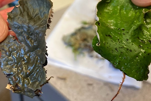
Two species of foliose (leaf-like) lichens from the genus Peltigera. In the species on the left (P. canina), the only photobiont is a cyanobacterium from the genus Nostoc, making it an example of bi-membered symbiosis. In the species on the right (P.aphthosa), on the other hand, the primary photobiont is a green alga (which is why the thallus is so green when wet). In this case, Nostoc is a secondary photobiont contained only in the cephalodia—the dark, wart-like structures on the surface. With two photobionts plus the mycobiont, this is an example of tri-membered symbiosis.
Lichens are classified based on their overall thallus shape. They can be foliose (leaf-like), fruticose (shrubby), or crustose (forming a crust on rocks or other surfaces). Lichens that grow on trees are epiphytic, while those that live on rocks are saxicolous; lichens that live on top of mosses are muscicolous, and ground-dwelling lichens are terricolous. Much of Miadlikowska’s research is on a group of cyanolichens (lichens with cyanobacteria partners) from the genus Peltigera. She works on the systematics and evolution of this group using morphology-, anatomy-, and chemistry-based methods and molecular phylogenetic tools. She is also part of a team exploring biodiversity, ecological rules, and biogeographical patterns in cryptic fungal communities associated with lichens and plants (endolichenic and endophytic fungi). She has been involved in multiple ongoing NSF-funded projects and also helping graduate students Ian, Carlos, Shannon, and Diego in their dissertation research. She spent last summer collecting lichens with Carlos and Shannon and collaborators in Alberta, Canada and Alaska. If you walk in the sub basement of the Bio Sciences building where Bio201 and Bio202 labs are located, check out the amazing photos of lichens (taken by Thomas Barlow, former Duke undergraduate) displayed along the walls! Notice Peltigera species, including some new to science, described by the Duke lichen team.
Lichens have value beyond the realm of research, too. “In traditional medicine, lichens have a lot of use,” Miadlikowska says. Aside from medicinal uses, they have also been used to dye fabric and kill wolves. Some are edible. Miadlikowska herself has eaten them several times. She had salad in China that was made with leafy lichens (the taste, she says, came mostly from soy sauce and rice vinegar, but “the texture was coming from the lichen.”). In Quebec, she drank tea made with native plants and lichens, and in Scandinavia, she tried candied Cetraria islandica lichen (she mostly tasted the sugar and a bit of bitterness, but once again, the lichen’s texture was apparent).
In today’s changing world, lichens have another use as well, as “bioindicators to monitor the quality of the air.” Most lichens can’t tolerate air pollution, which is why “in big cities… when you look at the trees, there are almost no lichens. The bark is just naked.” Lichen-covered trees, then, can be a very good sign, though the type of lichen matters, too. “The most sensitive lichens are the shrubby ones… like Usnea,” Miadlikowska says. Some lichens, on the other hand, “are able to survive in anthropogenic places, and they just take over.” Even on “artificial substrates like concrete, you often see lichens.” Along with being very sensitive to poor air quality, lichens also accumulate pollutants, which makes them useful for monitoring deposition of metals and radioactive materials in the environment.
Dr. Scott LaGreca with some of the 160,000 lichen specimens in Duke’s herbarium.
LaGreca, like Miadlikoska, is a lichenologist. His research primarily concerns systematics, evolution and chemistry of the genus Ramalina. He’s particularly interested in “species-level relationships.” While he specializes in lichens now, LaGreca was a botany major in college. He’d always been interested in plants, in part because they’re so different from animals—a whole different “way of being,” as he puts it. He used to take himself on botany walks in high school, and he never lost his passion for learning the names of different species. “Everything has a name,” he says. “Everything out there has a name.” Those names aren’t always well-known. “Some people are plant-blind, as they call it…. They don’t know maples from oaks.” In college he also became interested in other organisms traditionally studied by botanists—like fungi. When he took a class on fungi, he became intrigued by lichens he saw on field trips. His professor was more interested in mushrooms, but LaGreca wanted to learn more, so he specialized in lichens during grad school at Duke, and now lichens are central to his job. He researches them, offers help with identification to other scientists, and is the collections manager for the lichens in the W.L. and C.F. Culberson Lichen Herbarium—all 160,000 of them.
The Duke Herbarium was founded in 1921 by Dr. Hugo Blomquist. It contains more than 825,000 specimens of vascular and nonvascular plants, algae, fungi, and, of course, lichens. Some of those specimens are “type” specimens, meaning they represent species new to science. A type specimen essentially becomes the prototype for its species and “the ultimate arbiter of whether something is species X or not.” But how are lichens identified, anyway?
Lichenologists can consider morphology, habitat, and other traits, but thanks to Dr. Chicita Culberson, who was a chemist and adjunct professor at Duke before her retirement, they have another crucial tool available as well. Culbertson created a game-changing technique to identify lichens using their chemicals, or metabolites, which are often species-specific and thus diagnostic for identification purposes. That technique, still used over fifty years later, is a form of thin-layer chromatography. The process, as LaGreca explains, involves putting extracts from lichen specimens—both the specimens you’re trying to identify and “controls,” or known samples of probable species matches—on silica-backed glass plates. The plates are then immersed in solvents, and the chemicals in the lichens travel up the paper. After the plates have dried, you can look at them under UV light to see if any spots are fluorescing. Then you spray the plates with acid and “bake it for a couple hours.” By the end of the process, the spots of lichen chemicals should be visible even without UV light. If a lichen sample has traveled the same distance up the paper as the control specimen, and if it has a similar color, it’s a match. If not, you can repeat the process with other possible matches until you establish your specimen’s chemistry and, from there, its identity. Culberson’s method helped standardize lichen identification. Her husband also worked with lichens and was a director of the Duke Gardens.
Thin-layer chromatography plates in Dr. LaGreca’s office. The technique, created by Dr. Chicita Culberson, helps scientists identify lichens by comparing their chemical composition to samples of known identity. Each plate was spotted with extracts from different lichen specimens, and then each was immersed in a different solvent, after which the chemicals in the extracts travel up the plate . Each lichen chemical travels a characteristic distance (called the “Rf value”) in each solvent. Here, the sample in column 1 on the rightmost panel matches the control sample in column 2 in terms of distance traveled up the page, indicating that they’re the same species. The sample in column 4, on the other hand, didn’t travel as far as the one in column 5 and has a different color. Therefore, those chemicals (and species) do not match.
LaGreca shows me a workroom devoted to organisms that are cryptogamic, a word meaning “hidden gametes, or hidden sex.” It’s a catch-all term for non-flowering organisms that “zoologists didn’t want to study,” like non-flowering plants, algae, and fungi. It’s here that new lichen samples are processed. The walls of the workroom are adorned with brightly colored lichen posters, plus an ominous sign warning that “Unattended children will be given an espresso and a free puppy.” Tucked away on a shelf, hiding between binders of official-looking documents, is a thin science fiction novel called “Trouble with Lichen” by John Wyndham.
The Culberson Lichen Herbarium itself is a large room lined with rows of cabinets filled with stacks upon stacks of folders and boxes of meticulously organized lichen samples. A few shelves are devoted to lichen-themed books with titles like Lichens De France and Natural History of the Danish Lichens.
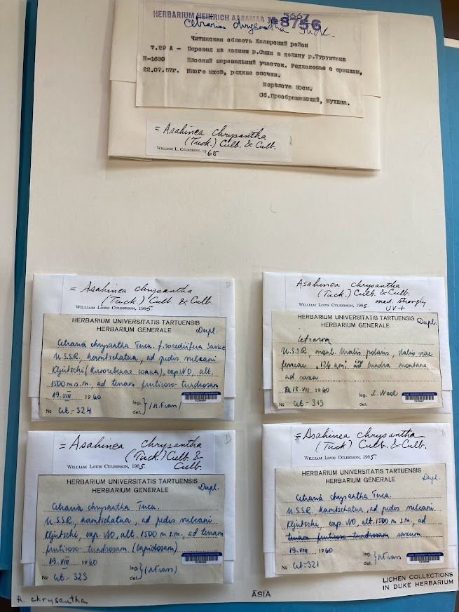
Each lichen specimen is stored in an archival (acid-free) paper packet, with a label that says who collected it, where, and on what date. (“They’re very forgiving,” says LaGreca. “You can put them in a paper bag in the field, and then prepare the specimen and its label years later.”) Each voucher is “a record of a particular species growing in a particular place at a particular time.” Information about each specimen is also uploaded to an online database, which makes Duke’s collection widely accessible. Sometimes, scientists from other institutions find themselves in need of physical specimens. They’re in luck, because Duke’s lichen collection is “like a library.” The herbarium fields loan requests and trades samples with herbaria at museums and universities across the globe. (“It’s kind of like exchanging Christmas presents,” says LaGreca. “The herbarium community is a very generous community.”)
Duke’s lichen collection functions like a library in some ways, loaning specimens to other scientists and trading specimens with institutions around the world.
Meticulous records of species, whether in databases of lichens or birds or “pickled fish,” are invaluable. They’re useful for investigating trends over time, like tracking the spread of invasive species or changes in species’ geographic distributions due to climate change. For example, some lichen species that were historically recorded on high peaks in North Carolina and elsewhere are “no longer there” thanks to global warming—mountain summits aren’t as cold as they used to be. Similarly, Henry David Thoreau collected flowering plants at Walden Pond more than 150 years ago, and his samples are still providing valuable information. By comparing them to present-day plants in the same location, scientists can see that flowering times have shifted earlier due to global warming. So why does Duke have tens of thousands of dried lichen samples? “It comes down to the reproducibility of science,” LaGreca says. “A big part of the scientific method is being able to reproduce another researcher’s results by following their methodology. By depositing voucher specimens generated from research projects in herbaria like ours, future workers can verify the results” of such research projects. For example, scientists at other institutions will sometimes borrow Duke’s herbarium specimens to verify that “the species identification is what the label says it is.” Online databases and physical species collections like the herbarium at Duke aren’t just useful for scientists today. They’re preserving data that will still be valuable hundreds of years from now.
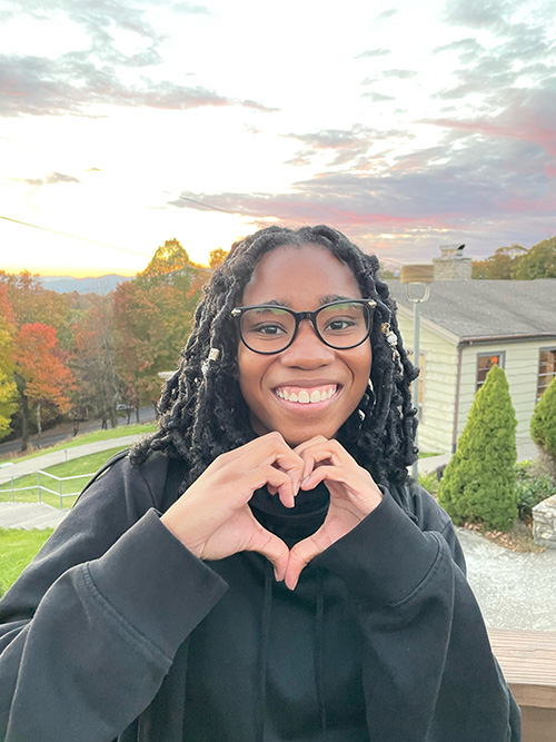
Hello, my name is Jakaiyah Franklin, and I am a sophomore here at Duke University. In terms of my major, I am undecided, but I do know my passion lies in biology, science communication, and environmental science.
Outside of classes, I am the treasurer for the Duke Chapter of the NAACP and LLC leader for the Stem Pathways for Inclusion, Readiness, and Excellence (SPIRE) program. Last year I was the stage manager for two Hoof n Horn productions.
This is the Rocky Horror Picture Show company after finishing our last show.
This year, I will start a research position along with this research blogging position.
In a more personal sense, I am the youngest of three and a proud aunt. Right now, I say I am from Texas, even though I have lived in Georgia, South Carolina, Germany, and presently North Carolina. If someone ever asked me, I would say that Germany holds my most memorable memories; however, I have grown into a better version of myself in each place I have lived. Other than school, I like to read and watch House of the Dragon and the earlier seasons of The Game of Thrones. I prefer to study outside or in a place where natural light is abundant. I also love learning new things pertaining to science, specifically infectious diseases.
My view of Muhuru Bay, Kenya, where I spent the summer after my first year at Duke. That’s Lake Victoria in the distance.
I find diseases fascinating, and I believe they are our natural predators. I want to be able to not only understand them, but also, I want to help prevent them. If one were to have a favorite type of disease to study, mine would be zoonotic diseases. They are interesting because the act of a virus being able to jump from a host like a rat to a human is captivating to me.
After graduating from Duke, I want to earn a master’s in public health or a Ph.D. in epidemiology, virology, or infectious disease to feed my curiosity about diseases. However, before I can even decide what Ph.D. or master’s I want to earn, my current goal is to decide on my major.
Jakaiyah Franklin
I do like to think ahead, so, for my very distant career, I know I want to be able to see infectious diseases in both the lab and in the places where they are infecting populations. I want my research to be digestible for the general population because, as seen with both COVID and Monkeypox, science can be easily misinterpreted if not delivered appropriately. I want to prevent this occurrence from happening to me by learning more about science communication and actively improving my communication skills.
I hope this blogging position will expose me to infectious disease research or general public health research. With this new understanding of the research, I hope this position will also educate me on how to inform others so that they can enjoy and understand the science.
It may be hard to put your finger on it, but Duke often allows students to connect their classes to something more personal.
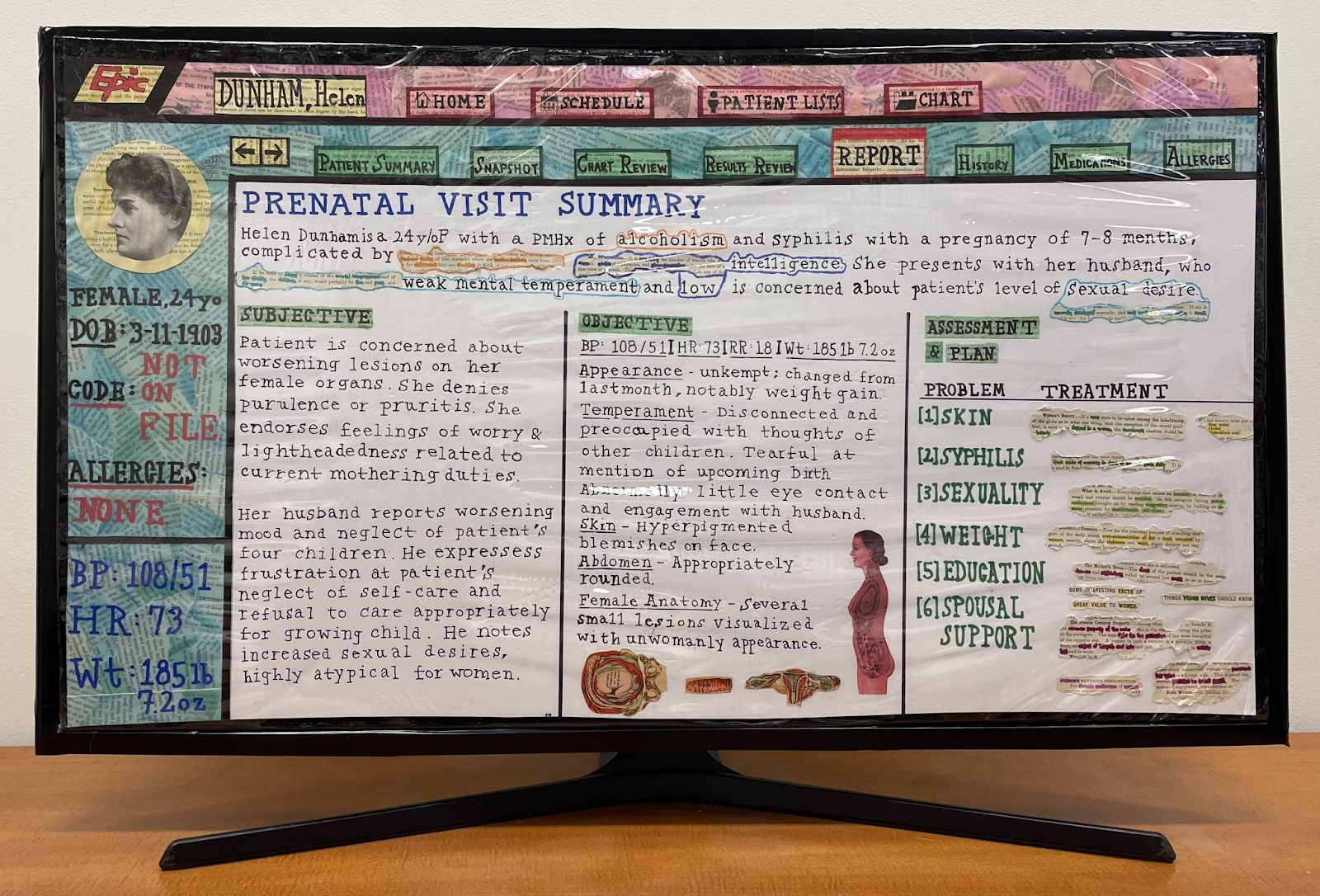
The university’s emphasis on interdisciplinary education is a major initiative that colors students’ academic experiences. While there are many examples of these connections between people, classes, fields, and departments, few so tangibly represent those connections like The SCOPES Project, which connects arts and humanities to medical education at Duke.
Beneath the Surface by Mihir Patel, 2022. Image Courtesy of SCOPES.
Art and medicine can exist in entirely different worlds. They can appeal to different people and tell different stories. But why be simple when you can be, well… stunning? They can be integrated to form something powerful, and that’s precisely what SCOPES leadership members Isa DeLaura, Raluca Gosman, Mason Seely, David Stevens, and Lindsay Olson aimed to do.
“It is encouraging as an upperclassman who previously participated in this program to see rising students continue the tradition of incorporating the humanities into medical practice,” Mason Seeley says. The generational aspect of the project seems to contribute to its personality; participants bring their own perspectives to their work only to walk away with dozens more.
“Having a creative outlet has helped me process interactions with patients and the difficulties of the profession, and celebrate happy moments as well,” says Isa DeLaura.
“The goal is to give artists creative freedom to explore their relationships with their patients with whatever medium and in whatever style works best for them. As such, every year the feel is entirely based on the decisions of the artists.”
Isa DeLaura, MS3+
David Stevens insists that the artists “resist… forces of depersonalization in compelling and beautiful ways.”
The project is inspired and supported by yet another interdisciplinary Duke initiative called APPLE (Appreciating Patient Perspectives through Longitudinal Encounters), which connects medical students with patients living with chronic illnesses. The artists/medical students/empaths-in-training then attended multiple creative workshops and developed art pieces to reflect their patients’ personal experiences. But this year’s 6th annual SCOPES exhibition looks a bit different from past years’ (which are conveniently available online for your viewing pleasure).
Having attended many an art opening myself, I am unashamed to say that much of my enjoyment comes from the cheeseplates (and the excitement in the air, but that’s besides the point). Some exhibitions opt for a traditional charcuterie, some marked Kirkland Signature and others displayed on a handmade butcherblock. The point of fingerfoods is to encourage the attendees to stand up, walk around, and interact with the masses. But it also encourages attendees to “just stop by,” making the affair all the less intimate.
Following limitations on group gatherings Duke enforced during COVID, the SCOPES team decided to apply their newfangled interdisciplinary/revolutionary/innovative thinking to the art opening itself: They held a banquet.
“I loved the way this turned out,” says DeLaura. “It was very personal and made for great discussion and comradery.”
Fences, Rivers, Walls by Taylor Yoder, 2022. Image Courtesy of SCOPES.
“SCOPES has provided an opportunity to reflect on my experiences as a first-year medical student while also exploring new ways to combine various art forms to create my vision,” says Taylor Yoder, who created Fences, Rivers, Walls, pictured above. “I hope to continue shooting film throughout my medical education and career.”
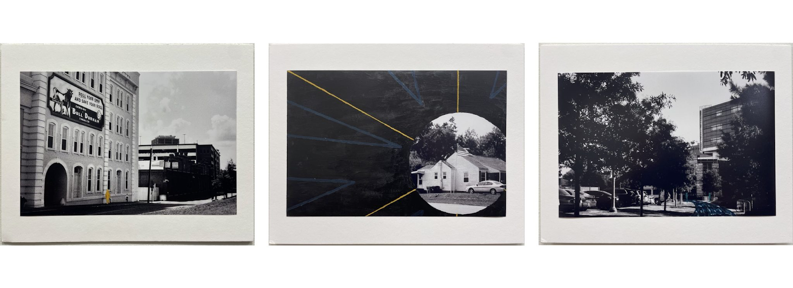
I was particularly (although wrongfully) surprised about the variety in the exhibit. While the artists attended the same workshops and worked with patients through the same program, they took radically different approaches to their creations. Esme Trahair, a second-year medical student, was a humanities major in undergrad. Her piece combines historical perspectives with modern (although antiquated) mechanisms, emphasizing “the importance of remembering and learning from historical, outdated medical teachings.”
For the Record by Esme Trahair, 2022. Image Courtesy of SCOPES.
The work features a variety of perspectives, but also some clear motifs that could be key takeaways for future medical providers. Like Yoder, artist Kreager Taber explores the patient’s value of “home.” Exploring these motifs could allow for more personal, “upstream” healthcare.
This year’s SCOPES exhibition is held in the Mars Gallery in the Duke University Hospital Concourse. It is an initiative of the Trent Center for Bioethics, Humanities & History of Medicine at the Duke University School of Medicine. It will be on display August 9-September 29, and available for viewing online at this link.
P.S. If you are an MS1 student interested in participating in SCOPES, I have a link for you!
New research shows that lemurs and their food trees are tightly linked in ecological networks, and that the extinction of lemurs will have cascading effects on ecosystem functions.
The Critically Endangered black-and-white ruffed lemur, Varecia variegata, is one of the lemurs that eats the most fruit. When they consume the fruit, they pass the whole far from the mother tree, effectively aiding in seed dispersal. Photo credit: Laura De Ara
Lemurs are the primates found only on Madagascar. They are unique in many ways, and like many organisms, they fit in complex ecological networks. These networks include interactions between lemurs and their food trees. Many interactions are beneficial, or mutualistic; for example, lemurs eat the fruits of trees and disperse their seeds, providing a critical service to the trees. If lemurs go extinct — 98% of species are threatened with extinction due to human activities — the links in the ecological network will be severed, with potentially negative impacts on the trees.
Research published in the Journal of Animal Ecology by Ph.D. student Camille DeSisto, of the Duke University Nicholas School of the Environment, and James Herrera, from the Duke Lemur Center, shows how tightly linked lemurs and trees are in their interaction networks, and the negative impacts of extinction on network resilience. If lemurs do in fact disappear, many trees will be left without a way to disperse their seeds, and may not be able to reproduce effectively.
DeSisto and Herrera used advanced techniques in social network analysis, including exponential random graph models, to test which traits of lemurs and trees predict their probability of interaction. The lemurs with the highest probability of interactions with trees were large, fruit-eating species with a short gestation length, occurring in arid habitats, and with a threat status of Least Concern. Closely related plants were more likely to interact with the same lemur species than distantly related plants, but closely related lemurs were not more likely to interact with the same plant genus.
Simulated lemur extinction tended to increase network structure in some properties, including connectance (% of realized interactions out of all possible interactions) and modularity (how many unique cliques or subcommunities form in the network), but decrease nestedness (the tendency for specialists which feed on only a few trees to be a subset of generalists which feed on many trees) and robustness (tolerance to future extinctions), compared to pre‐extinction networks. Networks were more tolerant to plant than lemur extinctions.
The silky sifaka, Propithecus candidus, is one of the most endangered primates in the world. Unlike some lemurs, the sifaka have antagonistic relationships with trees, eating leaves and prey on seeds, rather than pass them intact. Photo credit: Laura De Ara.
By simulating the loss of lemur and plant species, the authors could predict how network structure will erode over time if threatened lemurs and trees go extinct. The results showed that if the most well-connected lemurs in the network were to disappear, the percentage of trees with interactions would quickly decline, compared to scenarios in which lemurs were removed randomly or if the least-well connected lemurs went extinct. Given the threat status and geographic range size of lemurs, the percentage of trees that would lose their interacting lemurs would be greater than that expected if lemur extinctions were random.
The bamboo lemur, Hapalemur occidentalis, is a highly specialized species, eating mostly bamboo leaves. They do, however, occasionally eat fruits, and often spread the seeds effectively. Photo credit: Laura De Ara
Results also showed that if lemurs go extinct, the resilience of the networks to further disturbance would decrease. This indicates that the current links between lemurs and trees are critical to the stability of these complex ecological networks.
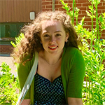
To prevent the loss of key ecosystem functions like seed dispersal, it is critically important to protect lemurs and trees, which depend so crucially on one another for survival. DeSisto is currently conducting field research in Madagascar, studying how well seeds germinate when eaten by lemurs. She created a tree nursery in the forest to grow the seeds obtained from lemur feces, and already has several species germinating. Interestingly, she is also showing how lemurs disperse the seeds of vines, which are an important yet understudied food source when tree fruits are not available. She will continue her research across multiple seasons, to determine how changes in plant phenology affect seed dispersal patterns.
Author Camille DeSisto and assistant Feno Telessy examine the seeds germinating from lemur feces.Amazingly, seeds from lemur feces are already germinating after only one month. Some tree seeds take months or even a year before germinating.
Many conservation programs are currently striving to safeguard Madagascar forests and the diverse species found only in these natural habitats. The Duke Lemur Center has an active conservation program in the northeast, called the DLC-SAVA Conservation Initiative. This program takes a community-based approach to conservation, partnering closely with local stakeholders and actors to develop projects that address the needs of both lemurs and people. By co-creating projects that include alternative and sustainable livelihood strategies, both nature and people benefit from conservation. Natural ecosystems provide important services for people, including locally, such as protecting watersheds and pollinators, as well as globally, such as carbon sequestration. Without the native forests, and the lemurs that call those forests home, people would lose the valuable and irreplaceable services forests provide.
CITATION: “Drivers and Consequences of Structure in Plant–Lemur Ecological Networks,” Camille DeSisto and James Paul Herrera. Journal of Animal Ecology, July 15, 2022. DOI: 10.1111/1365-2656.13776.
Lemurs like Ferdinand are leaf-eating machines, says Duke microbiome researcher Lydia Greene. Credit: Lydia Greene
DURHAM, N.C. — A jungle. A rainforest. A wetland. A wilderness. Researchers have used various metaphors to describe the complex, interconnected community of microbes (most of them bacteria) living inside your body, and all over it too.
If you were to count up all the trillions of cells inside and out, we are more bacteria than human. Fortunately, perhaps, the microbes that make a cozy home inside your nose, or clinging to your teeth, aren’t the same ones that are living behind your ear or busily multiplying in your belly button.
The same is true for our distant primate cousins the lemurs, particularly in their guts, say researchers Lydia Greene and Erin McKenney in a new study.
Lemurs rely on gut microbes to digest their leafy diets, explains Greene, a research scientist at the Duke Lemur Center. Microbes in lemurs’ GI tracts help ferment plant fiber, detoxify plant chemical defenses, and synthesize vitamins and nutrients that lemurs can’t make for themselves. Our own gut bugs do many of the same things for us.
Greene and McKenney study how lemur gut bacteria are shaped by what lemurs eat, how they evolved and the complexity of the route microbes travel through the body. They hope to better understand how these microorganisms keep lemurs healthy, or — when they’re out of balance — make them sick.
Researchers who do this kind of work spend a lot of time collecting poop. For good reasons, says McKenney, an assistant professor at North Carolina State University. Scientists can learn about lemurs by what they leave behind, and poop can be collected repeatedly without harming the animals. But for this study the team tried something different, made possible by a one-of-a-kind biobank:
When an animal dies at the Duke Lemur Center, veterinary staff determine the cause of death, and blood and tissue samples that might be important for research or education are collected and preserved.
Today, the collection holds thousands of samples, collected over decades from more than two dozen species of rare and endangered primates, which the center stores in super cold freezers in their headquarters in North Carolina. It’s a frozen ark maintained at up to minus 80 degrees Celsius, with redundant backup power.
In the event that any one of these species goes extinct, and the last living parts of them are gone, future generations will still be able to study the genetic and other information they left behind.
Using this bank, the team sampled several sites in the bowels of 52 deceased lemurs, including dwarf lemurs, aye-ayes, ruffed lemurs, bamboo lemurs, brown lemurs, ring-tailed lemurs and sifakas.
A trip through a lemur’s GI tract is a journey through a varied landscape. The long, twisting route from the stomach through the small intestine to the colon serves numerous functions, filtering, digesting, absorbing, detoxifying, fermenting.
Not all lemur guts work the same: Fruit-eaters, like ruffed lemurs, generally have short, simple guts. If you stretched them out, they’d be five times their body length — not much shorter than ours, relative to body size. Leaf-eaters like sifakas have more complex GI tracts with relatively longer colons and a leaf-fermentation pouch called a cecum. Their guts are the lemur champions — up to 16 times body length.
Having whole lemurs to study instead of just poo enabled the researchers to sample different regions of the gut to figure out what kinds of microbes were present in each spot. They used genetic sequencing technology to identify microbes and compare their relative abundance in different sites.
Sampling along the digestive tract, they found that different spots along this long, twisting pathway have their own communities of bacteria doing different kinds of jobs. The complex ecosystem lurking in a lemur’s small intestine, for example, isn’t the same as the microbial menagerie setting up camp in their colon.
Levels of biodiversity varied too. The stomach supports less microbial life because fewer species can tolerate its acidic digestive juices. But if the upper regions of the gut are a garden, the lower regions are more like a tropical rainforest. About two dozen types of bacteria were more abundant in the cecum and colon than elsewhere. Lemurs with relatively longer lower guts host the richest microbiomes, to better ferment high-fiber foods.
“We probably couldn’t have detected these relationships without such an extensive comparative dataset,” McKenney said.
“This kind of lemur research can really only be done at the Duke Lemur Center,” Greene said.
Rodelinda, a Coquerel’s sifaka lemur, munches on leaves at the Duke Lemur Center. Credit: Lydia Greene.
This research was supported by the National Science Foundation (DBI PRFB 1906416) and by a Duke Lemur Center Director’s fund.
CITATION: “Gut Site and Gut Morphology Predict Microbiome Structure and Function in Ecologically Diverse Lemurs,” Lydia K. Greene, Erin A. McKenney, William Gasper, Claudia Wrampelmeier, Shivdeep Hayer, Erin E. Ehmke, & Jonathan B. Clayton. Microbial Ecology, May 14, 2022. DOI: 10.1007/s00248-022-02034-4
Even before the COVID-19 pandemic, mental health challenges were the leading cause of poor life outcomes in young people. As many as 1 in 5 U.S. children aged 3 to 17 have a mental, emotional, developmental or behavioral disorder, according to the Centers for Disease Control and Prevention.
Now, that crisis has been exacerbated. Symptoms of depression and anxiety for children and adolescents have doubled during the pandemic.
Seventy percent of U.S. public schools reported an increase in the number of children seeking mental health services during the pandemic and many have struggled to meet the needs of those students, according to the latest federal data.
As the Biden administration and Congress consider policies and programs that could help curb these mental health challenges that children face, a group of Duke researchers may already have one answer.
Eighteen years after administering an intensive childhood intervention program called Fast Track, a group of Sanford School of Public Policy scholars has found that it not only proved effective at reducing conduct problems and juvenile arrests in childhood, it also improved family outcomes when the original children grow up and become parents themselves.
Their followup findings, which appeared in June in the Journal of Child Psychology and Psychiatry, show that girls who received the Fast Track intervention during first through 10th grades had improvements in several aspects of their family environments 18 years later.
Specifically, Fast Track reduced food insecurity in the mothers’ family environments, and lessened the mothers’ depression, alcohol and drug problems, and their use of corporal punishment.
“We knew the Fast Track early childhood intervention was successful at reducing aggressive behavior in childhood and criminal arrests in young adulthood,” said Drew Rothenberg, research scientist at the Duke Center for Child and Family Policy and lead author on the study.
“This research demonstrates that the early intervention doesn’t just benefit the children receiving the services,” he added. “It also improves the family environments those child form as adults, benefiting their own children. In other words, it looks like the effects of early intervention can ripple across generations.”
Drew Rothenberg
According to Rothenberg, the beneficial effects of Fast Track are just as large as those seen in prevention programs that only affect a single generation.
“Impressively, these beneficial effects were also almost as big as those seen immediately after the end of the Fast Track intervention 18 years earlier,” Rothenberg said. “Therefore, for mothers, Fast Track’s effects appear powerful across two generations of homes and are much longer-lasting than previous research suggested.”
“Surprisingly, the benefits of the Fast Track intervention on family environments formed as adults found for mothers did not extend to fathers,” said study co-author Jennifer Lansford, research professor at the Duke Sanford School of Public Policy and director of the Duke Center for Child and Family Policy.
Jennifer Lansford, Director of Duke Center for Child and Family Policy
“Even in contemporary society, women are still tasked with a greater proportion of child-rearing responsibility, and still more often called to create family routines and climate,” Lansford said. “Therefore, the beneficial Fast Track effects on reducing corporal punishment and increasing family food security may emerge only in mothers because mothers are still primarily responsible for the provision of parenting and procurement of resources for family meals, and consequently more likely to benefit from such intervention.”
Rothenberg said the findings suggest childhood mental health interventions can break maladaptive cycles and promote the development of healthy family environments when those children grow up and start their own families.
“With this evidence, we also demonstrate that investing in early childhood interventions won’t just pay off for today’s children but also for generations of children to come,” Rothenberg said.
Researchers surveyed 400 Fast Track participants who were now parents at age 34 about aspects of their current family environment. They wanted to assess whether parent substance use problems, depression, romantic partner violence, parent warmth, parent use of physical aggression and corporal punishment, family chaos, and food insecurity were better for adults who had participated in Fast Track as children than for adults who had been in the control group.
“We designed the Fast Track program to improve emotional awareness and interpersonal competence among children at high risk for peer conflict, antisocial and delinquent behaviors and life-course failure,” said study co-author Kenneth Dodge, the William McDougall Distinguished Professor of Public Policy Studies at the Duke Sanford School of Public Policy. Dodge is a member of the Conduct Problems Prevention Research Group that created the Fast Track program.
Participants had been drawn from high-risk elementary schools in Durham, North Carolina, Nashville, Tennessee, rural Pennsylvania and Seattle, Washington. Starting in first grade, students were randomly assigned to either receive Fast Track or be followed as a control group. Students who received the Fast Track intervention received social skills training, tutoring, and a social-emotional learning curriculum taught by teachers. Their parents received training in techniques to help the students manage their behavior.
The Fast Track project has been supported since 1991 by National Institute of Mental Health (NIMH) Grants R18MH48043, R18MH50951, R18MH50952, R18MH50953, R01MH062988, K05MH00797, and K05MH01027; National Institute on Drug Abuse (NIDA) Grants R01DA016903, K05DA15226, RC1DA028248, and P30DA023026; National Institute of Child Health and Human Development Grant R01HD093651; and Department of Education Grant S184U30002.
CITATION: “Intergenerational Effects Of The Fast Track Intervention on the Home Environment: A Randomized Control Trial,” William Andrew Rothenberg, Jennifer E. Lansford, Jennifer Godwin, Kenneth A. Dodge, William E. Copeland, Candice L. Odgers, Robert J. McMahon, Natalie Goluter, and Conduct Problems Prevention Research Group. Journal of Child Psychology and Psychiatry, June 15, 2022. DOI: 10.1111/jcpp.13648
Post by Sarah Brantley, communications director for Duke’s Center for Child and Family Policy
To many of us, cells are the building blocks of life, akin to bricks or Legos. But to biologist Regan Moore, a former Ph.D. student in Dan Kiehart’s lab at Duke, cells are so much more: they’re busy construction sites, machinery and materials moving about to build and shape the body. And now, new live imaging techniques make it possible to watch some of the nano-scale construction in action.
In time-lapse videos published this month, Moore, Kiehart and colleagues were able to peer inside cells as they filled a hole in the back of a developing fruit fly, a crucial step in the fly’s development into a larva. The process is coordinated with help from a thin mesh of protein fibers just under the cell surface, each one 10,000 times finer than a human hair. The fibers help the cells hold their shape, “kind of like rebar in concrete,” Moore said.
But unlike rebar, she added, “they’re constantly moving and changing.” Normally, features like these are too small and quick to see with conventional microscopes, which can only take a few images a second or are too out-of-focus. So Kiehart’s team used a technique called super-resolution fluorescence microscopy to track individual fibers with nanoscale resolution.
By watching the “rebar of the cell” at work during this hole-closing process in fruit flies, the researchers hope to better understand wound healing in humans, and what goes awry for children with birth defects such as cleft lip and spina bifida.
LEARN MORE: “Super-resolution microscopy reveals actomyosin dynamics in medioapical arrays,” Regan P. Moore, Stephanie M. Fogerson, U. Serdar Tulu, Jason W. Yu, Amanda H. Cox, Melissa A. Sican, Dong Li, Wesley R. Legant, Aubrey V. Weigel, Janice M. Crawford, Eric Betzig, and Daniel P. Kiehart. Molecular Biology of the Cell, July 15, 2022. DOI: 10.1091/mbc.E21-11-0537
What lies at the intersection of mathematics and biology? Freshly-minted math PhD Ruby Kim and her work on mathematically modeling human dopamine cycles.
Ruby Kim, recent Duke PhD graduate
Kim’s work has centered around her creation of a math model to predict how a person’s dopamine levels ebb and flow over the course of a day “to understand the general mechanics of how disruptions in the (biological) clock lead to disruptions in dopamine.”
She said there is a pretty long history of mathematicians using differential equations to see how different clock genes and proteins change over the course of a day’s circadian cycle. Yet, no previous models have connected the circadian clock – controlled by the brain’s suprachiasmatic nucleus – to dopamine levels. And Kim tells me that work suggesting dopamine changes throughout the day are likely controlled by the internal circadian clock itself is “relatively new.”
The first step in Kim’s work was validating scientists’– or “experimentalists,” as Kim dubs them – hypotheses about dopamine and dopaminergic enzyme cycling.
Many physiological processes are controlled via circadian rhythms and the internal clock in humans, as well as other organisms.
“But I’d like this work to help experimentalists go one step further and be able to test out hypotheses more easily.” For example, Kim says that her model has the potential to reveal other fascinating phenomena, such as how drug treatments or different genetic mutations may impact circadian rhythm or dopamine. This is thanks to the multifaceted layers of Kim’s model.
“From a mathematical perspective, the math model is very interesting. It has a lot of interesting dynamics,” she says. “Not only does it show nice, 24-hour rhythms, it shows both steady state behavior… but then also behavior that’s really wild – something called quasi-periodic behavior, where the internal clock is significantly different than the external 24-hour light-dark cycle.”
“This leads to oscillatory behavior that’s not periodic,” she says. These sorts of quasi-periodic behaviors have been observed in experiments and misunderstood, but they can be computed.
Kim emphasized the experimental and clinical implications of her work. Dopamine is involved in learning and motivation and is also linked a plethora of psychiatric conditions like Parkinson’s, ADHD, and schizophrenia. “Patients with these conditions often also experience circadian disruptions,” Kim says. “That’s a pretty big symptom.”
Kim began her academic career in her home state of California at Pomona College as a pre-med math major. “I had always been intrigued by human physiology. And math was one of the subjects I was also pretty drawn to. I just didn’t appreciate it much because throughout high school and the beginning of undergrad, I didn’t see any direct applications,” Kim told me.
The marriage of her love for math with her intrigue in biology actually began at Duke when Kim attended a mathematical biology workshop during the summer after her sophomore year. “I had never heard of math biology before that.”
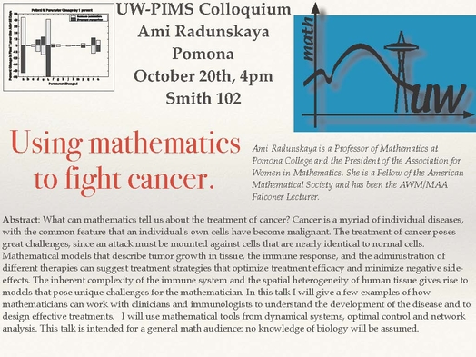
After working on a brief project to model sleep apnea in infants at the workshop, Kim returned to California and took up math modeling courses in her following semesters of undergrad. One of her professors, Ami E. Radunskaya, PhD, was extremely supportive and introduced Kim to a lot of “cool biological problems.” Kim went on to do research with Radunskaya, modeling tumor-immune interactions. This experience, Kim says, “kind of just threw me into academia.” The project gave way to an undergraduate thesis with Radunskaya that analyzed the long-term behavior of this tumor growth and treatment model.
Radunskaya then suggested that Kim pursue grad school. “I kind of applied on a whim,” Kim said, “It wasn’t something I had specifically imagined for myself.” Kim mentioned how no one from her home community had really ever gone to grad school and so it was not something she had ever “explicitly” thought about before.
An abstract of Radunskaya’s work on mathematical modeling and understanding tumor-immune interactions to address cancer.
In her search for a graduate program, Kim applied to math programs, as well as those that were interdisciplinary. “I ended up choosing Duke because I really liked my advisor,” Kim told me. While Kim’s advisor Michael Reed, PhD “does a lot of interesting math,” Kim wanted to work with him because math isn’t his focus – understanding “really complex biological systems using mathematical language is.”
“A lot of times you see people who do things at the intersection of math and biology that are more motivated from a mathematical standpoint … that’s just not what I’m interested in personally. I’m very interested in finding an interesting biological problem and then applying whatever mathematical tools I have.”
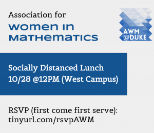
While at Duke, Kim was foundational to founding the university’s chapter of the Association for Women in Math (AWM). During her undergrad, Kim “had a really great experience with AWM,” finding both a community of women mathematicians and a network of women professors who were involved in the chapter.
At Duke, there wasn’t a chapter “but quite a few people who were interested in starting and being part of one.” This organization, which is open to people of any gender identity, heads mentorship programming that brings undergrads, grad students, postdocs, and professors together, organizes conferences, and contributes to their central focus of community building in math.
Example flyers of the events organized by the Duke chapter of the Association for Women in Mathematics.
Outside of her research, Kim spends most of her free time taking care of foster pups, which she describes as “extremely rewarding but also very tiring.” Her most recent foster, a four-month-old puppy, eavesdropped on our interview as he took a nap.
This fall, Kim will begin a post-doc with the University of Michigan’s math department as she “wanted to keep studying circadian rhythms with faculty who are really great in that area.”