Following the people and events that make up the research community at Duke

Students exploring the Innovation Co-Lab

Category: Lecture Page 15 of 20

Meat Glue — True to its Name

By Ashley Yeager

This is the third post in a four-part, monthly series that gives readers recipes to try in their kitchens and learn a little chemistry and physics along the way. Read the first post here and the second one here.

fish checkerboard

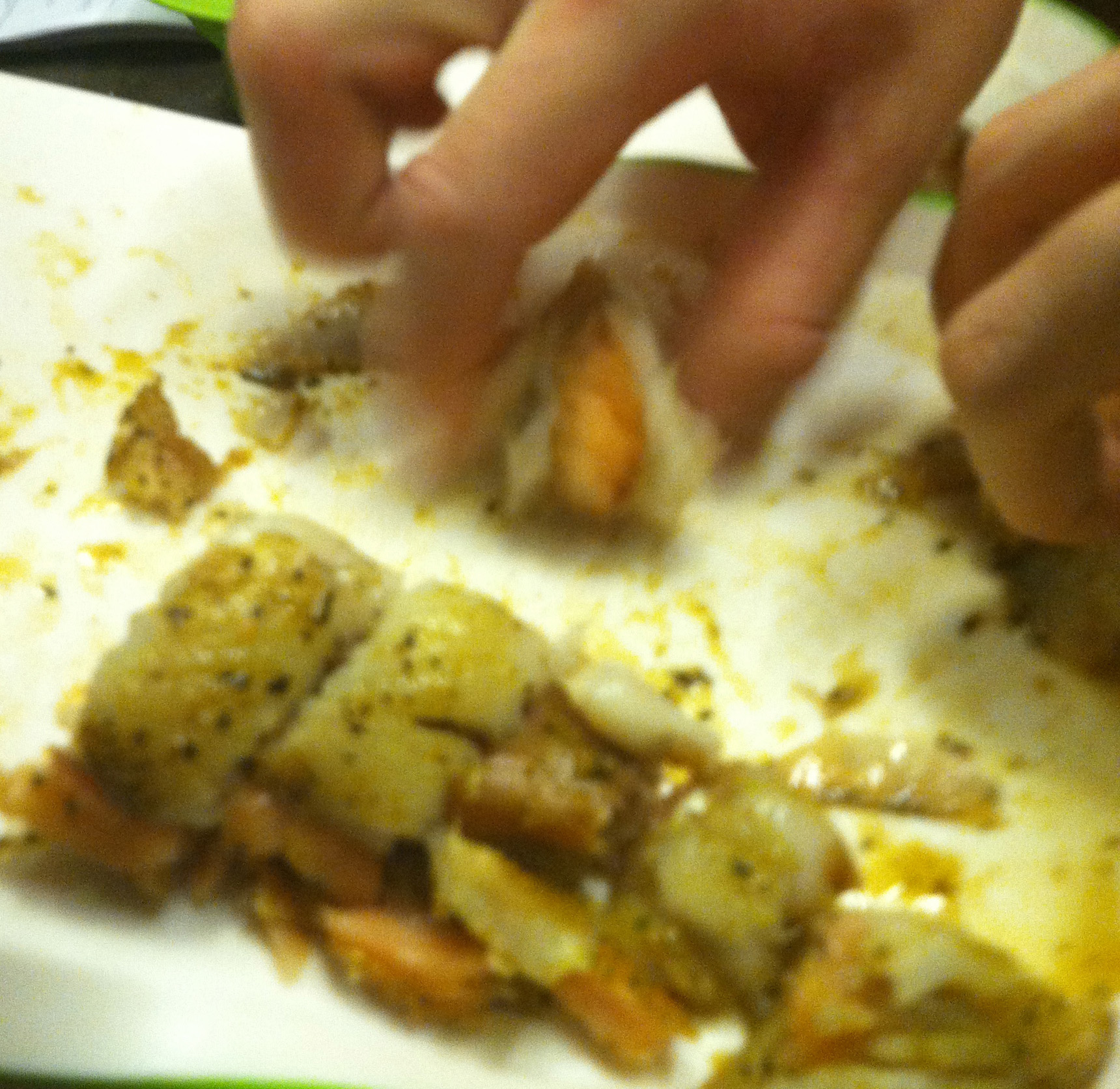
Students grab chunks of a fish “checkerboard” made from salmon and flounder cubes. Credit: Ashley Yeager, Duke.

Braided steak and checkerboard fish may sound exotic. But, freshmen in the Chemistry and Physics of Cooking had no fear fingering the meaty masterpieces into their mouths.

The students made this food art – one literally a braid of three steak strips and the other a combination of salmon and flounder cubes – using a molecule called transglutaminase, also known as meat glue.

In 2012, the media roasted meat glue’s reputation, branding it a dirty little secret meat vendors use to stick together cheap cuts of beef, lamb, chicken or fish and then sell as premium cuts.

“In this class, we’re not using the molecule to be dishonest. We’re using it to be creative,” said physical chemist Patrick Charbonneau, who leads the freshman seminar along with chef Justine de Valicourt and teaching fellows Mary Jane Simpson and Keely Glass.

During a lecture, Glass explained how meat glue — an enzyme that speeds chemical reactions — forms covalent bonds between some of the amino acids that make up the proteins in meat and meat substitutes. With just a sprinkle of the enzyme, which comes in a powder form, chefs can then weave together beef cuts, form game-piece patterns from fish or even bind beans, seeds and other ingredients into a veggie burger that doesn’t crumble after the first bite.

“Meat glue is like a lot of modern ingredients. It comes from industry, and you can use it to make industrial food,” like chicken nuggets, de Valicourt said. “But when you master it, you can use it in a very creative and delicious way.”

Chefs often use the fundamentals of chemistry and physics to shape other foods, such as chocolate. “We’re doing the same to shape meat,” Charbonneau said, explaining that the students used transglutaminase in lab to create beautiful, and delicious, combinations of meat far superior to chicken nuggets and other industrial food typically made with the enzyme.

To make your own meat masterpieces, try the following recipe:

Materials:

1 long sheet of plastic wrap OR a bowl
1 cutting board
1 knife
2 latex gloves for each person
1 mask for each person
1 meat grinder (optional)
1-3 gallon-sized Ziploc bags
1 scale

Ingredients:

1 portion fish, chicken, beef OR vegetarian protein (ie black beans and sunflower seeds)
10 g meat glue powder (available online here)

Instructions:

Gluing meat chucks together –

1. Choose meats
2. Place meat on plastic wrap
3. Choose meat pattern – braid or stack
4. Season meat with salt and pepper
5. Put on gloves and mask and measure 10 g of meat glue using the scale
6. Sprinkle meat glue on sides of meat you want to connect
7. Fold meat into desired pattern
8. Place meat in Ziploc bag
9. Refrigerate for 6 hours
10. Cook meat as you would any other time

Making meat patties –

1. Choose meats, grind in meat grinder, and mix in a bowl (Or, buy ground meat and mix)
2. Season meat with salt and pepper
3. Put on gloves and mask, then measure 10 g of meat glue using the scale
4. Add meat glue to meat and knead until fully mixed
5. Separate into two portions (or more for patties) and seal each in a Ziploc bag
6. Roll with rolling pin, if desired
7. Refrigerate for 6 hours
8. Cook meat as you would any other time

Finding Consciousness

By Nonie Arora

Brain scans of various disorders of consciousness. Credit: Wiki Commons

Can we be certain whether a patient is minimally conscious or in a persistent vegetative state?

What kinds of rights do minimally conscious patients have?

How should minimally conscious patients be treated?

Scientists, ethicists, lawyers and physicians asked these questions at the Finding Consciousness workshop at Duke in January 2013.

Recently, neuroscientists have devised methods to detect consciousness in patients with severe brain injury who may not appear to be aware of themselves and others. But as the science develops so do new ethical dilemmas.

Patients with severe brain injury are often written off, despite growing scientific evidence of potential improvement, said Joseph Fins  from Cornell University. Fins gave the annual Nancy Weaver Emerson Lecture sponsored by the Trent Center for Bioethics, Humanities & History of Medicine as part of the workshop, and he focused on the application of neuroethics to the minimally conscious state.

Fins believes that family members of patients are often forced to make decisions about withholding or withdrawing care without complete, understandable information. They are compelled to consider organ donation, even prematurely. In his work, Fins interviews family members of brain injury patients. In one conversation, a mother of a patient described an interaction with a neurologist who called the patient “basically an organ donor now” and said, “He doesn’t have the reflexes of a frog.”

Then, the neurologist urged the mother to consider organ donation — all within 72 hours of the injury. Fins called for patients and family members to be treated with more sensitivity and respect.

Jeremy Fins. Credit: Trent Center for Bioethics, Humanities, and History of Medicine

The vegetative state has been seen as medical futility, and the paradigm was “once you’re vegetative, you’re done,” Fins said. However, physicians in the field have begun to see families and patients who have looked vegetative, but then suddenly showed some level of response to stimulus.

While some patients become permanently vegetative, others can become minimally conscious, Fins said, referencing a study where about 40 percent of patients who were diagnosed as vegetative were actually minimally conscious.

“This is unconscionable, but that’s where we are,” he said, adding that much of the disparity could come from disinterest, neglect and marginalization of these patients. People would not accept this level of misdiagnosis in cancer or diabetes care, he said.

It is our obligation to give voice to minimally conscious patients as a basic civil right, Fins said, especially as better methods of identifying these patients and stimulating recovery are likely to come in the future.

Close Encounters of the Twitter Kind

By Ashley Yeager

Astrophysicist Katie Mack and other researchers are starting to join Twitter to do better science. Image courtesy of: mediabistro.com

Before launching into dark matter’s effects on particle physics in the early universe, astrophysicist Katie Mack of the University of Melbourne in Australia took a little detour Wednesday to talk about Twitter.

The social media tool is helping her “do better science and learn about new science,” she said during her Jan. 30 seminar at Duke.

The talk materialized from a tweet she had posted a few days ago about attending ScienceOnline, an annual, Raleigh-based conference for scientists and communicators talking and writing about science on the Internet.

Duke physicist Mark Kruse, who joined Twitter in October after the 2012 Council for the Advancement of Science Writers meeting, saw Mack’s tweet about coming to the Triangle and then contacted her to see if she would like to speak about her research.

She said yes, obviously, and explained during her talk that the invitation, as well as the other networking she has done on Twitter, got her to thinking about why all physicists (and scientists) should use the site.

@AstroKatie shares her top reasons scientists should be on Twitter. Credit: Katie Mack, U. of Melbourne.

Here is a paraphrased list of her top five reasons:

1. You can see what scientific breakthroughs people are getting excited about.
2. You can keep track of science discoveries outside of your field.
3. You can share your work with a broader audience.
4. You can connect with other scientists in and outside your field, building your professional network.
5. You can connect and share your work with the public.

Clearly Mack’s invitation to speak at Duke illustrates her third point about Twitter. Now, she said, she looks forward to attending her first ScienceOnline meeting to build on those points and learn new ways of using the tool to connect with other scientists and science enthusiasts.

You can follow Mack at @astrokatie, Kruse at @markckruse and ScienceOnline at @ScienceOnline (or #scio13) if you’re already on Twitter.

And, if you’re a Duke researcher not yet on Twitter but want to be, check it out here, then contact the university’s news office if you’ve got questions.

Designing Microbial "Factories" Rationally

By Pranali Dalvi

Using microbes to manufacture chemicals is starting to be cheaper and greener than traditional chemistry. And their feedstock is sugar, not oil.

Source: 2010 Agricultural Biotechnology International Conference

On Friday, Dr. Michael Lynch spoke to an engaged audience about how microbes have ushered in a new era in metabolic and genetic engineering. Lynch is the co-founder and CSO of OPX Biotechnologies, a Colorado-based company that makes bio-based chemicals and fuels from microbes. OPXBIO microbes produce fatty acids from hydrogen and carbon dioxide. In turn, the fatty acids are used to make cleaners, detergents, jet fuel, and diesel.

Lynch said it’s easier to understand the genetic circuits and enzymatic pathways of microbes, thanks to  much cheaper DNA sequencing. What we still lack though, is an understanding of how to rationally design complex biological systems – likely because we fail to recognize the interplay among an organism’s genotype, phenotype, and environment.

It’s a complex set of factors that go into making phenotypic traits such as color, size, or shape.

“In an industrial setting [phenotypes] are equivalent to metabolism or higher production of the product of interest,” Lynch said. “In a clinical setting, [phenotypes] could be virulence or pathogenesis.”

One approach to understanding how phenotypes are controlled has been through functional genomics.

Let’s say we take a population of wildtype microorganisms and introduce genetic modifications in a controlled way. Next, we selectively screen for the phenotype of interest and compare the sequence of this phenotype to the wildtype to pinpoint the genetic mutations that made the difference.

Comparing phenotypes one at a time is inefficient, though. Lynch wanted to find a way to speed up this process.

“We wanted a process or technology or toolkit that evaluates all of your genes in parallel in a single experiment for the phenotype of interest,” Lynch explained.

Lynch found his inspiration in microbial biofilms, extracellular polysaccharide matrices that grow quickly.

OPXBIO’s Efficiency Directed Genome Engineering (EDGE) technology platform, Source: opxbio.com

Lynch’s studies revealed that microbial cultures grown in enriched media made biofilms, while those in minimal media did not. In a process known as destructional mutagenesis, Lynch and his colleagues then knocked out biofilm-making genes to identify what genes cause the biofilm phenotype in enriched medium but prevent it in minimal medium.

Lynch saw the individual microbial systems as factories that he can genetically modify to produce chemical compounds in biofilms – specifically, 3-hydroxypropionic acid – that can be chemically converted to commercially relevant compounds such as acrylic.

Scientists at OPXBIO have cracked the code for making acrylic from sugar.  They give sugar feedstocks to genetically modified bacteria, whose enzymes convert the sugar into acrylic molecules. Acrylic has broad commercial applications in paints, adhesives, diapers, detergents, and even fuel – a $10 billion global market.

What makes humans so unique?

By Pranali Dalvi
Human and chimpanzees are very similar genetically despite the stark differences in their outward appearances. So it must be just a very small portion of human genes that are responsible for everything from our upright posture to our ability to sing. What makes humans so unique?

On Jan. 14, Duke Professor of Biology Greg Wray spoke about his group’s work on the genetic and molecular processes that contribute to our uniquely human physiology and brains as a part of the Computational Biology and Bioinformatics Seminar Series.

“Humans are not the best model organisms since there is a limit to what you can do genetically and mentally. You can’t really make a human knockout (but sometimes, nature makes it for you),” Wray said.

Still, humans are immensely important to study for practical reasons. We have uniquely human courses of disease in part due to our physiological, cognitive, and mechanical properties. Also, we’re just intrinsically curious about our own bodies.

According to Wray, the answer to human uniqueness is our regulome, the genes, mRNAs, proteins, and metabolites that regulate which genes are turned on when.

This graph shows the two major shifts in diet (meat-rich diet and grain-based diet) that likely contributed to our divergence from chimpanzees and thus differential gene expression. Source: Greg Wray

One prevailing hypothesis is that human forerunners likely began diverging from chimps about 2 million years ago when we took on a meat-rich diet in the savannah. The ancestors of chimpanzees retreated to the rainforest to eat a diet consisting mostly of fruits. Our meat-rich diet seems to coincide with an increase in brain size. And today we metabolize fats much differently than chimpanzees.

Wray’s lab studied the effects of dietary changes on five tissue samples – the cerebral cortex and cerebellum of the brain, liver, fat, and skeletal muscle. What seems to have changed in chimps versus humans are genes related to neural functioning, development, and metabolism. For instance, 31 of 61 genes involved in insulin signaling are operated differently in chimps and humans. These differences in gene expression may also explain why humans are uniquely susceptible to diet-related illnesses like type II diabetes.

On the other hand, genes involved in the transcription, translation and replication of DNA, RNA processing and protein localization haven’t changed in chimps versus humans.

Fat cells also behave differently in humans versus chimps. Wray’s lab took adult stem cells from adipose tissue in both chimps and humans and challenged them with either more oleic acid (the main fatty acid in a meat-heavy diet) or more linoleic acid (the dominant fatty acid in a grain-based diet). The enzymes involved in fatty acid synthesis were more common in human adipose tissues. Wray believes that the increased fatty acid synthesis is probably responsible for building and fueling a larger human brain.

Another major shift in diet occurred during the agricultural revolution, which introduced omega-6 fatty acids into our diet along with pro-inflammatory compounds. Wray explains that the increase in grains from the shift in diet likely contributes to chronic pro-inflammatory diseases in humans, such as atherosclerosis.

“Understanding our metabolic history from an evolutionary context can potentially give us insight into some pretty prominent health concerns,” says Wray.

A Call For Action: Genetic Testing Before Prescriptions

By Prachiti Dalvi

Structure of Codeine

Codeine is an opioid pain medication; but if you are a poor metabolizer of a particular enzyme (CYP2D6), you will experience no pain relief from this drug. However, if your doctor could administer something called pharmacogenetic testing, she would know to simply give you morphine (an active metabolite of codeine) instead. For now, this kind of testing isn’t available.

Mary Relling, PharmD

Mary V. Relling, PharmD, the Chair of Pharmaceutical Sciences at St. Jude’s Children Hospital spoke about the need to implement pharmacogenetic testing on Thursday, January 10. A number of  tests have recently emerged that are ready for prime time. When we know that some drugs may have adverse effects for people with  particular genetic phenotypes, it is unethical to prescribe these drugs without knowing the patient’s genetic status.

However, Relling said there are a number of barriers to integrating pharmacogenetic tests into clinical care: fragmentation of our healthcare system, a focus on sick-care rather than disease prevention, a lack of evidence for clinical utility or cost-effectiveness, complex underlying lab results, and a lack of a centralized system for recording patient information.

The best way to break through these barriers is to conduct testing preemptively, Relling said. We can simply take drop of blood when the baby is born and run genetic tests. “Genetic tests are lifetime results. It makes sense to have it in the background, just as we know a patient’s age, weight, sex, etc.,” Relling said. The barriers discussed above can be avoided to a certain extent at St. Jude’s because they have adopted a team approach to patient care and a 100% electronic system for recording patient records.

The growing affordability of genotyping makes using preemptive pharmacogenetic testing more feasible, she said. The cost of sequencing one or two genes in the past will now produce results for 225 genes. Two years ago, the Clinical Pharmacogenetics Implementation Consortium (CPIC) studied how to migrate pharmacogenetic testing from the laboratory into routine patient care. They looked for gene-drug pairs associated with potential risks of life-threatening toxicity, serious adverse effects, or lack of effectiveness. Eleven of the genes CPIC determined met the threshold for high-risk were found to have profound effects on 33 drugs.

Relling said approximately 48% of patients receiving drugs at St. Jude’s received orders for at least one of those pharmacogenetically high-risk medications.

She said the question now is how to use genetic test results rather than whether a genetic test should be ordered. In the coming years, we will have to address how to maintain the fine balance of providing the clinician with enough information to treat the patient and overwhelming the patient with genetic testing results that are difficult to interpret.

This lecture was a part of the Genomics and Personalized Medicine Forum sponsored by the Duke Institute for Genome Sciences and Policy (IGSP).

An Evening with Dr. Siddhartha Mukherjee

By Pranali Dalvi
Cancer is the uncontrolled growth of cells. We take this idea for granted today, but the definition of cancer evaded us for many years.
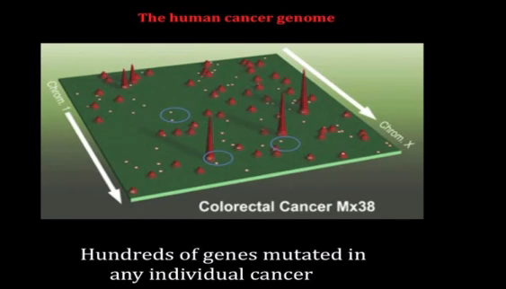
In a talk on on Dec. 6, the Pulitzer Prize-winning author, physician, and cancer researcher Dr. Siddhartha Mukherjee, took his audience on a journey through the archives of medicine to build a bird’s eye view of cancer and how we define the disease today. The presentation was part of the Weaver Memorial Lecture, hosted every other year in memory of William B. Weaver, a 1972 Duke graduate.

“The entire history of our encounter with cancer really consists of four major discoveries, of which we’re experiencing and living the fourth,” Mukherjee said.

Phase I: A Disease of Cells

The first discovery was that cancer is a disease of cells. In the late 1800s, the idea that cancer is a dysregulated growth of our own cells was a deeply radical idea. Scientists at the time and earlier insisted that all diseases in the human body could be explained by either an excess or a deficit in one of four fluids – black bile, yellow bile, blood and phlegm. Making no exception, Roman physician Galen posited that cancer, too, resulted from an excess of black bile in the body.

Andreas Vesalius, the founder of modern human anatomy, overthrew Galenic tradition by disproving the existence of black bile, which forced surgeons and early cancer scientists to seek a different explanation for cancer. That explanation came from Rudolf Virchow, who examined cancerous tissue microscopically and realized that all cancers had a commonality – the overabundance of cells. This conception of cancer drew in surgeons: if cancer originated from a single cell, it could be eliminated by surgically removing the cancerous cluster.

Scientists also developed radiation therapy to destroy cancer. Unfortunately, many who received radiation therapy for cancer ended up contracting cancer. Biologists were perplexed: how could X-rays which killed cells also be responsible for the abnormal growth of cells in cancer? Was there an interaction via the environment that was inducing cancer in cells? This question remained a mystery for over 70 years.

Another way to kill cells was chemotherapy, which emerged when mustard gas, a war gas, was found. Scientists added it to their arsenal of surgical and radiation treatment for cancer.

Phase II: A Disease of Genes

Still, these cancer treatments were all empirical; scientists had no biological understanding of the mechanism of the disease. They hypothesized that the empirical strategy in conjunction with chemotherapy, radiation, and surgery would bring a cure to all cancers by the summer of 1979. However, that summer came and went, forcing scientists to explore the mechanism of cancer before planning their next attack.

“The number of cancers diagnosed increased at the same time the number of deaths [due to emerging treatments] decreased, creating a cancer society – a society in which cancer became more visible in our public consciousness,” Mukherjee said.

The increased presence of cancer pressed for a mechanistic understanding of cancer at the gene level. The idea, first proposed in 1976, that cancer was a disease of genes was revolutionary yet disappointing. People had hoped that cancer was a virus or something foreign but the idea that cancer was in our own cells was terrifying – the enemy was our own body.

Phase III: A Disease of Genomes

The tall peaks in this map of human prostate cancer represent genes commonly mutated in cancer. The smaller peaks are rarely mutated genes, and the small dots are genes mutated in a single cancer patient. Credit: Johns Hopkins University.

In 1990, the definition of cancer changed once again as it was discovered to be a disease of genomes. Not just one gene but many genes are mutated in cancer, a depiction of the disease painted by the work of Bert Vogelstein among others. As multiple genes in our genome regulate normal cells, multiple genes must be mutated to cause cancerous cells.

While some genes are mutated in cancer patients across the board, there are mutations unique to each individual, too. The problem is that the tumors look identical under the microscope. Mukherjee compares this phenomenon to the fact that every single human face has a common anatomy but is still quite different.

“The challenge as you sequence cancer genomes is that there is great diversity and therefore you reach the frightening corollary that every breast cancer is unique in the same way that every woman who has breast cancer is unique,” Mukherjee said.

With such variation, how do we remain optimistic about a cure?

Mukherjee offered acute promyelocytic leukemia (APL) as an example. Once known as the most threatening variant of acute leukemias, APL is now the most curable variant. One gene was identified that was common to all APL variants and one medicine – retinoic acid – was successful in treating this disease.

Phase IV: An Organismal Disease

As of 2010, cancer has been reconceived as an organismal disease: the human is simultaneously the site of the cancer, its prevention, and cure.

“Our next step is to understand the physiology of cancer – not just the cell biology, not the gene biology, nor the genome biology – but the physiology of cancer,” Mukherjee reminded us.

Despite the disease’s high level of complexity, scientists have new tools of computation to process data they previously could not, leading to the belief that cancer is a pathway disease. It’s not just genes and genomes that are mutated in cancer, it’s the cells’ language that drives those pathways and the resulting abnormalities. That language is the focus of scientists new cancer investigations and another piece of the devastating disease’s biography.

The new blood diamond is your cell phone

by Ashley Mooney

There is an African proverb that says “when the elephants fight, the grass suffers.”

In the Democratic Republic of the Congo, the elephants are militias and the grass is the women, said John Prendergast, co-founder of Enough Project, an organization that fights to end genocide.

Congolese rape victims assemble outside of a peace hut. Courtesy of Wikimedia Commons.

Prendergast, who spoke at Duke Nov. 29, said the DRC is now the home of the deadliest war since World War II. The conflict has been created in part by large corporations seeking a variety of natural resources within the region throughout the past 150 years. Currently, the Congo is the main source of gold, tantalum, tin and tungsten, which are used to power electronics such as cell phones, laptops and digital cameras.

“Congo is now the most dangerous war because powerful corporations have come to [the country] for the last few centuries to take whatever they want, and structured the state to facilitate that,” he said. His talk was part of the Ferguson Family Distinguished Lectureship series on the Environment and Society.

The nation is currently riddled by struggles between the Congolese armies, militias and other groups from bordering nations Rwanda and Uganda. Many of the groups utilize brutal tactics throughout mineral-smuggling networks, and, Prendergast said, use sexual violence at the center of their methodology.

“[There has been] no other war in the world where the link between our consumer appetites and sexual violence is so direct,” he said. “All of these groups use rape as a means of social control… They target women to humiliate and destroy the will of the community.”

Prendergast has dedicated himself to the pursuit of peace in the region for over 30 years and has lobbied several companies – including Apple – to use free-trade models of mineral trade.

“Unless international capital or profit-seeking capital is regulated in some way, it will trample all over human rights,” he said.

Prendergast credited Duke’s student body for leading the nation in the Conflict-Free Campus Initiative, which 115 schools are involved in.

The way to create peace, he said, is to pressure the United States government to encourage the United Nations and other countries to support “an African-led peace process in Congo,” which deals with the root causes of the issue.

“We aren’t going to solve all of the Congo’s problems sitting here – we aren’t going to solve them in the United States or Europe,” he said. “But we can play a major role in supporting the Congolese to find those solutions.”

He added that until everyone is more aware of the root cause – the demand for phones, laptops and other electronics – the conflict will not end.

“When you log onto your laptops tonight, remember they wouldn’t be so cheap without minerals from the Congo,” he said. “When you answer your cell phone or make a call, remember… all of the women of the Congo who have survived sexual attacks.

From the basement, female physicists shaped Duke and German science

By Ashley Yeager

Google Doodle honors physicist Hedwig Kohn who fled Nazi Germany

Google Doodle honors physicist Hedwig Kohn who fled Nazi Germany

Physicist Hedwig Kohn‘s brother was murdered in a Nazi concentration camp in 1941.

Yet, when she trained young German physicists at Duke University a little more than 10 years later, she bore no resentment against them. Those students later returned to Germany and helped educate the country’s students in quantum mechanics.

Kohn fled Nazi Germany with the help of several prominent scientists in 1940, teaching first at the Women’s College in Greensboro, now UNC–Greensboro, and then at Wellesley College in Massachusetts. In 1952, she retired from teaching and accepted a research associate position working with physicist Hertha Sponer at Duke.

“It’s important that Kohn’s and Sponer’s tenure at Duke not be forgotten,” said physicist Brenda Winnewisser, an adjunct professor at The Ohio State University. The women’s lives and their research helped shape the physics department’s early encouragement of women interested in science.

Winnewisser, who earned her Ph.D. in physics at Duke in 1965, spoke briefly about Sponer and mostly about Kohn during a Nov. 28 physics colloquium. During her talk, Winnewisser recounted Kohn’s history, explained how she saved Kohn’s letters and photographs from destruction and described how she is using the archived information to write Kohn’s biography, a book called Hedwig Kohn: A Passion for Physics.

In her lab, which was in the subbasement of the Duke physics building, Kohn measured the absorption features and concentrations of atomic species in flames. The research was a continuation of what she had worked on from 1912 until 1933, when the Nazis stripped her of her privilege to do research and teach because of her being Jewish and female.

Still, the Nazis couldn’t take away the quality or importance of her work, which had a resurgence in citations in the 1960s as researchers began to test rocket designs and study plasmas, Winnewisser said. She added that Kohn also had an “indirect impact on improving quantum mechanics education in Germany after World War II.”

Three of the four physicists Kohn mentored at Duke returned to Germany to teach at prominent universities, bringing with them what they had learned from Kohn about flames, absorption and also quantum mechanics. “Kohn gave them the technical basis for successful careers,” Winnewisser said.

Her biography of Kohn, who died in 1964, is slated for release by Biting Duck Press in the spring of 2014.

What To Expect When You're Expecting the Nobel Prize

By Karl Leif Bates

Photo Illustration by Jonathan Lee, Duke News

Duke’s soon-to-be Nobel Laureate in Chemistry, Robert Lefkowitz, is off to Sweden next week to pick up his prize and to shake King Carl Gustav’s hand  — probably more than once.

But first, he has to visit President Obama at the White House, say a few words at the Swedish embassy, and do about a half-million other photo ops.

“It has been even more intense than I expected,” Lefkowitz said  in a hurried conversation on Tuesday.

His Nov. 29 visit to DC will be “an amazingly intense day,”  starting with a symposium and Q&A session at the Swedish embassy, followed by a 45-minute visit with the President and other American laureates in the Oval Office, then a reception at Blair House and maybe a trip to Capitol Hill. He’s been invited anyway;  he ‘s not sure he can go. Then it’s back to the embassy for a black tie dinner where he is to give remarks before 130 people or so, including Senators, members of the US Supreme Court and other Washington A-Listers.

Friday it’s back to campus, where Lefkowitz speaks to the Duke University  Board of Trustees meeting in the morning and then joins the board for a social event at Hart House in the evening.  Saturday, his synagogue honors him.  Sunday he packs.

“And then Stockholm? Fuhgeddaboudit.”

Guests raise a toast to Alfred Nobel at the 2011 banquet. (Nobel Foundation 2011)

Lefkowitz’s  sojourn in the Swedish capitol includes a whole week of Nobel Festival events leading up to the Monday, Dec. 10 award ceremony.  Among other things, he is to  give a formal half-hour lecture for posterity and visit a local high school.  There’s also the matter of a 5-minute toast at a white-tie dinner with the King of Sweden,  which his co-laureate Brian Kobilka was only too glad to let him handle.

“They said 3 minutes, but I watched 15 of them online and the mean was 5 minutes. So mine is 4:45.”

On Monday, Dec. 10 — the 116th anniversary of Alfred Nobel’s death — Lefkowitz will formally receive the medallion, a certificate, and “a document confirming the Nobel Prize amount” with his colleague and former student Kobilka in a white-tie and tails ceremony in the lavish Stockholm Concert Hall.

The Swedish Royal Family: (left to right) Queen Silvia, King Carl XVI Gustaf, Crown Princess Victoria, Prince Carl Philip and Prince Daniel. (Nobel Foundation 2011)

Laureates each receive only 14 tickets to this event, which is fewer than Lefkowitz has family members, unfortunately.  But even though they can’t get tickets, many Lefkowitz and Kobilka alumni from all over also will be coming to Stockholm, just to be close to it. They’ll have their own reception elsewhere during the week, Lefkowitz said.  And then on Dec. 11, there’s yet another white-tie dinner with the King and Queen — in the royal palace this time.

WHERE TO SEE IT

If you weren’t one of the lucky 14 people to get a ticket from Bob, Duke is hosting a viewing party for the live webcast of the Nobel ceremony from 10:30 a.m. to Noon on Monday, Dec. 10. in Schiciano Auditorium A&B.  (White tie and tails are optional.)

You can also tune in wherever you might be that morning at http://nobelprize.org.  The prize committee has not decided yet whether the 90-minute Nobel Banquet Highlights program will be made available on the web. It will be broadcast on Swedish television.

Learn more about Lefkowitz’s research and mentorship on Duke Today’s special site.


Here’s the hardware, baby: Linus Pauling’s Chemistry medal from 1954.

Page 15 of 20

Powered by WordPress & Theme by Anders Norén