Following the people and events that make up the research community at Duke

Students exploring the Innovation Co-Lab

Category: Cancer Page 5 of 6

Documenting Medicine to Understand Patients

Guest post by Clara Colombatto, T’15

The daily life of a doctor is filled with reports, numbers, and forms. Opportunities to sit down with patients and listen to their stories are rare. Yet, “most of the information practitioners need to care for patients is contained in their stories,” says Dr. John Moses, a Duke pediatrician who founded the Documenting Medicine program two years ago.

medical bracelets

A still from Dr. Tera Cushman’s “How to See the Forest

Mike and Patsy holding hands

Mike and Patsy from “I Will Go With You,” by Dr. Lisa Jones.

The innovative idea that he had with photographer Liisa Ogburn, an instructor at the Center for Documentary Studies, was to encourage medical residents to document their experiences and gain insight into patients’ stories to become better doctors.  Eleven Duke resident physicians, one physician and one physician assistant gathered on June 5 to share documentary projects they completed this year.

“The best attending physicians are both vast repositories of data and good storytellers – people who can bring the numbers and the patient into focus at the same time” says anesthesiologist Tera Cushman, one of the filmmakers. In her documentary How to See the Forest and the Trees, she presents both readings of medical records and interviews of patients Annie and Olivia.

Works produced in Documenting Medicine are objective reports of current issues in healthcare, but also powerful learning tools: residents enhance their understanding of patients to represent their perspective in the best way possible.

In Spectrum, Dr. Kathleen Dunlap interviews the parents of her patient Isaac, and discovers the deep changes and contrasting emotions beyond a simple and almost mechanical diagnosis of autism.

Dr. Lisa Jones tells us a patient’s journey after her time at the hospital in I Will Go with You: Patsy’s Mission to Educate Others about Colorectal Cancer Screening.

These stories also reveal aspects of patients’ lives that are crucial in recovery beyond treatments and prescriptions: in Welcome to Crazy Camp by Dr. Stephanie Collier, Tom, a patient at Duke Hospital, tells us about the importance of patients’ sincere care and profound respect for each other in a psychiatric clinic. The most supportive and understanding friend for him was a young friend who  “made a whole lot of sense except for this one little part of her life where she thought the government had placed something in her brain that was trying to control her.”

Documenting Medicine is now accepting applications for the coming year, and is open to medical residents, but also to anyone working in healthcare who wishes to tell a medical story.

Tracking the cell transitions that cause cancer

By Ashley Mooney

Courtesy of Tristan Bepler.

Researchers think that for cancer to develop, damaged cells have to undergo certain transitions that cause them to spread, or metastasize.

Junior Tristan Bepler, a biology and computer science major, is testing this hypothesis, studying two types of cell transitions scientists have linked to the spread of cancer. He works in the lab of Mariano Garcia-Blanco, professor of molecular genetics and microbiology, and looks at the mesenchymal-to-epithelial transition, or MET, and the epithelial-to-mesenchymal transition, or EMT. Mesenchymal cells are more motile, while epithelial cells tend to be fixed in rows.

The hypothesis is that when epithelial cells form tumors, like in colon, prostate or breast cancer, the cells at the edge of the tumor have to turn to mesenchymal cells for the cancer metastasize. Then, “the mesenchymal cells can leave the tumor, get into the blood stream and spread around your body,” Bepler said.

In order for cells to latch onto cells in new locations, they have to transition back to epithelial cells through MET. Bepler’s research focuses on whether MET or EMT are necessary for metastasis.

Most of Bepler’s daily activities involve culturing cells and handling the rats the lab uses in for the research.

The scientists grow tumors in the rats, they inject them under the skin on their flank. “When we’re growing tumors, we have to measure the size of the tumors and weigh the rats to make sure they’re not gaining too much weight,” Bepler said. “Once the tumors get too large, we have to sacrifice the rats and dissect them. We collect their tumors and their lungs, then we can section them and look at fluorescence, which is how we track MET and EMT.”

Although the project is in the basic sciences, it has the potential for clinical use. If EMT is necessary for diseased cells to spread, drugs that block the transition may be effective in treating certain types of cancer. Clinical application is still a long way down the road, though, Bepler said.

Choosing to study metastasis, rather than viruses, which the lab also investigates, “really wasn’t driven by a desire to study cancer at the time,” Bepler said. “I really didn’t know anything, so I decided I would do the cancer side.”

A Durham, N.C. native, Bepler began his lab work the summer before his freshman year. “When you first start working in the lab, you basically work as a lab tech. Your mentor says, ‘do this experiment,’ and you do the experiment,” he said. “It’s sometimes hard to feel like what you’re doing is important or like you’re really involved in the project because you’re just working as a lab tech, you’re not intellectually involved.”

“The key is to get into the biology of what’s going on or think about the experiments and then it becomes a lot more interesting, because after a while you come up with good ideas for experiments, and you become a little more independent and can do your own experiments,” he said.

Bepler added that he prefers working with rats, as opposed to mice, because they are more friendly and do not bite as often.

Blasting away glioblastomas

By Ashley Mooney

The purple area of this brain is a glioblastoma tumor.

Some undergraduates get to see the fruits of their lab labor early in their careers.

Junior Anirudh Saraswathula, a biology major and neuroscience minor, has been doing research at Duke since his first week on campus.

He started as a work-study student in professor of cell biology Blanche Capel’s lab, but said the basic sciences were not his true passion. Now, Saraswathula works on translating basic research with the Duke Brain Tumor Immunotherapy Program.

“A lot of what I do in the lab involves looking at protocols that are used in basic science research and trying to apply them to what we’re doing here,” he said. “So a lot of it is going to be culturing cells from patients, and then doing a variety of tests depending on what it is that I want to do.”

He is currently studying immune-system therapy for glioblastoma, a type of malignant brain tumor. By reprogramming a patient’s T-cells, researchers can direct the immune system to fight glioblastoma. Although Saraswathula was not involved in developing the treatment, he is working to evaluate the treatment’s mechanism and its long-term effects on the immune system.

“One of the reasons that brain tumors are so devastating (with treatment they can extend survival to about 18 months) is that they’re just so recurrent,” he said. “These types of tumors also change who you are as a person because of where they happen.”

Saraswathula’s day-to-day work involves culturing tissue, using flow cytometry — a technique used to sort cells, detect biomarkers and engineer proteins — and PCR, which copies DNA.

Saraswathula is also studying the quality of T-cell responses to different clinical trials and understanding whether certain types of B-cells are repressing the function of the tumor vaccine.

“Those projects are focused on future trials. How can we improve, how can we modify these therapies to better improve the immune system’s response in order to fight these tumors,” he said.

Although he began his research just for the experience of doing it, Saraswathula said that applicability is now what is most important to him.

“If I discover some obscure gene in stem cells, there’s not going to be any real application there for maybe 30 years,” he said. “With my current research, if I find something, [in] the next trial a few years from now, there will be a patient getting the drug, and I would have had a contribution to that.”

An Evening with Dr. Siddhartha Mukherjee

By Pranali Dalvi
Cancer is the uncontrolled growth of cells. We take this idea for granted today, but the definition of cancer evaded us for many years.

In a talk on on Dec. 6, the Pulitzer Prize-winning author, physician, and cancer researcher Dr. Siddhartha Mukherjee, took his audience on a journey through the archives of medicine to build a bird’s eye view of cancer and how we define the disease today. The presentation was part of the Weaver Memorial Lecture, hosted every other year in memory of William B. Weaver, a 1972 Duke graduate.

“The entire history of our encounter with cancer really consists of four major discoveries, of which we’re experiencing and living the fourth,” Mukherjee said.

Phase I: A Disease of Cells

The first discovery was that cancer is a disease of cells. In the late 1800s, the idea that cancer is a dysregulated growth of our own cells was a deeply radical idea. Scientists at the time and earlier insisted that all diseases in the human body could be explained by either an excess or a deficit in one of four fluids – black bile, yellow bile, blood and phlegm. Making no exception, Roman physician Galen posited that cancer, too, resulted from an excess of black bile in the body.

Andreas Vesalius, the founder of modern human anatomy, overthrew Galenic tradition by disproving the existence of black bile, which forced surgeons and early cancer scientists to seek a different explanation for cancer. That explanation came from Rudolf Virchow, who examined cancerous tissue microscopically and realized that all cancers had a commonality – the overabundance of cells. This conception of cancer drew in surgeons: if cancer originated from a single cell, it could be eliminated by surgically removing the cancerous cluster.

Scientists also developed radiation therapy to destroy cancer. Unfortunately, many who received radiation therapy for cancer ended up contracting cancer. Biologists were perplexed: how could X-rays which killed cells also be responsible for the abnormal growth of cells in cancer? Was there an interaction via the environment that was inducing cancer in cells? This question remained a mystery for over 70 years.

Another way to kill cells was chemotherapy, which emerged when mustard gas, a war gas, was found. Scientists added it to their arsenal of surgical and radiation treatment for cancer.

Phase II: A Disease of Genes

Still, these cancer treatments were all empirical; scientists had no biological understanding of the mechanism of the disease. They hypothesized that the empirical strategy in conjunction with chemotherapy, radiation, and surgery would bring a cure to all cancers by the summer of 1979. However, that summer came and went, forcing scientists to explore the mechanism of cancer before planning their next attack.

“The number of cancers diagnosed increased at the same time the number of deaths [due to emerging treatments] decreased, creating a cancer society – a society in which cancer became more visible in our public consciousness,” Mukherjee said.

The increased presence of cancer pressed for a mechanistic understanding of cancer at the gene level. The idea, first proposed in 1976, that cancer was a disease of genes was revolutionary yet disappointing. People had hoped that cancer was a virus or something foreign but the idea that cancer was in our own cells was terrifying – the enemy was our own body.

Phase III: A Disease of Genomes

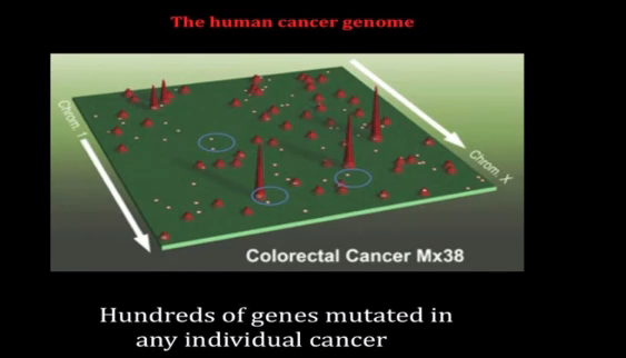
The tall peaks in this map of human prostate cancer represent genes commonly mutated in cancer. The smaller peaks are rarely mutated genes, and the small dots are genes mutated in a single cancer patient. Credit: Johns Hopkins University.

In 1990, the definition of cancer changed once again as it was discovered to be a disease of genomes. Not just one gene but many genes are mutated in cancer, a depiction of the disease painted by the work of Bert Vogelstein among others. As multiple genes in our genome regulate normal cells, multiple genes must be mutated to cause cancerous cells.

While some genes are mutated in cancer patients across the board, there are mutations unique to each individual, too. The problem is that the tumors look identical under the microscope. Mukherjee compares this phenomenon to the fact that every single human face has a common anatomy but is still quite different.

“The challenge as you sequence cancer genomes is that there is great diversity and therefore you reach the frightening corollary that every breast cancer is unique in the same way that every woman who has breast cancer is unique,” Mukherjee said.

With such variation, how do we remain optimistic about a cure?

Mukherjee offered acute promyelocytic leukemia (APL) as an example. Once known as the most threatening variant of acute leukemias, APL is now the most curable variant. One gene was identified that was common to all APL variants and one medicine – retinoic acid – was successful in treating this disease.

Phase IV: An Organismal Disease

As of 2010, cancer has been reconceived as an organismal disease: the human is simultaneously the site of the cancer, its prevention, and cure.

“Our next step is to understand the physiology of cancer – not just the cell biology, not the gene biology, nor the genome biology – but the physiology of cancer,” Mukherjee reminded us.

Despite the disease’s high level of complexity, scientists have new tools of computation to process data they previously could not, leading to the belief that cancer is a pathway disease. It’s not just genes and genomes that are mutated in cancer, it’s the cells’ language that drives those pathways and the resulting abnormalities. That language is the focus of scientists new cancer investigations and another piece of the devastating disease’s biography.

A Passion for Research

By Prachiti Dalvi

Akash Shah, Trinity ’13

“Research enables me to think about a question that excites me and helps patients,” says Trinity senior Akash Shah.  A biology major, philosophy minor, and a candidate for the Genome Sciences and Policy Certificate, Akash became interested in genomics as a freshman in the Genome Focus. Originally from Fullerton, CA, Akash was drawn to Duke because of its its immense biomedical research enterprise. He also loved the fact that at Duke, the medical school, law school, and business school were on the same campus as the undergraduate campus.

Intrigued by the research his professor Dr. Hunt Willard was conducting, he asked to get involved. His work in Dr. Willard’s lab dealt with artificial human chromosomes. More specifically, he was working with others in the lab to identify which regions of the chromosome would be deleted when transformed into human cells.

Now, Akash works in the Nevins Lab, where he looks at candidate genes in the epidermal growth factor receptor (EGFR) pathway: an important pathway in many cancers. When growth factors bind to the external portion of the receptor, the receptor becomes activated. Side effects of receptor activation include tumor growth and metastasis. When scientists target genes associated with this pathway, they can increase tumor cells’ sensitivity to pathway inhibitors and better prevent tumor cell reproduction.

http://www.youtube.com/watch?v=zE4BkAw_lL4

The advent of computational genomics has allowed for major advances in the field. Fifteen to twenty years ago, cloning genes was considered a PhD project, and now, it is something an undergraduate can do.

Akash’s favorite aspect of research at Duke is its collaborative nature. Faculty members work with another and across departments. His research is not limited to labs at Duke. In fact, he as also worked with professors at UCLA and Harvard. The culture of research varies from one university to the next; thus, Shah encourages undergraduates to do research at different institutions. “It gives you a chance to succeed in different cultures.”

When he is not in the lab, Akash enjoys playing cricket and exploring local restaurants with friends. During his time at Duke, he has been involved with numerous organizations, and has become an integral part of the Genome Research and Education Society (GRES). During his sophomore year, he founded a program in which undergraduates shadowed other undergraduates doing genomics research. In order to make research more accessible to undergraduates, Akash has helped organize career talks, including MD/PhD information sessions. After graduating from Duke in the spring, Akash hopes to begin medical school, and eventually pursue a career in academic medicine so he can continue conducting research. He has worked extensively in cancer genomics research and hopes to explore cancer stem cells in the future.

Crossing the Valley of Death

By Nonie Arora

David Adams shared his concerns about the output of America’s drug development pipeline at last month’s Science and Society Journal Club at the Institute for Genome Science and Policy (IGSP).

When it costs more than $1 billion to bring a drug to market with many failures along the way, “many (drug) companies are focused on de-risking,” according to Adams, an Associate Professor of Medicine in the Division of Medical Oncology.

Adams said the pharmaceutical industry seems to be counting on biotech companies and academia to help drugs cross the “Valley of Death” in anticancer drug development, but that has yet to happen.

While the rate of publication and quantity of scientific data continue to increase rapidly, not enough funding is being devoted to the “translational” research, which helps an idea make it from the lab to the clinic, Adams said.

Example of a tissue chip project sponsored through NCATS to improve drug safety. Credit to NCATS website.

The pharmaceutical industry has cut down on translational research, Adams said. Essentially, companies don’t want to take on projects unless they have a very high probability of success.

He also said that the National Institutes of Health (NIH) is generally risk adverse and tends to fund projects that represent “the next logical step,” rather than going for higher-risk, but potentially higher-gain ideas. The National Center for Translational Sciences (NCATS), established by the NIH director Francis Collins last year, is a promising step, Adams said.

When researchers design their cancer studies, many times they “don’t really take into account tumor physiology because we live in this era of molecular biology,” Adams said. He believes accounting for tumor physiology is very important. For example, doctors are rarely able to monitor how much drug makes it to a tumor, but translational research could solve this problem, said Adams.

Adams said drug development is also hampered by a human tendency toward  “technological lock-in.”


Personal Genome Machine Sequencer Credit: Benchmarks, a publication of the National Cancer Institute

“Why do we not change the way that we do things when there is compelling evidence to do so?”  He argued that:

1) people resist change in research and clinical environments because they are creatures of habit and convenience,

2) academics and clinicians often operate in silos rather than multidisciplinary teams that would enhance communication, and

3) we are obsessed with technology, as evidenced by genomics.

Adams said a new area to watch out for is theranostics: compounds that combine a therapeutic and diagnostic in the same formulation. Another area is miniaturization of electronics to permit real-time measurement of drug activity in and around tumors. An implantable radiation dose monitoring system is already commercially available and implantable sensors for management of diabetes are in the pipeline.

Ultimately, greater emphasis on translational research and breaking technological and organizational lock-in may help us cross this “Valley of Death,” he said.

Igniting U.S. health care's 'escape fire'

The film Escape Fire explores what's fanning the flames of the health care debate. Credit: escapefiremove.com

By Ashley Yeager

Imagine lighting a match to protect yourself from the flames of a fire.

It’s probably not the first thing you would think to do to stop from being burnt. But when there’s no other escape, the technique works. In 1949, Robert Wagner Dodge became living proof when he lived through the Mann Gulch fire by setting his surroundings on fire.

Now, his actions have become a metaphor for drastic ways government and industry should change U.S. health care before it too burns everything in its path.

Escape Fire: The Fight to Rescue American Healthcare showcases the health care system’s metaphorical blaze. The award-winning documentary, described as the “Inconvenient Truth” of the health care debate, opens nationwide in October. But members of the Duke community saw it for free on Wed., Sept. 19. They were also able to participate in a post-film panel discussion, which fleshed out a few potential escape fires for the health care industry.

“I think working with one patient at a time can help everyone become healthier,” said Annie Nedrow, a primary care physician and the associate director of Duke Integrative Medicine, which sponsored the event. But as the film points out, the current system rewards doctors for the number of patients they see, not the amount of time they spend with each person or the plans they may develop to help that patient prevent and manage disease.

Ironically, almost 75 percent of health care costs are spent on preventable diseases. The film, directed by Matthew Heineman and Susan Froemke, illustrates this statistic through anecdotes, where a band-aide, pill or even invasive surgery, such as inserting a stent to relieve heart blockages, provides immediate relief but does nothing to address the underlying causes of illnesses – typically diet and lifestyle.

Preventable diseases are 75% of our health care costs. Credit: escapefiremovie.com

“There’s got to be a shift in our culture, one where we actually have access to safe parks to exercise, healthy food, and the time to eat it,” said Adam Perlman, Duke Integrative Medicine’s executive director. He also agreed with Nedrow that a new system should invest more in primary care and health promotion, rather than disease treatment.

To set an example and test the feasibility of such a system, Duke Integrative Medicine has opened a primary care practice that limits the number of patients each physician sees so the doctors can spend more time with each patient and create a more holistic approach to that person’s health.

Perlman said that health coaching could be another important aspect of correcting the healthcare system. He explained that doctor X might tell a patient to lower his blood sugar, doctor Y then tells him to lower his blood pressure, and all the patient really wants to do is dance at his daughter’s wedding. “A health coach helps the patient reach those bigger goals by connecting the dots and helps them execute the plan to get there,” he said.

The film and remarks prompted many audience members to question what it would take to change the current system. Nedrow, who said she has been inspired by books on creativity and innovation, suggested that it was dialogues like the one they were having that could ignite change to repair the broken model of health care or create a new system.

More innovation, however, may mean that more people need to step into the fire and strike a match, rather than run and try to dodge the flames.

Varmus Encourages Provocative Questions

By Nonie Arora

“Provocative questions,” the important but non-obvious ones, the questions to be answered by technology that doesn’t  exist yet, are one focus of Nobel Laureate Harold Varmus‘s storied career these days.

What environment factors change the risk of various cancers when people move from one geographic area to another?

Why are different tissues so dramatically different in their tendency to develop cancer?

How does obesity contribute to cancer risk?

Harold Varmus, Director of NCI Source: cancer.gov

Varmus, director of the National Cancer Institute, visited Duke April 12 to address  the Duke Medical Scientist Training Program 2012 Symposium and to share some of these provocative questions with a full house in Love Auditorium.

Varmus said we are in a period of rapid scientific change because “clinical research and basic research are mingled in a way that is extraordinary.” And while he acknowledged recently flat NIH budgets, he said, “let’s not worry about budgets, let’s worry about opportunities.”

For example, he cited one such scientist who looked for important opportunities, Renato Dulbecco (1914-2012), a virologist from Caltech who died recently. Dulbecco advocated for a systematic approach to sequencing the human genome as early as 1986 despite the many naysayers in the biology community. Varmus called Dulbecco, “a visionary who saw beyond the technology of the day” as he encouraged his audience to think in the spirit of Dulbecco: boldly.

Varmus spoke of how the “precision medicine” of genomics may lead to more accurate diagnoses and a new taxonomy of disease. He made the distinction clear between precision medicine and personalized medicine claiming that even his father practiced personalized medicine because he knew his patients well.

Cancer deaths are rising globally, especially in less developed countries, according to Varmus. He explained how open exchange of information with cancer centers around the world is important to solving new cancer challenges. He described how cancers are more frequently related to infectious agents in the developing world, like the Epstein-Barr virus’s relationship to Burkett’s lymphoma, and the implications for research.

To fund important but non-obvious questions in cancer research, he has launched the “Provocative Questions” Project. These questions are meant to build on specific advances and address broad issues. Because researchers can be more risk-averse when funding levels are lower, this project hopes to fund intriguing questions that would otherwise remain unfunded and unanswered.

These questions need answers. Who better to answer them than the Duke MD/PhD candidates in the audience.

Sugar-coated cells not like Peanut M&M's

By Nonie Arora

Carolyn Bertozzi. Source: Wikimedia commons

UC Berkeley professor Carolyn Bertozzi spoke about sugar coated cells and peanut M&Ms following the unveiling of the English translation of Hertha Sponer’s biographyon April 5.

Hertha Sponer (1895-1968), a noted scientist who studied quantum mechanics, physics, and chemistry, was the first woman on the Duke Physics faculty. Bertozzi called it a privilege to “celebrate the life, career, and legacy of Hertha Sponer.”

Bertozzi discussed how scientists used to think of sugars on the surfaces of cells like the candy coating on peanut M&Ms – only there to serve as a protective barrier. Now, she says people appreciate the diverse information stored in the sugars as an important diagnostic tool.

These cell surface sugars, called glycans, can give us information about a cell state. In fact, human blood groups are determined by cell surface glycans. The glycans specific to each blood group are not much different chemically, but according to Dr. Bertozzi, “the human immune system is exquisitely specific to recognize functional groups. If someone who is blood type A is given type B blood, they will have a massive reaction.”

Tamiflu Source: Wikimedia Commons

Glycans are relevant to many different areas of medicine, from stem cell biology to viral infection. Drugs for influenza viruses were developed by taking advantage of glycan chemistry. Bertozzi explained that in the pharmaceutical industry, better understandings of binding to cell-surface sugars enabled creation of Relenza and Tamiflu.

Bertozzi also described how X-ray crystallography was used to understand the binding of the enzyme and sugars and that Hertha Sponer made valuable contributions to that field.

Cell surface sugars also differ between healthy and diseased cells. Cancer cells have a different profile of cell surface sugars than normal cells. For example, polysialic acid (PSA) which is usually only in neurons in brains, pops up frequently on tumor cells. According to Bertozzi, detecting the levels of the acid would allow scientists to see tumors without invasive surgery. Bertozzi’s lab has been working on several projects to develop this method for tumor and cancer imaging.

Solving cells' social networks

By Ashley Yeager

Rick Durrett is looking at the math underlying the most challenging biological problems. Credit: Les Todd, Duke Photography.

“Cancers are complicated systems, and you want to figure out how they work,” says mathematician Rick Durrett in a Q&A in the latest editions of the Proceedings of the National Academy of Sciences.

Durrett, who was recently elected to the Academy, came to Duke in the summer of 2010 and has been collaborating with researchers to here, using the tools of probability theory to look for ways to study cancer questions through computer simulations rather than large experimental trials.

One of the greatest challenges is to understand how cells use social networking to survive and adapt to their environment. Durrett has been working with biologists to describe mathematically how individual cells secrete chemicals that affect the behavior of nearby cells.

“This may be important for the spread of cancers. It’s an interesting area, and one I’m learning more about now,” he said in the PNAS interview.

You can read the full interview here.

Page 5 of 6

Powered by WordPress & Theme by Anders Norén