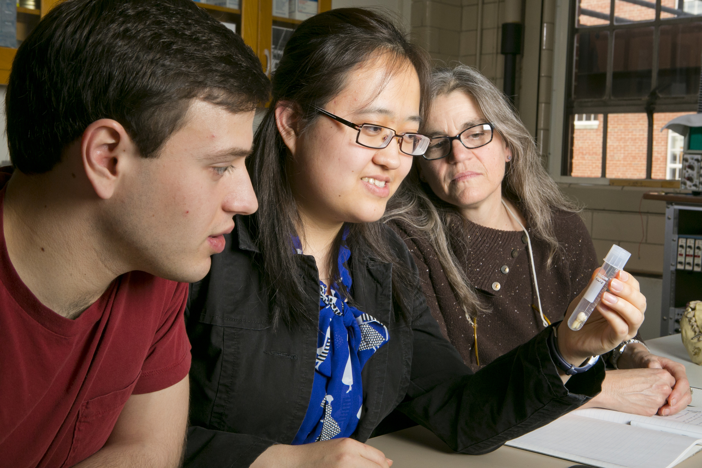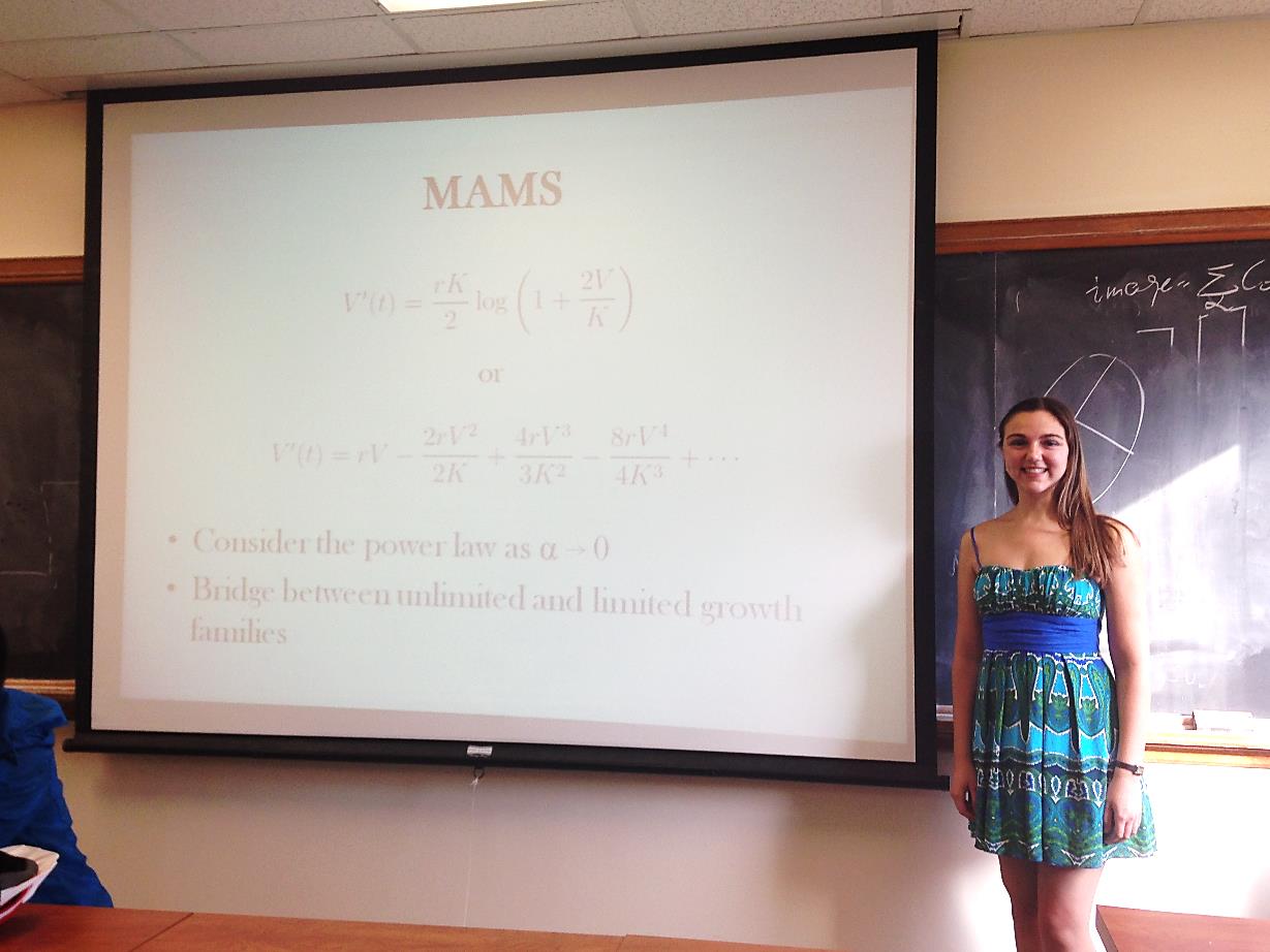
Anne Talkington with the MAMS function
By Olivia Zhu
Anne Talkington, an undergraduate Mathematics student under the auspices of Richard Durrett, attempts to gain a quantitative grasp on cancer through mathematical modeling. Historically, tumor growth has only been measured in vitro (in a laboratory setting); however, Talkington looks at clinical data from MRIs and mammograms to study how tumors grow in vivo (in the human body).
Talkington is primarily interested in how fast tumors grow and if growth is limited. To analyze these trends, Talkington extracted two time-point measurements of tumor size — one at diagnosis and one immediately before treatment — and compared their change to a variety of mathematical functions. She studied unlimited functions, including the exponential, the power law, and the 2/3 power law, which represents growth limited by surface area, as well as limited functions, including the generalized logistic, which has an upper growth limit, and the Gompertz. Her favorite function is an unlimited function that she created called the Modified Alternating Maclaurin Series, or MAMS, which she originally intended to model microbial growth.
Talkington also examined various types of cancer: breast cancer, liver cancer, tumors of the nerve that connect the ear to the brain, and meningioma, or tumors of the membranes that surround the brain and spinal cord. She expected growth rates among clinical groups to be constant, but she did not generalize between the groups due to demographic bias and other confounding factors.
Ultimately, Talkington found that breast cancer and liver cancer grew exponentially, while tumors of the meninges or vestibulocochlear nerve grew according to the 2/3 power law. Talkington’s work in model-fitting cancer growth will facilitate the administration of effective treatment, which is often growth-stage dependent.