Following the people and events that make up the research community at Duke

Students exploring the Innovation Co-Lab

Category: Visualization Page 7 of 10

Finding Order in Insect and Orc Swarms

Ouellette's model of insect swarming

Ouellette’s model of insect swarming

By Olivia Zhu

Dr. Nicholas Ouellette looks for the organization in disorder.

Ouellette, associate professor in the mechanical engineering department at Yale University, studies collective motion in animal systems. On February 17, he presented his models of swarming of Chironomus riparius, the non-biting midge, as part of Duke’s Physics Colloquium. Ouellette ultimately hopes to pin down fundamental laws of biology through his physics research.

In the lab, Ouellette has found that Chironomus insects swarm in a columnar, teardrop shape in the center of their container. They only live in their flying state for two to three days, during which they mate, lay eggs and die. During this period, swarming affords them protection from predators and the opportunity to mate.

Ouellette and his lab have devised various methods of modeling the insects’ swarming. They found that the insect density remains constant, and that the “scattering,” or collisions of insects, mirrors that of an ideal gas over long periods of time. Interestingly, the graph of individual insect speed follows a Maxwell-Boltzmann distribution, even though the lab did not track the usual factors that create such a distribution, like temperature.

The most pressing question Ouellette would like to answer is which factors create a swarm—he has determined that close insect-insect repulsion contributes to swarming, but distant insect-insect attraction does not. To pursue this question, Ouellette is testing how many insects it takes to make a swarm.

Wildebeest stampede modeled in The Lion King

Wildebeest stampede modeled in The Lion King

Other animals that exhibit collective motion are mackerel, wildebeests and starlings. Some familiar examples of collective motion modeling are visible as the Orcs storm the castle in Lord of the Rings and as the wildebeests charge the canyon in The Lion King.

Learn to Fly a Drone in Three Minutes

By Erin Weeks

Missy Cummings has accomplished a lot of difficult things in her life — she was one of the Navy’s first female pilots, after all — but being a guest on The Colbert Report, she said, was hard.

Cummings told the story of her journey from Naval lieutenant to media drone expert last week at the Visualization Friday Forum seminar series in a talk (video archived here) titled “Designing a System for Navigating Small Drones in Tight Spaces.”

Missy Cummings joined Duke as an associate professor of mechanical engineering and materials science last semester

Missy Cummings joined Duke as an associate professor of mechanical engineering and materials science last semester.

Last semester, Cummings moved her renowned Humans and Automation Lab from MIT to Duke University. She’s wasted no time immersing herself in the new university and volunteered for the semester’s first seminar to introduce herself and her lab’s latest work to Duke’s visualization community.

Cummings’ research over recent years has centered on the development of a smartphone interface through which, she said, anyone can learn to pilot a one-pound drone in three minutes. The technology could be a boon to the U.S. Army, which now issues smartphones to its personnel and mostly relies on cumbersome, gas-powered drones.

The lab tested the technology by asking volunteers to maneuver a drone through an obstacle course both in the field — where they learned wind and cold temperatures are not a drone’s friend — and in simulated environments.

One of the things they discovered in both cases was that individuals who performed well in a spatial reasoning test were more likely to complete the obstacle course. Moreover, these performances tended to be gendered, with men scoring higher than women in spatial reasoning. Interestingly, Cummings noted, other studies have shown women tend to perform better piloting drones in long-term, “boring” scenarios with little action.

Cummings is interested in teasing out the reasons for these results, which could have significant implications for the U.S. Army or companies one day interested in hiring drone pilots.

As Stephen Colbert confirmed, you may be able to fly a drone with three minutes’ training, but that doesn’t mean you can fly it well.

Cummings talks to a full house at the Visualization Friday Forum on January 24.

Cummings talks to a full house at the Visualization Friday Forum on January 24.

New Course Offers Lessons from Lasering Priceless Art

Duke graduate student Tana Villafana and chief conservator at the NC Museum of Art William Brown stand over The Crucifixion (inset). (Photo: Martin Fischer)

Duke graduate student Tana Villafana and chief conservator at the NC Museum of Art William Brown stand over The Crucifixion (inset). (Photo: Martin Fischer)

By Erin Weeks

A group of chemists at Duke University has gained recognition in recent years for shooting lasers at medieval artwork — technology that allows a harmless peek at the many layers and materials in a painting and offers insight into long gone eras and artists. Now, Duke students will have the chance to learn from this pioneering work at the intersection of chemistry and art history in a new course on the science of color.

The course coincides with the publication of the first scientific measurements from the laser work, reported Jan. 20 in the Proceedings of the National Academy of Sciences.

“The images we have now are enormously better than a year ago,” said Warren S. Warren, head of the lab performing the imaging and the James B. Duke professor of chemistry. He and fellow Duke authors, grad student Tana Villafana and associate research professor Martin Fischer, have not only demonstrated the technology works — they’ve shown it works at an incredible level of detail, telling the difference, for example, between nearly identical pigments.

But lasering The Crucifixion by Puccio Cappano was just the start, as the team envisions countless more cultural applications of the technology. Given enough funding and manpower, they could visualize ancient scrolls of text too fragile to unroll, reveal the bright colors that once adorned Greek statues, learn the secrets of China’s terracotta warriors, and even detect the beginnings of pigment degradation in aging artwork.

There are talented people in art conservation, Warren said, whose work could benefit from more advanced technology, and there are talented people at the cutting-edge of laser science looking for meaningful ways to apply their inventions. For the past several years, Warren’s lab has brought these people together.

Now, he hopes to accomplish something similar with students at Duke. Warren, Fischer, and another chemistry instructor, Adele DeCruz, are teaming up to teach “The Molecular, Physical, and Artistic Bases of Color” in the second half of spring semester.

The class will visit the Nasher Museum of Art, the North Carolina Museum of Art in Raleigh, and possibly even the National Gallery of Art in Washington, D.C, to learn first-hand from art conservators and working artists. Students can expect to learn about how humans have used and made pigments over the millennia; how color works at a molecular level; and the basics of how human vision, microscopes, cameras, and lasers all see or image color.

Students can register for the half course, CHM 590, until the add/drop deadline for classes on January 22. “Students should not be scared off by the course number,” Warren said. “The prerequisite is one college-level science course, and the intent is to make both the science and artistic components accessible to a broad audience.”

Funding for the research was provided by National Science Foundation grant CHE-1309017.

CITATION: “Femtosecond pump-probe microscopy generates virtual cross-sections in historic artwork.” Tana E. Villafana, William P. Brown, et al. Proceedings of the National Academy of Sciences, Jan. 20, 2014. Doi: 10.1071/pnas.1317230111

Pretty pictures show lemurs responding to changing climate

Guest Post by Sheena Faherty, Biology Graduate Student 

Madagascar’s much-adored and fuzzy lemurs might be “sweated out” of habitats by warming environments under global climate change. Or will they?

A team of researchers at the Duke Lemur Center is employing high-tech heat cameras used in  fire fighting, sports medicine and cancer diagnostics to take “glowing” rainbow pictures of lemurs and their forest surroundings. The results look similar to a child’s coloring project gone rogue.

A mother and baby Coquerel's Sifaka at the Lemur Center in thermograph and visible light. (Leslie Digby)

A mother and baby Coquerel’s Sifaka at the Lemur Center in thermograph and visible light. (Leslie Digby)

This technology, known as infrared thermography, is a camera that allows researchers to detect surface temperatures of lemurs and their hang-outs in the forest—at different depths and heights—and on varying surfaces such as the ground, leaves, and tree trunks.

Combining these data with records of where an animal prefers to spend time, the researchers can begin to determine what temperatures make lemurs most happy.

Leslie Digby, an associate professor in the Department of Evolutionary Anthropology, and her students want to see  how the lemurs are changing their behavior to warm-up on cool days, and cool-down on warm days without having to shiver or sweat.

This sounds rather like a lizard basking on a rock during a sunny day to warm his cold-blooded body up, but lemurs aren’t cold-blooded. They shouldn’t have to do this.

It turns out that even though lemurs are warm-blooded, they can conserve precious energy by channeling their inner Buddha — using sunning behaviors, just like lizards, to fine-tune core body temperatures.

Digby’s team is trying to understand why some species have seemingly restricted territories, even without obvious geographical barriers like mountain ranges or rivers. They suspect temperature plays a part.

“We know that primate species ranges have been very different in the past, so understanding how flexible these animals are, or [are] not, to temperatures can help us understand these larger scale impacts [of changing climate]”, says Digby.

Figuring out how animals respond to alterations in their environment, like rising temperatures, can help scientists anticipate species’ survival in the face of globally changing climates. And knowing which areas of the forest are preferred by lemurs, could help direct conservation efforts, like reforesting parts that have been cut down, or preserving those areas that have not.

Changing temperatures will undoubtedly have major impacts on lemur home ranges in the future, potentially altering them until the animals  are forced into an area outside their thermal limits. By gearing her research toward understanding the thermal tolerances of lemurs, Digby is doing her part to protect the vulnerable lemurs.

A ringtailed lemur striking the classic belly-warming Buddha pose in one of the natural enclosures at Duke Lemur Center. (David Haring)

A ringtailed lemur striking the classic belly-warming Buddha pose in one of the natural enclosures at Duke Lemur Center. (David Haring)

Teaching Young Scientists the Elements of Design

by Erin Weeks

Ten visiting undergraduate researchers spent the summer sharpening their science communication skills at Duke. They came from around the country to chemistry and engineering labs to participate in a National Science Foundation program called Chemistry and Applications of Smart Molecules and Materials and to learn the principles of ‘molecule-to-material’ research.

While the students spent most of their days in the lab, they were also tasked with creating a visual representation to explain some aspect of their summer research—once at the beginning of the summer, and once again at the end, after feedback and instruction on the basics of good visual design. The process was designed to help the students understand their research, their roles as scientists, and the importance of science communication.

“You want to catch peoples’ eye, but you want to be fairly simple and easy to interpret,” said chemistry professor and department chair Stephen Craig. Craig and project co-leader, associate chemistry professor Kathy Franz, discussed their project at a visualization seminar series last week (Nov 1).

As for the visual don’ts, Craig advised the students to skip abstract art and avoid anything flashy or over the top. In addition to the images, the students practiced explaining their research in strictly timed three-minute talks.

“We wanted them to give that elevator pitch, that three-minute pitch,” said Franz, so that the students would be able to “communicate to their peers what their project for the summer was going to be.”

Duke professor Jane Richardson first visualized protein as ribbon-like (Courtesy Wikimedia)

Duke professor Jane Richardson first visualized protein as ribbon-like (Courtesy Wikimedia)

When Franz was a student, she was never trained how to make her research graphics clear and intelligible. But as a chemist, she knew the significance of effective visuals. Take, for example, the structure of proteins, which were first visualized as ribbon-like in 1980 by Duke biochemist Jane Richardson. These days, Franz said, she and generations of biology students only picture protein as a ribbon.

“The way people represent scientific results changes the way we imagine it,” Franz said.

New App May Help Protect Wild Dolphins

By Ashley Yeager

A screenshot from a new app, the Nai'a Guide, which provide info about eco-friendly dolphin-watching tours in Hawai'i. Credit: Demi Fox, Lenfest.

A screenshot from a new app, the Nai’a Guide, which provide info about eco-friendly dolphin-watching tours in Hawai’i. Credit: Demi Fox, Lenfest.

Traveling to Hawai’i sometime soon?

If so, you’re probably excited to experience spinner dolphins in the wild. If not, you can still dream about it. And now, there’s an app for that.

Scientists at Duke’s Marine Lab in Beaufort, N.C. have released the Nai’a Guide — a new iPad app that teaches users about wild Hawai’i spinner dolphins and how to see the animals without harming them. Tourists can use the app to plan an eco-friendly tour to experience the dolphins.

“If we can harness the power presented by mobile technology for conservation and responsible tourism, we have the chance to reach a wide audience and really make a difference for these animals,” says Demi Fox, a postgraduate researcher at the Lenfest Ocean Program who developed the app, along with Duke marine biologist Dave Johnston.

Nai’a is the Hawaiian word for dolphin. The Nai’a Guide explores the biology and ecology of spinner dolphins with photos, videos and sound clips. It also describes sustainable dolphin-based tourism practices outlined NOAA’s Dolphin SMART program.

With the Nai'a Guide, users can learn about spinner dolphins and their habits. Credit: Demi Fox, Lenfest.

With the Nai’a Guide, users can learn about spinner dolphins and their habits. Credit: Demi Fox, Lenfest.

Designed by Fox and developed by an online company called Kleverbeast, the Nai’a Guide also connects tourists with sustainable tour operators so everyone can make more responsible decisions when going to see spinners.

“The principles advocated within the Naia Guide could also be useful for dolphin-based tourism in other places, and with other species. Many of these best practices are generalizable,” Johnston says.

He and other scientists are concerned about human interaction with wild dolphins and other species worldwide. In Hawai’i, the main concern is that spinner dolphins rest during the day in the same shallow bays that people use for snorkeling, kayaking and swimming. Many tourists misinterpret the dolphins’ close proximity and curiosity for playfulness and try to swim with and even ride the animals while they are sleeping.

Intense and consistent human interactions could affect the dolphins’ health over time, Johnston says. The negative effects may also threaten the animals, a resource the state uses to draw tourists to the islands. As a result, he and colleagues at Murdoch University’s Cetacean Research Unit have been tracking spinner populations and monitoring their interaction with people in the Hawai’i island bays.

Researchers study dolphins in boats and high on the cliffs of Hawai'i Island, which is covered in the new app. Credit: Demi Fox, Lenfest.

Researchers study dolphins in boats and high on the cliffs of Hawai’i Island, which is covered in the new app. Credit: Demi Fox, Lenfest.

Scientists “can do all the science in the world, but until we share our findings broadly and in an accessible way, we will not effect serious change,” Fox says. She included the team’s research in the app so users can better understand researchers’ concerns about human-dolphin interactions and can make more informed decisions when choosing a dolphin tour.

“My hope is that the app will serve as an ecological conscience,” she says.

The app, available in Apple’s iTunes Store, can also be found on Twitter @NaiaGuide and on its website, http://www.naiaguide.org.

Documenting Medicine to Understand Patients

Guest post by Clara Colombatto, T’15

The daily life of a doctor is filled with reports, numbers, and forms. Opportunities to sit down with patients and listen to their stories are rare. Yet, “most of the information practitioners need to care for patients is contained in their stories,” says Dr. John Moses, a Duke pediatrician who founded the Documenting Medicine program two years ago.

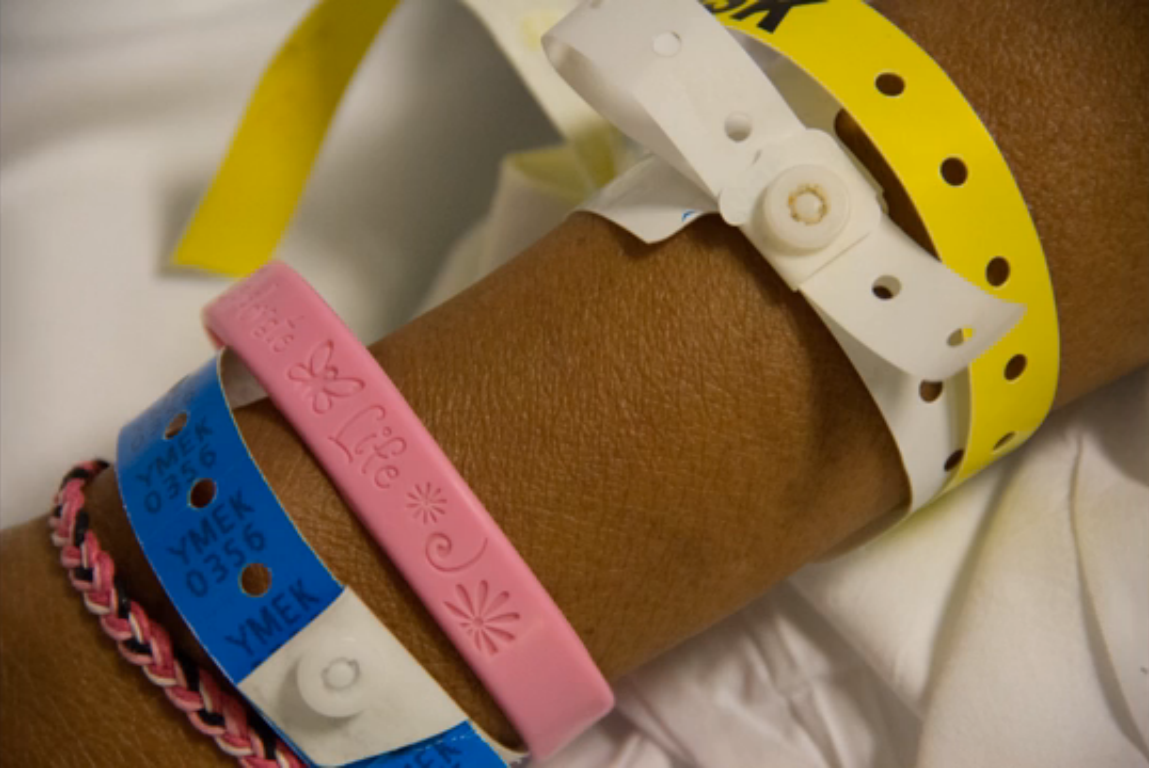
medical bracelets

A still from Dr. Tera Cushman’s “How to See the Forest

Mike and Patsy holding hands

Mike and Patsy from “I Will Go With You,” by Dr. Lisa Jones.

The innovative idea that he had with photographer Liisa Ogburn, an instructor at the Center for Documentary Studies, was to encourage medical residents to document their experiences and gain insight into patients’ stories to become better doctors.  Eleven Duke resident physicians, one physician and one physician assistant gathered on June 5 to share documentary projects they completed this year.

“The best attending physicians are both vast repositories of data and good storytellers – people who can bring the numbers and the patient into focus at the same time” says anesthesiologist Tera Cushman, one of the filmmakers. In her documentary How to See the Forest and the Trees, she presents both readings of medical records and interviews of patients Annie and Olivia.

Works produced in Documenting Medicine are objective reports of current issues in healthcare, but also powerful learning tools: residents enhance their understanding of patients to represent their perspective in the best way possible.

In Spectrum, Dr. Kathleen Dunlap interviews the parents of her patient Isaac, and discovers the deep changes and contrasting emotions beyond a simple and almost mechanical diagnosis of autism.

Dr. Lisa Jones tells us a patient’s journey after her time at the hospital in I Will Go with You: Patsy’s Mission to Educate Others about Colorectal Cancer Screening.

These stories also reveal aspects of patients’ lives that are crucial in recovery beyond treatments and prescriptions: in Welcome to Crazy Camp by Dr. Stephanie Collier, Tom, a patient at Duke Hospital, tells us about the importance of patients’ sincere care and profound respect for each other in a psychiatric clinic. The most supportive and understanding friend for him was a young friend who  “made a whole lot of sense except for this one little part of her life where she thought the government had placed something in her brain that was trying to control her.”

Documenting Medicine is now accepting applications for the coming year, and is open to medical residents, but also to anyone working in healthcare who wishes to tell a medical story.

Students Create Multimedia Ocean Conservation Text

By Ashley Yeager

This screenshot shows one of the opening page of of Johnston's new iBook. Image courtesy of Dave Johnston, Duke.

This screenshot shows one of the opening pages of a chapter in Johnston’s new iBook. Image courtesy of Dave Johnston, Duke.

Duke marine biologist Dave Johnston and his students are back in business on iTunes.

They’ve just released The View From Below, a free iBook for middle school students and teachers that uses multimedia and classroom exercises to discuss overfishing, marine debris, climate change, invasive species and other issues related to marine conservation.

This is Johnston’s second digital textbook. His first was Cachalot, an iPad textbook covering the latest science of marine mammals like whales, dolphins and seals. Experts contributed the text, images and open-access papers.

The View From Below, however, is a bit different.

Undergraduate students in Johnston’s Marine Conservation Service Learning class wrote the book using Apple’s iBooks authoring tool. Johnston and Tom Schultz, Director of the Marine Conservation Molecular Facility at Duke’s Marine Lab, edited it.

“There are a lot of people exploring the use of the iBooks platform for student-generated content, among other development platforms,” Johnston says. “I don’t think we’ve seen many that focus on marine science yet though, and I’m pretty sure it’s the first marine conservation textbook written by students on the iTunes store.”

Johnston says the class chose to use the iBooks software because the technology is free, easy to use and provides “great templates to get things going quickly.” The software also works well because Duke’s Marine Lab has an iPad loaner program, making the tablet the platform of choice for developing and testing the textbook.

The middle school that the service learning class works with also has access to iPads for students and instructors, so the audience was there for the iPad format, Johnston adds.

His students chose to write the book as the class project to spur learning and discussion about some of the most serious problems facing Earth’s oceans.

“As the text indicates, all life on earth is ultimately supported by the ocean, so we need to take care of it,” he says.

Chocolate's crisp crack comes from chemistry

By Ashley Yeager

This is the final post in a four-part, monthly series that gives readers recipes to try in their kitchens and learn a little chemistry and physics along the way. Read the first post here and the second one here and the third one here.

chocolate-bunny

This bunny must have been made from quality chocolate. His ears are already gone. Credit: Waponi, Flickr.

When you snap off and savor the ears of a chocolate bunny this Sunday, say a quick thanks to science.

“The essence of science is to make good chocolate,” said Patrick Charbonneau, a professor of chemistry and physics at Duke.

He explained that cocoa butter, one of the main ingredients in chocolate, can harden into six different types of crystals. All six types are made of the same molecules. But, at the microscopic level, the types have distinct molecular arrangements, which lead to differences in the crystals that form.

“The problem with chocolate is that only two of these types have good texture when eaten,” Charbonneau told students in the Chemistry and Physics of Cooking.

He teaches the freshman seminar with chef Justine de Valicourt and chemistry graduate students Mary Jane Simpson and Keely Glass.

During class, students looked at and tasted chocolate containing only the good-tasting crystal types and some that also contained the less favorable ones. The first had that signature sheen and snap of quality chocolate and melted evenly when left on the tongue. The latter pieces were dull, melted with the slightest touch and left a sandy texture on the tongue.

The demonstration showed that the different types of chocolate crystals melt at different temperatures. By carefully controlling the chocolate as it cools, chocolate-makers can create mixtures of only the favorable crystal types.

The process, called tempering, takes chocolate through a series of heating and cooling steps. The initial cooling step forms many of the chocolate crystal types, including the dull, unfavorable ones. Warming the mixture a little — to about 31°C (87°F) — melts the unfavorable crystals but not the best-tasting ones.

As the mixture cools again, the remaining, favorable crystals “seed” the chocolate so that good-tasting crystals form preferentially throughout, ensuring good chocolate structure and taste.

Students got a chance to test the science in lab later that evening, and judging by the number of mouths (and faces) covered with chocolate, it’s safe to say the science was successful.

If you’re looking to try it out — or save a poor bunny’s ears — here’s the recipe.

Tempering chocolate:

Materials:
1 small, microwave safe bowl
1 big bowl
1 spatula
2 scraper spatulas
1 chocolate mold
parchment paper
cooking thermometer

Ingredients:
250 g Dark Chocolate or 250 g Milk Chocolate (about 1 1/3 cups)

Filling:
60g white chocolate (about 1/4 cup)
60g yogurt (a little less than 1/4 cup)

Instructions:

1. Place milk or dark chocolate in the small bowl.
2. Heat the bowl in 30-second intervals in a microwave (stirring after each) until the chocolate is melted. Note: The milk chocolate should take about 1.5 minutes and the dark chocolate about 2 minutes to melt.
3. Once heated, pour half the liquid chocolate onto a clean marble or stone counter. The chocolate puddle should be the size of a medium pancake. (Note: If there is not stone or marble surface, another technique is to melt less chocolate and then add good tempered chocolate in it to lower the temperature.)
4. Spread the pancake portion out in ribbons using the scraper spatula. Bring the chocolate back together into a mound repeatedly for 5 minutes, until it starts to solidify.
5. Put the chocolate back in the original heating bowl. Adding the cooler chocolate will cool the rest of the liquid to the right temperature.
6. Mix the cold and hot chocolate.
7. Check the temperature of the chocolate. (Dark: 31-32°C/88-89.5°F; Milk: 29-30°C/84-86°F).
8. Dip the parchment paper in the mixture of the “hot” and “cold” chocolate. If it cools on the parchment paper and is uniform and shiny, then it’s ready.
9. Pour chocolate into mold.
10. To make stuffed chocolate candies, flip the mold to empty excess chocolate.
11. Turn it back, scrap the excess of chocolate off the surface. Let the thin layer of chocolate in the mold crystallize.
11. Melt white chocolate. Mix it with yogurt. Cool to room temperature.
12. Add filling to 2/3 of the mold cavities, and then pour more tempered chocolate on top.
13. Level the chocolate with a scraper and scrape off excess.
14. Let it rest for few minutes at 20°C (68°F) or put it in the fridge.
15. Pop candies from mold and enjoy.

Duke Math Makes Final Four

By Ashley Yeager

This NCAA bracket is based on the quality of the school's math department. Courtesy of: Jordan Ellenberg, UW-Madison.

A few mathematicians made their NCAA bracket based on the quality of universities’ math departments. Courtesy of: Jordan Ellenberg, UW-Madison.

If NCAA basketball championships were won with math, Duke would move on to the Final Four.

At least, that’s what Jordan Ellenberg, a University of Wisconsin mathematician, and his friends think.

They didn’t use complex algorithms to make their bracket, but picked their winners based on the quality of each school’s math department.

With that ranking scheme, Harvard would win it all, with Cal second and UCLA and Duke rounding out the Final Four.

“Of course, these judgments are for entertainment only, and were produced by a group, so if you find any of the picks absurdly wrong, those were the ones I didn’t make,” Ellenberg wrote on his blog, where he posted the picks.

Sadly, the bracket isn’t doing so well as March Madness moves forward.

But it is a fun way to learn more about the math departments around the country and how well their quality does, or does not, correlate with the quality of the schools’ basketball teams.

Special thanks to Duke mathematician Jonathan Mattingly for pointing the bracket out to us.

Go Duke!

Page 7 of 10

Powered by WordPress & Theme by Anders Norén