Following the people and events that make up the research community at Duke

Students exploring the Innovation Co-Lab

Category: Cancer

Cancer drug shortage is ‘a real nightmare’

By Ashley Mooney

A national shortage of the cancer drug methotrexate is causing concern for Duke doctors and their patients, and is part of a larger national trend of shortages in cancer drugs and antibiotics.

“It’s been a real nightmare—it’s a very real shortage,” said Dr. David Rizzieri of Duke Medicine’s Cellular Therapy Division. “It’s part of a bigger [drug shortage] problem, not just a methotrexate problem.”

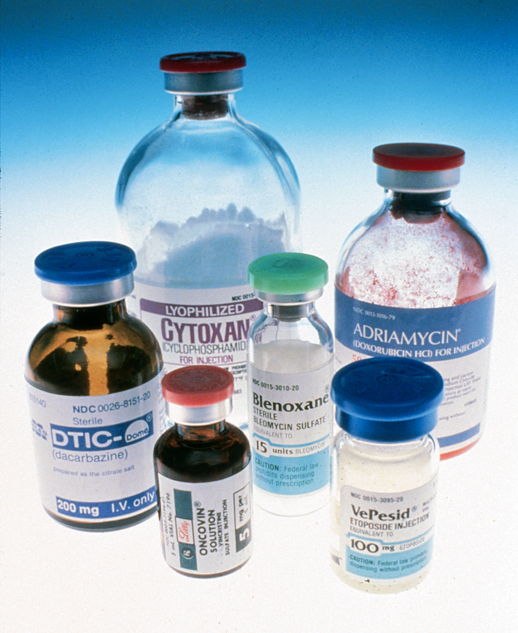
Six bottles of different cancer drugs. Methotrexate is not pictured. Image courtesy of the National Institutes of Health.

Ben Venue Laboratories—the largest manufacturer of methotrexate—temporarily closed its manufacturing facility in Bedford, Ohio, because it could not guarantee product safety, according to an article from the New York Times. Shortfalls have also been caused by prescription drugs going generic and becoming less profitable, according to an article from WRAL.

Rizzieri said members of the Duke community and others have approached Congress and national advocacy groups, but not much can be done about the lack of medication. Shipments from other countries are currently providing American hospitals with the drugs.

Issues of quality control and cost make the duration of drug shortages unknowable, Rizzieri said.

“The shortages have been at variable length with the drugs over the last couple of years,” he said. “Sometimes we’ve had shipments arrive from China and be held up at the docks [while] the drugs were tested to see if they were at the right quality to be released in the United States.”

A shipment of methotrexate will soon arrive in the United States, with enough medication to last the entire nation about a month, according to the New York Times article.  But the shortage is still not solved.

Rizzieri said patients are left with few options for care.

When his patients need medicine that is in low supply, Rizzieri said he often calls his colleagues first and if they have the proper drug, he sends his patient there to be treated. If nobody else has the right medication, a different regiment of chemotherapy may be used in its place. In some instances, he said, patients may need to travel out of the country to buy a different version of the drug.

”There are a number of good people working very hard to find drugs, but the issues of quality concern and cost remain,” he said. “With that the duration of these shortages are unknowable.”

Chaos puts a path on nanoparticles

By Ashley Yeager

Shaquille O'Neal towers over Ralph Stefanelli, the doctor who delivered him. Credit: Rich Krauss.

At just over seven feet tall, Shaquille O’Neal is easy to spot in crowd. But the individual virus structures that give him, and us, a cold aren’t so easy to see.

They’re harder to spot because they, unlike Shaq, are much smaller than the wavelengths of light we use to see them. But, physicists at Duke are now designing a technique that could give scientists a way to sense these tiny viruses and other nanoparticles and even capture their path when they’re on the go.

The team describes the new sensing system in the Dec. 16 issue of Physical Review Letters.

The new detector will compliment the few existing ways to track tiny objects, says physics graduate student Seth Cohen. He says that currently detectors can sense the presence of nanoparticles and that biomedical researchers have used virus-sized objects to tag specific parts of human cells.

“Ultimately, I would like to see this new system used to map out the dynamics inside of a living cell, using nanoparticle tags on the cell’s internal structures,” he says.

That can’t be done right now because the internal structures and other nanoparticles are smaller than 100 nanometers. Our eyes see wavelengths of light between 400 to 700 nanometers, and Shaq, by comparison, is 2.16 billion nanometers. We see Shaq because he has a lot more nanometers than those found in the wavelengths of light we use to see. But, we can’t see virus or cell structures because they’re size is far below that limit.

To overcome the problem, Cohen and his colleagues based their new sensing system on two physical properties, wave chaos and nonlinear dynamics. The proposed apparatus is shaped like a stadium, where multiple reflections of radio-frequency waves fill the entire cavity and take many different paths, a form of wave chaos. The stadium-shaped cavity and multiple-path reflections also allow the physicists to suspend the information flowing through the system, forming a nonlinear delayed feedback loop.

virus structure of a cold

The virus structure, shown here, that causes the common cold is only 20 nanometers across. Credit: Michael Taylor, TurboSquid.com

In initial experiments, the team used a pair of stationary broadband antennas to track a small container filled with water, which also scatters the radio waves broadcast into the cavity. Working out the geometry of the cavity, along with all of the possible reflection angles of light, the team was able to pinpoint the object in the cavity and track it as it moved. In other words, by combining wave chaos and nonlinear dynamics, the physicists could track an object that was much smaller than the wavelengths scientists used to see it.

The team is now designing a new version of the system using laser light and a microcavity, which is only a few hundred micrometers in size and can be constructed on silicon chips. The system, which will work in the visible wavelengths of light, will provide a new and simple technique for tracking small nano-scale objects, such as viruses. Cohen will report on his progress on this sensing system in May at the Experimental Chaos and Complexity Conference in Ann Arbor, Michigan.

Citation: Subwavelength Position Sensing Using Nonlinear Feedback and Wave Chaos. Seth D. Cohen, Hugo L. D. de S. Cavalcante, and Daniel J. Gauthier. Phys. Rev. Lett. 107, 254103 (2011). DOI: 10.1103/PhysRevLett.107.254103

A sip or quick dip could change your DNA

By Ashley Yeager

Micronuclei in mammalian cells form when DNA undergoes stress and not all the material makes it into the two new nuclei of a dividing cell. Credit: CRIOS.

As a long-time swimmer, I was a bit disturbed when EPA scientist David DeMarini said he had scientific evidence showing that extra time in the water could damage my DNA or even raise my risk for bladder cancer.

The damage, he said, comes from leftover chemicals from the treatment process in which bromine and chlorine are used to kill E. coli and other bacteria in drinking, bath and swimming water.

There are at least 600 of these chemicals, called disinfection byproducts or DPBs, released into the water after treatment, and DeMarini has spent more than a decade identifying them and how they interact with the molecules in our bodies.

Through his research, he has shown that many of the DPBs, whether ingested, inhaled or absorbed through our skin, can change our DNA. Yet, only 11 DPBs, all from drinking water, are regulated in the U.S., and none are regulated in any of the other developed countries, DeMarini said during a Nov. 11 Integrated Toxicology & Environmental Health seminar at Duke.

No one really thought about pool water until about five years ago because “people always thought swimmers weren’t at risk for anything. They thought, ‘swimmers are healthy, so why waste our time studying them,’ ” DeMarini said.

That assumption changed in 2007. Researchers in Spain found, based on interviews, that swimmers had a 1.6-fold increase for bladder cancer. Then, in 2010, DeMarini and his colleagues showed that after a 40-minute workout, swimmers’ cells created micronuclei, suggesting damage was done to the DNA so that another nuclei formed as the cell began to divide.

Together, the teams were able to identify a specific gene that makes some individuals more susceptible to DNA damage from DPBs, further increasing their cancer risks. About 28 percent individuals in the U.S. have this gene.

That doesn’t mean we should stop swimming, bathing or drinking municipal water though, DeMarini stressed. He said that the known benefits of drinking, bathing with or swimming in chlorinated water are still much greater than the potential health risks from DPBs.

“You’re naïve, though, if you think that the environment you live in is pristine,” he said. “It’s not.”

But to keep pools a little cleaner and reduce the burden of disinfection byproducts, he suggested not peeing in the water and showering before taking a dip. Drinking pool water is not a good idea either.

Science Under the Stars

Building on earlier successes with K-12 classroom outreach and a huge appearance at the 2010 USA Science and Engineering Festival, Duke University students and faculty are inviting Triangle-area families to join them for an evening of interactive science demonstrations called SCIENCE UNDER THE STARS.

USA Science and Engineering Fest

Duke students wowed kids and grownups alike at last year's national science festival in Washington DC.

The October 19 festival will include hands-on, all-ages activities from Chemistry, Physics, Biology, Engineering, Genomics, Environmental Science,  Microbiology, Immunology, and Molecular Genetics.

SCIENCE UNDER THE STARS will be from 6 p.m. to 8 p.m. on Wednesday, Oct. 19, on the front lawn of the French Family Science Center on Duke’s West Campus.

At 7:30, the chemists will stage a spectacular grand finale — not quite fireworks, but close!

Free parking is available in the Chemistry parking lot at Research Drive and Towerview, and overflow parking will be available in the Bryan Center structure on Science Drive as well.

RAIN DATE – Thursday, Oct. 20.

For more information contact Kenneth Lyle, PhD at kenneth.lyle@duke.edu


Detecting disease with sound

By Becca Bayham

Most people experience ultrasound technology either as a pregnant woman or a fetus. Ultrasound is also employed for cardiac imaging and for guiding semi-invasive surgeries, largely because of its ability to produce real-time images. And Kathy Nightingale, associate professor of biomedical engineering, is pushing the technology even further.

“We use high-frequency sound (higher than audible range) to send out echoes. Then we analyze the received echoes to create a picture,” Nightingale said at a Chautauqua Series lecture last Tuesday.

According to Nightingale, ultrasound maps differences in the acoustic properties of tissue. Muscles, blood vessels and fatty tissue have different densities and sound passes through them at different speeds. As a result, they show up as different colors on the ultrasound. Blood is more difficult to image, but researchers have found an interesting way around that problem.

“The signal from blood is really weak compared to the signal coming from tissue. But what you can do is inject microbubbles, and that makes the signal brighter,” Nightingale said.

Microbubbles are small enough to travel freely throughout the circulatory system — anywhere blood flows. Because fast-growing tumors require a large blood supply, microbubbles can be particularly helpful for disease detection.

Like most other electronics, ultrasound scanners have gotten smaller and smaller over the years. Hand-held ultrasounds “are not as fully capable as one of those larger scanners, just as with an iPad you don’t have as many options as your computer or laptop,” Nightingale said. However, the devices’ portability has earned them a place both on the battlefield and in the emergency room.

Nightingale’s research explores another aspect of ultrasonic sound — its ability to “push” on tissue at a microscopic scale. The amount of movement reveals how stiff a tissue is (which, in turn, can indicate whether tissue is healthy or not). It’s the same concept as breast, prostate and lymph node exams, but allows analysis of interior organs too.

“We can use an imaging system to identify regions in organs that are stiffer than surrounding tissue,” Nightingale said. “That would allow doctors to look at regions of pathology (cancer or scarring) rather than having to do a biopsy or cut someone open to look at something.”

Exploring the "last frontier" of our genome

human centromeres

In this image of a human cell, the centromeres are the pink spots and the blue "sticks" are chromosomes. Image courtesy of Karen Hayden, Duke.

Guest post by Viviane Callier, Duke biology

The human genome first appeared in print in 2001. But scientists aren’t done yet. There’s part of our DNA that geneticists have yet to assemble a sequence for: the centromeres.

Centromeres are necessary for chromosomes to segregate during cell division so that each new cell receives a complete copy of the genome. If chromosome segregation does not occur correctly, the resulting cells could die or become cancerous.

The sequence of centromeres remains one of the mysterious regions in the human genome because these areas are made of highly repetitive DNA sequences called satellite DNAs, said Karen Hayden, a recent graduate of Hunt Willard’s lab in Duke’s IGSP.

Centromere sequences are currently represented as gaps or spaceholders in the genome. Hayden, however, has developed a new strategy to study these elusive arrangements of DNA.

To study genomic material, scientists first break it into small pieces and sequence them. Then, much like a puzzle, they reassemble the pieces into the full sequence.

But when highly repetitive DNA, such as is found in centromeres, is broken into pieces, the parts of the puzzle look strikingly similar. As a result, scientists have trouble knowing if they have truly reassembled the pieces into the original sequence.

Using computational methods and studying the centromere sequences in the lab, however, Hayden was able to solve the puzzle and determine sequence arrangement in human centromeres. She also created a database to analyze the variations in among centromere sequences in the human genome.

Hayden said she hopes that the experiments she designed, along with the database of sequences, will provide the tools to study whether certain centromere sequences are more highly associated with diseases, such as cancer and birth defects.

This fall she will go to the Segal Lab at the Weizmann Institute in Israel to model the physical properties of centromeric sequences and study if centromeric sequences play a role in the centromere function. She then plans to continue her work in David Haussler’s lab at UC Santa Cruz.

Page 6 of 6

Powered by WordPress & Theme by Anders Norén