Following the people and events that make up the research community at Duke

Students exploring the Innovation Co-Lab

Category: Biology Page 18 of 32

New Blogger: Lola Sanchez-Carrion

Hi! My name is Lola Sanchez-Carrion and I am currently a sophomore at Duke pursuing a double major in Biology and Global Health. I was born in New York, and raised between Miami and Lima, Peru. It was in Lima that I developed a passion for global health, and a strong understanding of the implications scientific research can have on communities like the one I lived in.

New Blogger Lola Sanchez-Carrion is a sophomore in pre-med.

New Blogger Lola Sanchez-Carrion is a sophomore in pre-med.

Throughout high school, I did volunteer work with “TECHO,” an NGO that works towards mitigating poverty by building emergency relief homes, improving health systems, and encouraging political advocacy in developing regions of South America. By working with this organization and interacting with communities on a personal level, I began taking greater notice of global health issues and the need to address them.

I was so moved by my experiences with TECHO that I wrote an article about it for an online publication for international schools, and in doing so another interest emerged: a desire to write about all things health/science-related. I wrote for my high school’s “Environmental Science Blog,” a medium through which student writers showcased conferences and events taking place on campus and around Lima regarding environmental activism. I organized a conference on climate change at my high school to instigate conversations on scientific topics relevant to those of my generation. I realized the power that one’s words, written and verbal, had on teaching and inspiring others, particularly those outside the realms of the “scientific community.”

I am currently on the pre-med track at Duke, but am still very much open to the idea of following a scientific career that does not entail pursuing a medical degree. My courses in Global Health, particularly classes taught by Dr. Broverman and Dr. Whetten, have allowed me to recognized the infinite opportunities that exist through research at Duke, and how tangible the impact from research really is.

I hope that by writing for the Duke Research Blog, I will get to experience this research hands-on, meet the interesting students and faculty behind the cutting-edge work, and share it with other members of the Duke community so that they too can experience that impact.

Apart from my work with the Duke Research Blog, I am a tour guide on campus and am a member of Duke’s WISER Club, an organization that works towards empowering and educating women in rural Kenya.

New Blogger on the Scene: Meg Shieh

Hi everyone! My name is Meg Shieh, and I’m currently a Duke froshling. Where I come from cannot be answered in the standard 5-second or two- to three-word answer: I was born and raised in La Verne, California for almost thirteen years. The summer before 9th grade, my family and I moved to Kaohsiung, Taiwan; I’ve lived there for the past four years.

In Taiwan, I met my high school IB HL Chemistry teacher and Science Club advisor, Dr. Marilou Gallos. She was my greatest mentor in high school and piqued my interest in the sciences. I was the Science Club President, so I spent most of my time in her lab researching and conducting test drives on projects for members to complete. My school was also undergoing Leadership in Energy and Environmental Design (LEED) certification by the U.S. Green Building Council (USGBC) at the time. I was made the sole student representative on our LEED Committee, so I was privy to the inner workings of the certification and construction processes. Besides giving school tours regarding LEED certification to visitors, I also wrote and published my first article on the USGBC website.

dsc00655

Resting by a river in the Qilian Mountains

The summer before senior year, my IB HL Biology teacher, Mr. Robert Oddo, took me and eleven of my classmates to China on an Operation Wallacea research expedition. On our first day in the Qilian Mountains, my group was selected to go up into the mountains. We woke up at 5 AM and needed to be at the top of the mountains by 7 AM in order to observe blue sheep or bharal. I had never hiked before, much less climbed to the top of a mountain at more than 3,200 km above sea level. Based on the confusion amongst the guides, I surmised that they had never been up mountains in this area before. We ended up following the extremely narrow goat paths. Once we reached the top, I sunk to the ground and observed the blue sheep skull a ways down and blue sheep excrement. We were too late. For the rest of the trip, I measured the size of a mouse, made bait, set small mammal traps, performed mist netting, and held birds in my hand. I realized that field research is something to look into.

My current interests lie in Chemistry and Biology, but I’m also interested in Psychology and Russian. I hope to be involved in enthralling research here at Duke, so I’m always on the lookout for opportunities!

Outside of academics, I enjoy reading murder-mystery novels, hanging out with friends, and basking in the lit-ness of the Brown Common Room. I love acting as an Admissions Ambassador and being a part of Club Figure Skating and Best Buddies.

I am so excited to be on the Research Blog Team and cannot wait to attend events, interview researchers, and share stories with all of you!

Aging Gracefully, and Cheaply, in a Small Space

The old joke is, “We’ve cured cancer several times — in mice!”

But the trouble with our favorite lab animal is that they aren’t nearly as close to humans as we had hoped.

Researchers who are working on human longevity obviously need a model organism — they can’t keep their funding going for 100 years to see how a person dies. And other primates aren’t ideal, either; they’re also pretty long-lived and expensive to house, besides.

microcebus mouse lemurs

Mouse lemurs at a lab outside Paris eagerly lap up their calories. Sometimes it’s great being in the control group. (CNRS photo)

So what if you had a primate that was relatively short-lived, say 13 years tops, and quite small, say 100 grams, a bit bigger than a mouse? Behold the Mouse Lemur, Microcebus, the smallest member of the primate family.

In a pair of presentations Friday during the Duke Lemur Center’s 50th Anniversary scientific symposium, gerontologists Fabien Pifferi of the French national lab CNRS, and Steven Austad, chair of biology at the University of Alabama-Birmingham (UAB), made their arguments for how well “le microcèbe” might work in studying aging in humans.

Pifferi works at one of two mouse lemur breeding colonies in France, which is housed in an elegant old chateau in Brunoy, a suburb southeast of Paris. There, a 450-member breeding colony of grey mouse lemurs produces about 100 pups a year, and the scientists have devised many clever, non-invasive ways to test their physical and mental abilities as they age.

“It seems like their normal aging is very similar to humans,” Pifferi said. But about 20 percent of the tiny lemurs follow a different trajectory, marked by the formation of brain plaques, atrophy of the brain and cognitive declines. It’s not exactly Alzheimer’s disease, he said, but it may be a useful scientific model of human aging.

Aging, UAB’s Austad began, is already the number one health challenge on the planet and will remain so for the foreseeable future. We need a good research model to understand not just how to achieve longevity, but how to live healthy longer, he said.

Filbert, a grey mouse lemur, was born at the Duke Lemur Center in June 2013, weighing less than two cubes of sugar. He should still be around in 2023 at least.

Filbert, a grey mouse lemur, was born at the Duke Lemur Center in June 2013, weighing less than two cubes of sugar.

Citing some early studies on using calorie restriction and rapamycin to increase longevity, Austad said mouse lemurs may be “a mid-way model between mice and humans.”

The CNRS colony at Brunoy tried to replicate a study on calorie restriction and longevity that had yielded mixed results in other animals. The mouse lemurs in the experimental condition thrived.

“I saw this colony last year,” Lemur Center Director Anne Yoder said. “The one remaining control animal was old and feeble and sort of pathetic. The four calorie-restricted animals were bouncing around, they were glossy.” Though suffering age-related blindness at that point, they were very much alive and frisky, Pifferi added.

“I think the mouse lemur is a great intermediate to do these sorts of studies,”  Austad said.

But, as you may imagine, some members of the lemur community who have dedicated their lives to preserving rare and critically endangered lemurs might struggle with the idea of  breeding up mouse lemurs to use as lab animals, even if the tests are non-invasive. Nobody asked hostile questions, but the discussion is sure to continue.

Karl Leif BatesPost by Karl Leif Bates

Lemur Poop Could Pinpoint Poaching Hotspots

DNA detective work aims to map the illegal pet lemur trade in Madagascar

Local business owners in Madagascar sometimes use ring-tailed lemurs to sell photo ops to tourists. Tourists visiting the country can easily support the illegal pet lemur trade unknowingly by paying to touch or have their picture taken with a lemur. Photo courtesy of the Pet Lemur Survey project (www.petlemur.com)

Businesses in Madagascar sometimes use ring-tailed lemurs to sell photo ops to tourists. Tourists visiting the country can easily support the illegal pet lemur trade unknowingly by paying to touch or have their picture taken with a lemur. Photo courtesy of the Pet Lemur Survey project (www.petlemur.com)

When Tara Clarke went to Madagascar this summer, she packed what you might expect for a trip to the tropics: sunscreen, bug spray. But when she returned seven weeks later, her carry-on luggage contained an unusual item: ten pounds of lemur droppings.

“That’s a lot of poop,” Clarke said.

A visiting assistant professor of evolutionary anthropology at Duke, Clarke and colleagues are analyzing DNA from lemur feces to pinpoint poaching hotspots in Madagascar’s pet lemur trade.

Pet lemurs are illegal in Madagascar, the only place on Earth where lemurs — the world’s most endangered primates — live in the wild.

More than 28,000 lemurs were taken from the wild and kept as pets on the island between 2010 and 2013 alone, surveys suggest.

Many pet lemurs are captured as babies, separated from their mothers and sold for less than two dollars apiece to hotels and restaurants to lure tourists, who pay to touch the animals and have their photo taken with them.

Anyone caught removing lemurs from the forest, selling them, or keeping them without a government permit can be fined and sentenced to up to two years in jail. But the laws are difficult to enforce, especially in remote villages, where rural poverty is common and law enforcement personnel may be few.

Clarke (left) and LaFleur (right) co-direct a nonprofit called Lemur Love that aims to protect ring-tailed lemurs and their habitat in southern Madagascar. Follow them at https://www.facebook.com/lemurloveinc/.

Primatologists Tara Clarke (left) and Marni LaFleur (right) co-direct a nonprofit called Lemur Love that aims to protect ring-tailed lemurs and their habitat in southern Madagascar. Follow them at https://www.facebook.com/lemurloveinc/.

In 2011, Malagasy officials began confiscating pet ring-tailed lemurs, the most popular species in the pet lemur trade, and handing them over to a non-governmental organization in southwestern Madagascar called Renalia, home of the Lemur Rescue Center.

About two dozen ring-tailed lemurs are currently being rehabilitated there in the hopes that many of them will one day be reintroduced to the wild.

But rounding up all the lemurs held illegally in private hands and taking them in would be nearly impossible, Clarke said. “There just isn’t a facility big enough, or the funding or the manpower.”

If we can figure out where the animals are being taken from the forest, Clarke said, we might be able to target those poaching hotspots and try to prevent them from becoming pets in the first place through education and outreach initiatives.

Ring-tailed lemurs live in southern Madagascar, an island nation off the coast of Africa. Map by Alex Dunkel.

Ring-tailed lemurs live in southern Madagascar, an island nation off the coast of Africa. Map by Alex Dunkel.

This summer, Clarke and biological anthropologist Marni LaFleur of the University of California, San Diego began collecting baseline samples of ring-tailed lemur poop from national parks and protected areas around southern and southwestern Madagascar, where ring-tailed lemurs live in the wild. They also collected samples from 19 ex-pets at the Lemur Rescue Center.

The samples are being shipped to the Primate Molecular Ecology Laboratory at Hunter College in New York for analysis.

There, with help from lab director Andrea Baden, the team will use DNA extracted from the wild samples to build a map of variation in ring-tailed lemur genes across their range.

By analyzing the DNA of the ex-pets housed at the Lemur Rescue Center and comparing it with their map, the researchers hope to pinpoint or rule out where the animals were first taken from the wild.

In addition to collecting feces, Clarke and LaFleur also worked with local guides to count ring-tailed lemurs in their natural habitat and estimate how many are left.

The pet trade isn’t the only threat to lemur survival. Over the past 40 years, logging, slash-and-burn agriculture, and charcoal production have reduced forest cover in southwestern Madagascar by nearly half.

“Their habitat is disappearing,” said Clarke, who has conducted field research in Madagascar since 2004.

Their 2016 census suggests that fewer than 2000 ring-tailed lemurs remain in the wild — a significant decline compared with the last census in 2000, when ring-tailed lemurs were estimated based on satellite images to number more than 750,000.

In every town the researchers visited they also passed out hundreds of posters about the illegal pet lemur trade as part of a nationwide education campaign called “Madagascar’s Treasure: Keeping Lemurs Wild,” which aims to raise interest in protecting the few wild populations that remain.

Lemur protection programs such as theirs can also benefit other threatened wildlife that share the lemurs’ forest habitat, such as the giant-striped mongoose and the radiated tortoise.

Keeping lemurs as pets isn’t unique to Madagascar. “There are thousands of lemurs in private hands in the U.S. too,” said Andrea Katz, curator at the Duke Lemur Center. Every year, the Duke Lemur Center gets phone calls from people in the U.S. looking for answers to questions about their pet lemurs’ health or behavioral problems.

“In some states it’s legal to have a pet lemur,” Clarke said. “You can find them online. You can find them in pet stores. A lot of times what happens is they reach sexual maturity and they get aggressive, and that’s when people call a zoo or a sanctuary.”

“Because you can see ring-tailed lemurs in zoos and movies people don’t think that they need our help. They don’t believe that they’re endangered. We’re trying to change that view,” Clarke said.

This research was supported by grants from the Margot Marsh Biodiversity Foundation and Conservation International’s Primate Action Fund.

These crowned lemurs are among more than 30 of the roughly 100 known lemur species in Madagascar that are affected by the pet lemur trade. Explore interactive data visualizations of pet lemur sightings in Madagascar by species, date and location at http://www.petlemur.com/data-visualization.html. Photo courtesy of the Pet Lemur Survey project (www.petlemur.com)

These crowned lemurs are among more than 30 of the roughly 100 known lemur species in Madagascar that are affected by the pet lemur trade. Explore interactive data visualizations of pet lemur sightings in Madagascar by species, date and location at http://www.petlemur.com/data-visualization.html. Photo courtesy of the Pet Lemur Survey project (www.petlemur.com)

Robin Smith


Post by Robin A. Smith

In Sync

DiTalia2
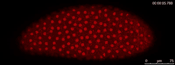
The dividing red spots in this time-lapse video belong to a busily developing fruit fly embryo. A fruit fly egg can divide into some 6,000 cells in just two hours —  faster division than cancer tumors. To watch them action, graduate student Victoria Deneke and assistant professor Stefano Di Talia tagged the nuclei with a protein that glows red. In a recent study, they show that the cells coordinate their rapid divisions via waves of protein activity that spread across the embryo. The waves help ensure that all the cells enter the next stage of development at the same time.

Duke graduate student Victoria Deneke has been awarded an international student research fellowship from the Howard Hughes Medical Institute.

Duke graduate student Victoria Deneke has been awarded an international student research fellowship from the Howard Hughes Medical Institute.

Starting September 2016, Deneke became one of 20 graduate students from 14 countries selected for an international student research fellowship from the Howard Hughes Medical Institute.

Three-year fellowship is designed to support outstanding international graduate students studying in the United States who are ineligible for fellowships or training grants through U.S. federal agencies.

Born in El Salvador, Deneke earned her undergraduate degree in chemical engineering from the University of Notre Dame before joining Stefano Di Talia’s at Duke in 2013.

Fellows must be nominated by their institution; participation is by invitation only. Deneke is only the second student at Duke to receive an HHMI International Student Research Fellowship since the program was established in 2011.

CITATION:  “Waves of Cdk1 Activity in S Phase Synchronize the Cell Cycle in Drosophila Embryos,” Victoria Deneke, Anna Melbinger, Massimo Vergassola and Stefano Di Talia. Developmental Cell, August 2016. http://dx.doi.org/10.1016/j.devcel.2016.07.023

Turning Duke Experiences into Science Fair Gold

Do we each have our own story about science fair? Mine is about that time my grandpa and I set fire to my parents’ garage while testing out the new corn stove we had built together. We were looking into cleaner fuels. It was a small fire, easily squelched, fortunately.

Katherine Yang presenting her poster

Katherine Yang presenting her poster

But in the rite of passage that is the science fair, two Duke-mentored high schoolers are not embarking on half-baked projects with non-scientific relatives like mine, but are instead blazing new trails in science with all of the high-end equipment and faculty mentoring that Duke has to offer.

Katherine Yang and Alisa Cui, of the North Carolina School of Science and Mathematics in Durham, are presenting their results in Phoenix this week in Intel’s International Science and Engineering Fair (ISEF), a prestigious annual science fair that convenes 1,700 of the best and brightest STEM students from around the world

Working in Qiu Wang’s group, Yang has discovered a potential new drug to treat cancer, focusing on a protein targeted called CARM1, which is known to cause breast and prostate cancers to grow uncontrollably.

Yang’s new molecule blocks CARM1. What’s more, in the process of narrowing her list of five candidates, she developed a new cell-based test that can inform the development of future screening tools for other CARM1 inhibitors.

Cui has worked in Jorg Grandl’s lab on the mechanism by which a family of proteins called Piezo ion channels allow cells to detect mechanical touch and eventually become desensitized to repeated stimulation and shut off. By recording the electrical activity of cells that express one type of Piezo, Cui determined that the channels do not use a particular type of shutdown mechanism that researchers had previously thought. Now, the group will move on to test another major mechanism.

NCSSM_Alisa Cui

Alisa Cui and her award-winning project.

On Friday, it was announced that Alisa had won a fourth place grand award in Cellular and Molecular Biology, which includes a $500 prize.

“I am very impressed by the impact Alisa made,” said Grandl, who is a member of the Duke Institute for Brain Sciences. “The data she collected helped starting a completely new line of research,” in understanding how these channels deal with repeated stimulations, such as vibrations.

Growing up, I was oblivious to the existence of international science fairs but my own experiences ignited a lifelong love for science. I can only hope that these young ladies felt something similar.

KellyRae_Chi_100Guest Post by Kelly Rae Chi

The Future of 3D Printing in Medicine

While 3D printers were once huge, expensive devices only available to the industrial elite, they have rapidly gained popularity over the last decade with everyday consumers. I enjoy printing a myriad of objects at the Duke Colab ranging from the Elder Wand to laptop stands.

One of the most important recent applications of 3D printing is in the medical industry. Customized implants and prosthetics, medical models and equipment, and synthetic skin are just a few of the prints that have begun to revolutionize health care.

3D printed prosthetic leg: “customizable, affordable and beautiful.”

Katie Albanese is a student in the Medical Physics Graduate Program who has been 3D printing breasts, abdominal skeletons, and lungs to test the coherent scatter x-ray imaging system she developed. Over spring break, I had the opportunity to talk with Katie about her work and experience. She uses the scatter x-ray imaging system to identify the different kinds of tissue, including tumors, within the breast. When she isn’t busy printing 3D human-sized breasts to determine if the system works within the confines of normal breast geometries, Katie enjoys tennis, running, napping and watching documentaries in her spare time. Below is the transcript of the interview.

How did you get interested in your project?

When I came to Duke in 2014, I had no idea what research lab I wanted to join within the Medical Physics program. After hearing a lot of research talks from faculty within my program, I ultimately chose my lab based on how well I got along with my current advisor, Anuj Kapadia in the Radiology department. He had an x-ray project in the works with the hope of using coherent scatter in tissue imaging, but the system had yet to be used on human-sized objects.

Could you tell me more about the scatter x-ray imaging system you’ve developed?

Normally, scatter in a medical image is actively removed because it doesn’t contribute to diagnostic image quality in conventional x-ray. However, due to the unique inter-atomic spacing of every material – and Bragg’s law – every material has a unique scatter signature. So, using the scattered radiation from a sample (instead of the primary x-ray beam that is transmitted through the sample), we can identify the inter-atomic spacing of that material and trace that back to what the material actually is to a library of known inter-atomic spacings.

Bragg diffraction: Two beams with identical wavelength and phase approach a crystalline solid and are scattered off two different atoms within it.

How do you use this method with the 3D printed body parts?

One of the first things we did with the system was see if it could identify the different types of human tissue (ex. fat, muscle, tumor). The breast has all of these tissues within a relatively small piece of anatomy, so that is where the focus began. We were able to show that the system could discern different tissue types within a small sample, such as a piece of excised human tissue. However, in order to use any system in-vivo, which is ideally the aim, you have to determine whether or not it works on a normal human geometry. Another professor in our department built a dedicated breast CT system, so we used patient scans from that machine to model and print an accurate breast, both in anatomy and physical size.


What are the three biggest benefits of the x-ray imaging system for future research? 

Main breast phantom used and a mammogram of that phantom with tissue samples in it

Main breast phantom used and a mammogram of that phantom with tissue samples in it

Coherent scatter imaging is gaining momentum as an imaging field. At the SPIE Medical Imaging Conference a few weeks ago in San Diego, there was a dedicated section on the use of scatter imaging (and our group had 3 out of 5 talks on the topic!). One major benefit is that it is noninvasive. There is always a need for a noninvasive diagnostic step in the medical field. One thing we foresee this technology being used for could be a replacement for certain biopsy procedures. For instance, if a radiologist finds something suspicious in a mammogram, a repeat scan of that area could be taken on a scatter imaging system to determine whether or not the suspicious lesion is malignant or not. It has the potential to reduce the number of unnecessary invasive (and painful!) biopsies done in cancer diagnosis.

Another thing we envision, and work has been done on this in our group, is using this imaging technique for intra-operative margin detection. When a patient gets a lumpectomy or mastectomy, the excised tissue is sent to pathology to make sure all the cancer has been removed from the patient. This is done by assessing whether or not there is cancer on the outer margins of the sample and can often take several days. If there is cancerous tissue in the margin, then it is likely that the extent of the cancer was not removed from the patient and a repeat surgery is required. Our imaging system has the potential to scan the entirety of the tissue sample while the patient is still open in the operating room. With further refinement of system parameters and scanning technique, this could be a reality and help to prevent additional surgeries and the complications that could arise from that.

What was the hardest or most frustrating part of working on the project? 

We use a coded aperture within the x-ray beam, which is basically a mask that allows us to have a depth-resolved image. The aperture is what tells us where the source of the scatter came from so that we can reconstruct. The location of this aperture relative to the other apparatus within our setup is carefully calibrated, down to the sub-millimeter range. If any part of the system is moved, everything must be recalibrated within the code, which is very time-consuming and frustrating. So basically every time we wanted to move something in our setup to make things better or more efficient, it was like we were redesigning the system from scratch.

 What is your workspace like?

Katie and the team at the AAPM (American Association of Physicists in Medicine) conference from this past summer in Anaheim, CA where she presented in a special session on breast imaging. From left to right: Robert Morris (also in the research lab and getting his degree in MedPhys), Katie, Dr. James Dobbins III (former program director and current Associate Vice Provost for DKU) and Dr. Anuj Kapadia, my advisor and current director of graduate studies in the program

Katie presented in a special session on breast imaging at the American Association of Physicists in Medicine conference this past summer in Anaheim, CA. From left to right: Robert Morris, also working in the lab; Katie; Dr. James Dobbins III, former program director and current Associate Vice Provost for Duke-Kunshan University; and Dr. Anuj Kapadia, Katie’s advisor and current director of graduate studies.

We have a working experimental lab within the hospital. It looks like any other physics lab you might come across- messy, full of wires and strange electronics. It is unique from other labs within the Medical Physics department because a lot of research that is done there focuses on image processing or radiation therapy treatment planning and can be done on just a computer. This lab is very hands-on in that we need to engineer the system ourselves. It is not uncommon for us to be using power tools or soldering or welding.

What do you like best about 3D printing? 

3D printing has become such a great community for creativity. One of my favorite websites now, called Thingiverse, is basically a haven for 3D printable files of anything you could ever dream of, with comments on the best printing settings, printers and inks. You can really print anything you want — I’ve printed everything from breasts, lungs and spines to small animal models and even Harry Potter memorabilia to add to my collection. If you can dream it, you can print it in three dimensions, and I think that’s amazing.


Anika_RD_hed100_2By Anika Radiya-Dixit


Why Testing Lemur Color Vision is Harder Than it Looks

Elphaba the aye-aye is not an early riser. A nocturnal primate with oversized ears, bulging eyes and long, bony fingers, she looks like the bushy-tailed love child of a bat and an opossum.

She would much rather sleep in than participate in Duke alum Joe Sullivan’s early morning vision tests.

“I can’t blame her,” said Sullivan, who graduated from Duke in 2015.

Elphaba is one of 14 aye-ayes at the Duke Lemur Center in Durham, North Carolina, where researchers like Sullivan have been trying to figure out if these rare lemurs can tell certain colors apart, particularly at night when aye-ayes are most active. But as their experiments show, testing an aye-aye’s eyesight is easier said than done.

Elphaba the aye-aye takes a vision test at the Duke Lemur Center in Durham, North Carolina. She’s getting encouragement from student researcher Joe Sullivan and technician Jennifer Templeton. Photo by David Haring.

Elphaba the aye-aye takes a vision test at the Duke Lemur Center in Durham, North Carolina. She’s getting encouragement from student researcher Joe Sullivan and technician Jennifer Templeton. Photo by David Haring.

Aye-ayes don’t see colors as well as humans do. While we have genes for three types of color-sensing proteins in our eyes, aye-ayes and most other mammals have two, one tuned to blue-violet light and another that responds to green.

In all animals, the eyes’ color-detecting machinery depends on medium to bright light. In a version of “use it or lose it,” the genes responsible for color vision in some nocturnal species have decayed over time, such that they see the world in black and white.

But in aye-ayes, research shows, the genes for seeing colors remain intact, and scientists at Duke and elsewhere are trying to understand why.

One possibility is the aye-aye’s color vision genes are mere leftovers, relics passed down from daylight-loving ancestors and no longer useful to aye-ayes today.

Or, the genes may have been preserved because color vision gives aye-ayes an edge. Wild aye-ayes live by eating fruit, nuts, nectar and grubs in the rainforests of Madagascar. Wouldn’t an animal that could distinguish the blue fruits of a favorite snack like the Traveler’s palm from the green of the surrounding foliage have an advantage?

Understanding what aye-ayes can see is no easy feat. One of the most common tests for colorblindness, the Ishihara, requires the subject to recognize and identify numbers hidden within a patch of colored dots of different sizes and brightness.

Aye-ayes don’t read numbers, so Sullivan tests for color vision using food and colored cards.

The first tests were simple enough. In a dimly lit enclosure, a trainer held up two cards: a white card and a black one.

Each time the aye-ayes chose the white card over the black one by reaching out and touching it with their hand, the animal got a peanut.

Even animals with no color vision can tell white from black, so Sullivan was confident they’d ace the test. But aye-ayes aren’t programmed to please. Just getting them to sit still, instead of running around their enclosure, was a challenge.

One aye-aye, 29-year-old Ozma who was born in the wild in Madagascar, never got the hang of even the most basic task, a warmup involving a single white card.

“That’s when I realized that aye-ayes don’t always play by my rules,” said Sullivan, who started working at the Duke Lemur Center as an undergraduate research intern in 2012.

After four months and 200 trials, all five of the aye-ayes in Sullivan’s study started picking the white card more often than not, with Merlin, Elphaba and Grendel passing the test at least 70 percent of the time.

Norman and Ardrey tended to reach for the card on their left, no matter what the color.

Sullivan isn’t giving up. Still working at the Duke Lemur Center post-graduation, now he’s trying to see if aye-ayes can distinguish a purplish card from a green one, in brighter light more similar to dawn or dusk.

So far, Merlin and Grendel are getting it right just over half the time, leaving Sullivan still unsure if the aye-ayes are choosing the cards by their colors or by some other cue.

“I came in thinking that the aye-ayes were going to play nice and do everything I wanted. That was so wrong,” Sullivan said. “Still, they’ve been very good sports.”

How do you give a lemur a vision test? Photo by David Haring, Duke Lemur Center.

How do you give a lemur a vision test? Photo by David Haring, Duke Lemur Center.

Post by Robin A. Smith Robin Smith

3-D Movies of Life at Nanoscale Named Best Science Paper of the Year

If you’ve ever wanted to watch a killer T cell in action, or see human cancer make new cells up-close, now is your chance.

A collection of 3-D movies captured by Duke biology professor Dan Kiehart and colleagues has won the 2015 Newcomb Cleveland Prize for most outstanding paper in the journal Science.

The paper uses a new imaging technique called lattice light-sheet microscopy to make super high-resolution three-dimensional movies of living things ranging from single cells to developing worm and fly embryos.

https://www.youtube.com/watch?v=fwzIUnKNw0s

Cutting-edge microscopes available on many campuses today allow researchers to take one or two images a second. But the lattice light-sheet microscope, co-developed by 2014 Nobel Prize winner Eric Betzig, lets researchers take more than 50 images a second, and in the specimen’s natural state, without smooshing it under a cover slip.

You can watch slender antennae called filopodia extend from the surface of a human cancer cell, or tiny rods called microtubules, several thousand times finer than a human hair, growing and shrinking inside a slide mold.

Daniel Kiehart and former Duke postdoctoral fellow Serdar Tulu made a video of the back side of a fruit fly embryo during a crucial step in its development into a larva.

Chosen from among nominations submitted by readers of Science, the paper has been viewed more than 20,000 times since it was first published on October 24, 2014.

The award was announced on February 12, 2016, at an award ceremony held during the annual meeting of the American Association for the Advancement of Science (AAAS) in Washington, D.C.

Winners received a commemorative plaque and $25,000, to be shared among the paper’s lead authors Eric Betzig, Bi-Chang Chen, Wesley Legant and Kai Wang of Janelia Farm Research Campus.

Read more: “Lattice light-sheet microscopy: Imaging molecules to embryos at high spatiotemporal resolution,” Chen, B.-C., et al. Science, October 2014. DOI:10.1126/science.1257998


Post by Robin A. Smith Robin Smith


One Small Worm, One Duke Senior, and One Big Conference

Duke senior Grace Lim isn’t grossed out by the innards of the tiny worm C. elegans. In fact, she finds them beautiful.

As a researcher in the David Sherwood Lab, she peers inside the transparent 1-millimeter creature under a microscope, watching for “cell invasion” — a process that occurs when one type of cell literally bursts into an area occupied by another type of cell.

Version 2

Grace Lim presenting the results of her research at the AAAS Annual Meeting on Saturday.

Last weekend, the aspiring developmental biologist had the opportunity to take her work to the national stage when she presented at the Student Poster Competition as part of the annual AAAS meeting in Washington, D.C.

“It’s been really exciting,” said Lim. “The researchers here are experts and it is great to learn about their projects. At the same time, I’ve met scientists from all different fields who have asked questions and provided insights that I didn’t expect.”

Cell invasion plays a key role in organism growth and development, Lim said. For example, a fertilized egg will use cell invasion to implant itself into the uterine wall. However, cell invasion can also occur in less desirable processes, like cancer and other diseases.

In her work, Lim created C. elegans mutants that lacked specific genes related to cell invasion. She then observed whether uterine cells in the growing mutants could still invade tissue in the vulva — a key milestone in the growth of the developing larva.

C. elegans is a good system to study because it is transparent, so you can watch these biological processes happening under a microscope,” she said.

C_elegans

The tiny transparent C. elegans. Photo courtesy of the National Human Genome Research Institute

Her experiments uncovered four new genes that appear to regulate cell invasion in C. elegans. In addition to presenting at the conference, Lim will also be writing up these results as an honors thesis.

Lim, who wants to pursue a graduate degree in biology after finishing up at Duke, says her favorite part of working in the Sherwood lab has been interacting with the graduate students. “We work together to come up with creative ways to solve problems, which is something you don’t always get to do in class,” she said.

And her favorite part of working with C. elegans?

“They have this amazing ability to control their metabolism,” she said. “We grow these worms in petri dishes, and when the plate fills up and they run of out food, they just stop growing. But if you take a few and put them on a new plate they grow again, as if nothing had happened.”

Post by Kara Manke

Kara J. Manke, PhD

Page 18 of 32

Powered by WordPress & Theme by Anders Norén