Following the people and events that make up the research community at Duke

Students exploring the Innovation Co-Lab

Tag: microbiome

What is it like to Direct a Large, Externally-Funded Research Center?

What are the trials and tribulations one can expect? And conversely, what are the highlights? To answer these questions, Duke Research & Innovation Week kicked off with a panel discussion on Monday, January 23.

The panel

Moderated by George A. Truskey, Ph.D, the Associate Vice President for Research & Innovation and a professor in the Department of Biomedical Engineering, the panelists included…

  • Claudia K. Gunsch, Ph.D., a professor in the Departments of Civil & Environmental Engineering, Biomedical Engineering, and Environmental Science & Policy. Dr. Gunsch is the director of the NSF Engineering Research Center for Microbiome Engineering (PreMiEr) and is also the Associate Dean for Duke Engineering Research & Infrastructure.
Dr. Claudia Gunsch
  • Yiran Chen, Ph.D., a professor in the Department of Electrical & Computer Engineering. Dr. Chen is the director of the NSF AI Institute for Edge Computing (Athena).
Dr. Yiran Chen
  • Stephen Craig, Ph.D., a professor in the Department of Chemistry. Dr. Craig is the director of the Center for the Chemistry of Molecularly Optimized Networks (MONET).
Dr. Stephen Craig

The centers

As the panelists joked, a catchy acronym for a research center is almost an unspoken requirement. Case in point: PreMiEr, Athena, and MONET were the centers discussed on Monday. As evidenced by the diversity of research explored by the three centers, large externally-funded centers run the gamut of academic fields.

PreMiEr, which is led by Gunsch, is looking to answer the question of microbiome acquisition. Globally, inflammatory diseases are connected to the microbiome, and studies suggest that our built environment is the problem, given that Americans spend on average less than 8% of time outdoors. It’s atypical for an Engineering Research Center (ERC) to be concentrated in one state but uniquely, PreMieR is. The center is a joint venture between Duke University, North Carolina A&T State University, North Carolina State University, the University of North Carolina – Chapel Hill and the University of North Carolina – Charlotte.

PreMiEr – not to be confused with the English Premier League

Dr. Chen’s Athena is the first funded AI institute for edge computing. Edge computing is all about improving a computer’s ability to process data faster and at greater volumes by processing data closer to where it’s being generated. AI is a relatively new branch of research, but it is growing in prevalence and in funding. In 2020, 7 institutes looking at AI were funded by the National Science Foundation (NSF), with total funding equaling 140 million. By 2021, 11 institutes were funded at 220 million – including Athena. All of these institutes span over 48 U.S states.

Athena, or the Greek goddess of wisdom, is a fitting name for a research center

MONET is innovating in polymer chemistry with Stephen Craig leading. Conceptualizing polymers as operating in a network, the center aims to connect the behaviors of a single chemical molecule in that network to the  behavior of the network as a whole. The goal of the center is to transform polymer and materials chemistry by “developing the knowledge and methods to enable molecular-level, chemical control of polymer network properties for the betterment of humankind.” The center has nine partner institutions in the U.S and one internationally.

MONET, like French painter Claude Monet

Key takeaways

Research that matters

Dr. Gunsch talked at length about how PreMiEr aspires to pursue convergent research. She describes this as identifying a large, societal challenge, then determining what individual fields can “converge” to solve the problem.

Because these centers aspire to solve large, societal problems, market research and industry involvement is common and often required in the form of an industry advisory group. At PreMiEr, the advisory group performs market analyses to assess the relevance and importance of their research. Dr. Chen also remarked that there is an advisory group at Athena, and in addition to academic institutions the center also boasts collaborators in the form of companies like Microsoft, Motorola, and AT&T.

Dr. Chen presenting on Athena’s partner institutions at Monday’s talk.

Commonalities in structure

Most research centers, like PreMiEr, Athena, and MONET, organize their work around pillars or “thrusts.” This can help to make research goals understandable to a lay audience but also clarifies the purpose of these centers to the NSF, other funding bodies, host and collaborating institutions, and the researchers themselves.

How exactly these goals are organized and presented is up to the center in question. For example, MONET conceptualizes its vision into three fronts – “fundamental chemical advances,” “conceptual advances,” and “technological advances.”

At Athena, the research is organized into four “thrusts” – “AI for Edge Computing,” “AI-Powered Computer Systems,” “AI-Powered Networking Systems,” and “AI-Enabled Services and Applications.”

Meanwhile, at PreMiEr, the three “thrusts” have a more procedural slant. The first “thrust” is “Measure,” involving the development of tracking tools and the exploration of microbial “dark matter.” Then there’s “Modify,” or the modification of target delivery methods based on measurements. Finally, “Modeling” involves predictive microbiome monitoring to generate models that can help analyze built environment microbiomes.  

A center is about the people  

“Collaborators who change what you can do are a gift. Collaborators who change how you think are a blessing.”

Dr. stephen craig

All three panelists emphasized that their centers would be nowhere without the people that make the work possible. But of course, humans complicate every equation, and when working with a team, it is important to anticipate and address tensions that may arise.

Dr. Craig spoke to the fact that successful people are also busy people, so what may be one person’s highest priority may not necessarily be another person’s priority. This makes it important to assemble a team of researchers that are united in a common vision. But, if you choose wisely, it’s worth it. As Dr. Craig quipped on one of his slides, “Collaborators who change what you can do are a gift. Collaborators who change how you think are a blessing.”

In academia, there is a loud push for diversity, and research centers are no exception. Dr. Chen spoke about Athena’s goals to continue to increase their proportions of female and underrepresented minority (URM) researchers. At PreMiEr, comprised of 42 scholars, the ratio of non-URM to URM researchers is 83-17, and the ratio of male to female researchers is approximately 50-50.

In conclusion, cutting-edge research is often equal parts thrilling and mundane, as the realities of applying for funding, organizing manpower, pushing through failures, and working out tensions with others sets in. But the opportunity to receive funding in order to start and run an externally-funded center is the chance to put together some of the brightest minds to solve some of the most pressing problems the world faces. And this imperative is summarized well by the words of Dr. Craig: “Remember: if you get it, you have to do it!”

Post by Megna Datta, Class of 2023

Rewilding the Gut

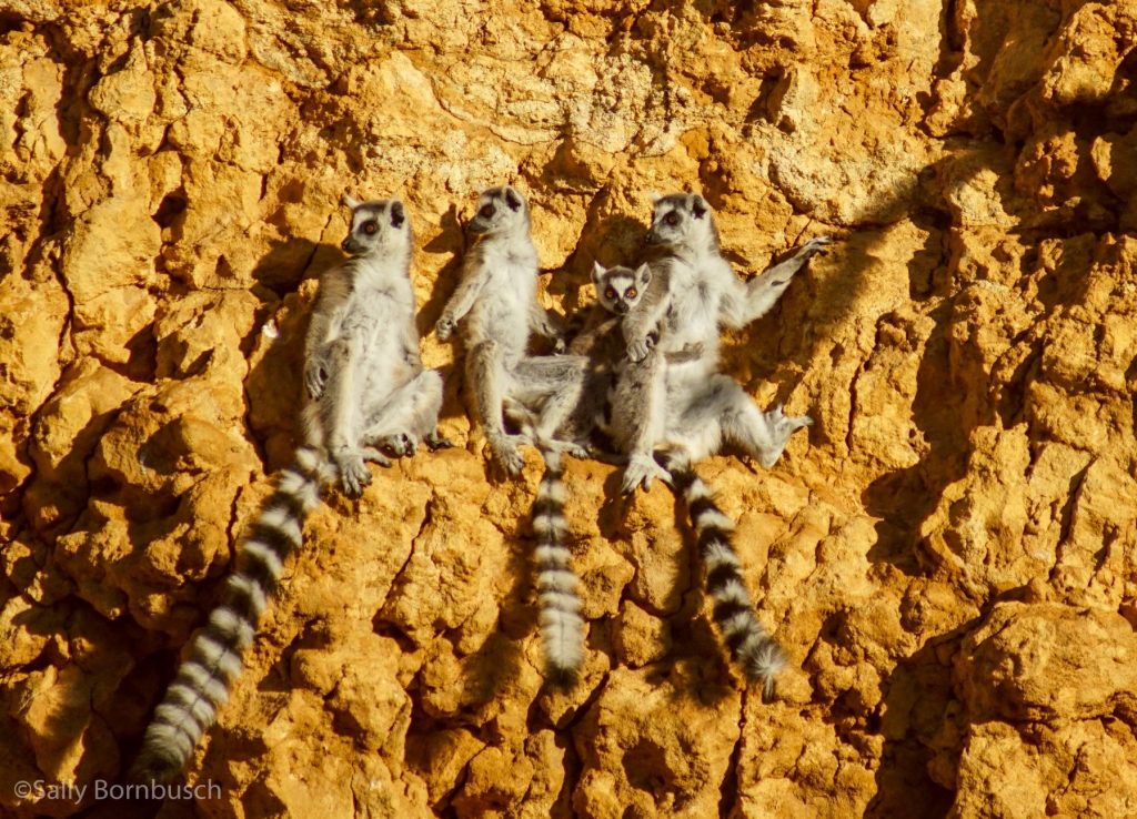
Processed foods and overuse of antibiotics can wreak havoc on the trillions of bacteria and other microbes that inhabit the gut. A new study of the gut microbiomes of lemurs looks at whether reconnecting with nature can help restore this internal ecosystem to a more natural state. Credit: Sally Bornbusch.

Modern life messes with the microbiome -– the trillions of bacteria and other microbes that live inside the body. Could reconnecting with nature bring this internal ecosystem back into balance?

A new study suggests it can, at least in lemurs. Led by Duke Ph.D. alumnus Sally Bornbusch and her graduate advisor Christine Drea, the research team collected fecal samples from more than 170 ring-tailed lemurs living in various conditions in Madagascar: some were living in the wild, some were kept as pets, and some were rescued from the pet and tourism industries and then relocated to a rescue center in southwestern Madagascar where they ate a more natural diet and had less exposure to people.

Collecting fecal samples in Madagascar

Then the researchers sequenced DNA from the fecal samples to identify their microbial makeup. They found that the longer lemurs lived at the rescue center, the more similar their gut microbes were to those of their wild counterparts. Former pet lemurs with more time at the rescue center also showed fewer signs of antibiotic resistance.

By “rewilding” the guts of captive animals, researchers say we may be able to better prime them for success, whether after rescue or before translocation or reintroduction into the wild.

This research was supported by grants from the National Science Foundation (1945776, 1749465), the Triangle Center for Evolutionary Medicine, Duke’s Kenan Institute for Ethics, the Margot Marsh Biodiversity Fund and Lemur Love.

CITATION: “Microbial Rewilding in the Gut Microbiomes of Captive Ring-Tailed Lemurs (Lemur catta) in Madagascar,” Sally L. Bornbusch, Tara A. Clarke, Sylvia Hobilalaina, Honore Soatata Reseva, Marni LaFleur & Christine M. Drea. Scientific Reports, Dec. 27, 2022. DOI: 10.1038/s41598-022-26861-0.

Robin Smith
By Robin Smith

Lemur Gut Isn’t One Ecosystem, It’s Many

Lemurs like Ferdinand are leaf-eating machines, says Duke microbiome researcher Lydia Greene. Credit: Lydia Greene

DURHAM, N.C. — A jungle. A rainforest. A wetland. A wilderness. Researchers have used various metaphors to describe the complex, interconnected community of microbes (most of them bacteria) living inside your body, and all over it too.

If you were to count up all the trillions of cells inside and out, we are more bacteria than human. Fortunately, perhaps, the microbes that make a cozy home inside your nose, or clinging to your teeth, aren’t the same ones that are living behind your ear or busily multiplying in your belly button.

The same is true for our distant primate cousins the lemurs, particularly in their guts, say researchers Lydia Greene and Erin McKenney in a new study.

Lemurs rely on gut microbes to digest their leafy diets, explains Greene, a research scientist at the Duke Lemur Center. Microbes in lemurs’ GI tracts help ferment plant fiber, detoxify plant chemical defenses, and synthesize vitamins and nutrients that lemurs can’t make for themselves. Our own gut bugs do many of the same things for us.

Greene and McKenney study how lemur gut bacteria are shaped by what lemurs eat, how they evolved and the complexity of the route microbes travel through the body. They hope to better understand how these microorganisms keep lemurs healthy, or — when they’re out of balance — make them sick.

Researchers who do this kind of work spend a lot of time collecting poop. For good reasons, says McKenney, an assistant professor at North Carolina State University. Scientists can learn about lemurs by what they leave behind, and poop can be collected repeatedly without harming the animals. But for this study the team tried something different, made possible by a one-of-a-kind biobank:

When an animal dies at the Duke Lemur Center, veterinary staff determine the cause of death, and blood and tissue samples that might be important for research or education are collected and preserved.

Today, the collection holds thousands of samples, collected over decades from more than two dozen species of rare and endangered primates, which the center stores in super cold freezers in their headquarters in North Carolina. It’s a frozen ark maintained at up to minus 80 degrees Celsius, with redundant backup power.

In the event that any one of these species goes extinct, and the last living parts of them are gone, future generations will still be able to study the genetic and other information they left behind.

Using this bank, the team sampled several sites in the bowels of 52 deceased lemurs, including dwarf lemurs, aye-ayes, ruffed lemurs, bamboo lemurs, brown lemurs, ring-tailed lemurs and sifakas.

A trip through a lemur’s GI tract is a journey through a varied landscape. The long, twisting route from the stomach through the small intestine to the colon serves numerous functions, filtering, digesting, absorbing, detoxifying, fermenting.

Not all lemur guts work the same: Fruit-eaters, like ruffed lemurs, generally have short, simple guts. If you stretched them out, they’d be five times their body length — not much shorter than ours, relative to body size. Leaf-eaters like sifakas have more complex GI tracts with relatively longer colons and a leaf-fermentation pouch called a cecum. Their guts are the lemur champions — up to 16 times body length.

Having whole lemurs to study instead of just poo enabled the researchers to sample different regions of the gut to figure out what kinds of microbes were present in each spot. They used genetic sequencing technology to identify microbes and compare their relative abundance in different sites.

Sampling along the digestive tract, they found that different spots along this long, twisting pathway have their own communities of bacteria doing different kinds of jobs. The complex ecosystem lurking in a lemur’s small intestine, for example, isn’t the same as the microbial menagerie setting up camp in their colon.

Levels of biodiversity varied too. The stomach supports less microbial life because fewer species can tolerate its acidic digestive juices. But if the upper regions of the gut are a garden, the lower regions are more like a tropical rainforest. About two dozen types of bacteria were more abundant in the cecum and colon than elsewhere. Lemurs with relatively longer lower guts host the richest microbiomes, to better ferment high-fiber foods.

“We probably couldn’t have detected these relationships without such an extensive comparative dataset,” McKenney said.

“This kind of lemur research can really only be done at the Duke Lemur Center,” Greene said.

Rodelinda, a Coquerel’s sifaka lemur, munches on leaves at the Duke Lemur Center. Credit: Lydia Greene.

This research was supported by the National Science Foundation (DBI PRFB 1906416) and by a Duke Lemur Center Director’s fund.

CITATION: “Gut Site and Gut Morphology Predict Microbiome Structure and Function in Ecologically Diverse Lemurs,” Lydia K. Greene, Erin A. McKenney, William Gasper, Claudia Wrampelmeier, Shivdeep Hayer, Erin E. Ehmke, & Jonathan B. Clayton. Microbial Ecology, May 14, 2022. DOI: 10.1007/s00248-022-02034-4

When the gut’s internal ecosystem goes awry, could an ancient if gross-sounding treatment make it right?

Lemur researchers make a case for fecal transplants to reduce the side effects of antibiotics. Photo by David Haring, Duke Lemur Center.

Dr. Cathy Williams knew something wasn’t right. The veterinarian had felt off for weeks after her 2014 trip to Madagascar.

At first she just felt bloated and uncomfortable and wasn’t interested in eating much. But eventually she developed a fever and chills that sent her to the emergency room.

When tested, doctors found that what she had wasn’t just a stomach bug. She was suffering from an infection of Clostridium difficile, a germ that causes severe diarrhea and abdominal pain and can quickly become life-threatening if not treated promptly.

“It was horrible,” Williams said.

The condition is often triggered when antibiotics disrupt the normal balance of bacteria that inhabit the gut, allowing “bad” bacteria such as C. difficile to multiply unchecked and wreak havoc on the intestines.

To get her infection under control, Williams asked her doctors if they could try an approach she and other veterinarians had used for decades to treat lemurs with digestive problems at the Duke Lemur Center. The procedure, known as a fecal microbiota transplant, involves taking stool from a healthy donor and administering it to the patient to add back “good” microbes and reset the gut.

At the time it was considered too experimental for clinical use in human cases like Williams’. She was prescribed the standard treatment and was sent home from the hospital, though she wouldn’t feel well enough to go back to work for another month. But now new research in lemurs is confirming what Williams and others long suspected: that this ancient if gross-sounding treatment can help an off-kilter gut microbiome get back to normal.

In a recent study in the journal Animal Microbiome, a research team led by Duke professor Christine Drea, former PhD student Sally Bornbusch and colleagues looked at the gut microbiomes of 11 healthy ring-tailed lemurs over a four-month period after receiving a seven-day course of the broad-spectrum antibiotic amoxicillin.

The lemurs were split into two experimental groups. One was a wait-and-see group, with continued follow-up but no further treatment after the antibiotics. The other group was given a slurry of their own feces, collected prior to antibiotic treatment and then mixed with saline and fed back to the same animal after their course of antibiotics was over.

“It sounds crazy,” Williams said. But she has used a similar procedure since the 1990s to treat illnesses in Coquerel’s sifaka lemurs, whose infants are known to eat their mother’s poop during weaning — presumably to get the microbes they’ll need to transition to solid food.

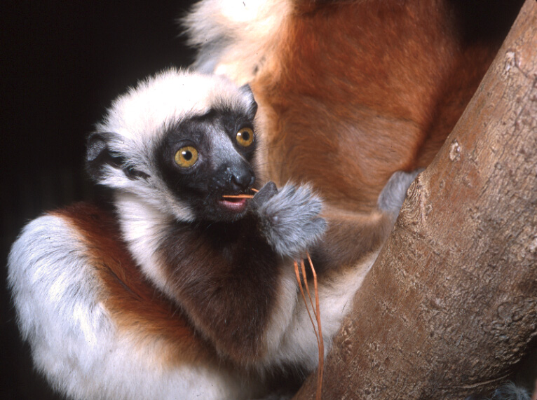
A baby Coquerel’s sifaka tries some of her first solid foods. Photo by David Haring.

Drea, Bornbusch and team used genetic sequencing techniques to track changes in the lemurs’ gut microbiome before, during and after treatment.

As expected, even a single course of antibiotics caused the numbers of microbes in their guts to plunge compared with controls, briefly wiping out species diversity in both experimental groups before returning to baseline.

“Antibiotics had dramatic effects, even in healthy animals,” Drea said.

But in terms of which types of bacteria bounced back and when, the patterns of recovery in the two groups were different. Lemurs that received the “poop soup” treatment started to stabilize and return to their pre-antibiotic microbiome within about two weeks. In contrast, the bacterial composition in the wait-and-see group continued to fluctuate, and still hadn’t quite returned to normal even after four months of observation.

This kind of therapy isn’t new. Reports of using fecal transplants to treat people suffering from food poisoning or diarrhea date back as far as fourth century China. The evidence for its effectiveness in captive settings has Bornbusch advocating for freezing stool at Smithsonian’s National Zoo, where she is now a postdoctoral fellow.

“If we can bank feces from animals when they’re healthy, that can be a huge benefit down the road,” Bornbusch said. “It can help the animals get better, faster.”

And now if any of her lemur patients were to get sick with C. difficile like she did, Williams said, “I would absolutely go with a fecal microbiota transplant.”

“People are put off by it,” Drea said, “But the disgust for this approach might actually have been holding up a fairly cheap and useful cure.”

Ring-tailed lemurs at the Duke Lemur Center in North Carolina. Photo by David Haring, Duke Lemur Center

This research was supported by the National Science Foundation (BCS 1749465), the Duke Lemur Center Director’s Fund, and the Duke Microbiome Center.

CITATION: “Antibiotics and Fecal Transfaunation Differentially Affect Microbiota Recovery, Associations, and Antibiotic Resistance in Lemur Guts,” Sally L. Bornbusch, Rachel L. Harris, Nicholas M. Grebe, Kimberly Roche, Kristin Dimac-Stohl, Christine M. Drea. Animal Microbiome, Oct. 1, 2021. DOI: 10.1186/s42523-021-00126-z.

By Robin Ann Smith

The Duke Dentist and her Research: Saving Children’s Teeth, One Tooth at a Time

Walking into our small meeting room with green scrubs and a white lab coat on, our special guest set her bag down in the front and stated “I fixed 60 teeth today and haven’t sat down since this morning.” To us, it sounds like a nightmare, but to Dr. Martha Ann Keels, working in her clinic and conducting dental research is a dream come true. 

Born and raised in North Carolina, Dr. Keels has kept her roots as she studied here at Duke. As a Duke undergrad, she received her bachelor’s degree in Chemistry and a minor in Art History, later choosing to become a pediatric dentist at UNC. It wasn’t long until she returned back to Duke to volunteer at Duke’s Children Hospital, and in 1986, she became the first pediatric dentist to get privileges to practice at Duke. She continues to run her own clinical practice alongside Duke Health System to this day, working for over 30 years!

“I get to feel the satisfaction that something I used my hands for helped alleviate pain in children,” Keels said. “I also get to watch them grow as they come in over the years. It feels super rewarding.”

With her passion and dedication, not only does she help those that enter her office, but she also conducts research on the side, wanting to help dentists all over.

Dr. Keels currently has her hands dirty with a major research project she has been working on for the past nine years. According to the National Institute of Dental and Craniofacial Research, 42% of children between the ages of two to eleven years old have at least one cavity in their primary teeth, and 23% of those children are untreated. With how high these numbers are, she and a group of other researchers are trying to develop tools that allow pediatricians and pediatric dentists to be able to identify high risk factors of cavities in children and care for them before they do occur; tools like questionnaires, surveys, and ‘top 5 predictors…’.

Table of percentages of children with cavities corresponding to age, sex, race, and poverty (National Institute of Dental and Craniofacial Research)

By observing a group of 1,300 children ever since birth, they have been analyzing all aspects of each child: collecting saliva, looking at biofilm (more commonly known as plaque), physical deformities in their teeth, and even social factors like parents’ dental experience. 

Despite the children still being fairly young, Dr. Keels reveals that a surprising amount of information has been found. “No one has ever looked at tight teeth– when your teeth are closely spaced– but we are seeing that it puts a child at high risk of cavities,” Keels said. She also adds that they have also begun to identify which types of bacteria help with reducing chances of getting a cavity, as well as bacteria that bring a high risk of creating a cavity.

 This also goes hand in hand with the microbiomes in our mouths. Dentists first believed that the microbiomes of the child’s caregiver affected the child’s microbiome, in the sense that their microbiomes would be similar from the beginning. Dr. Keels’s study says otherwise. It’s being shown that a child’s microbiome starts off as its own, unique microbiome, and it is over time that it begins to become similar to their caregiver’s microbiome.

With the vast amount of information already collected, Dr. Keels and her team continue to persevere, now wanting to push the study for another five more years. They want to start working with adolescents, wanting to also analyze mental states and how that might affect their dental hygiene and risks of cavities. 

Maybe in the near future, as you speak to your dentist at your next appointment, and they bring up a list of risk factors for cavities, who knows? That list or table could be coming from the one and only Dr. Martha Ann Keels.

Post by Camila Cordero, Class of 2025

Powered by WordPress & Theme by Anders Norén