Following the people and events that make up the research community at Duke

Students exploring the Innovation Co-Lab

Category: Medicine Page 1 of 25

Blowing the Whistle: Bioethics’ Role in Medicine

Sticky post

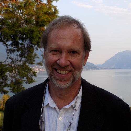
Before introducing the theme of his April 22 talk on abuses in medical research, University of Minnesota philosophy professor Dr. Carl Elliott discussed a literary device in detail. “Man-in-a-hole,” coined by the acclaimed American novelist Kurt Vonnegut, describes a story structure in which the main character, upon facing adversity, plunges into crisis yet overcomes this challenge at the finale. For instance, the famous works “Alice in Wonderland” and “The Wizard of Oz” are good examples of this literary device.  

Dr. Carl Elliott, professor of philosophy at the University of Minnesota Twin Cities

How does this connect to bioethics?  

Before answering this question, Elliott dove into his testimony investigating the case of Dan Markingson at the University of Minnesota. Almost twenty years ago, the university’s Department of Psychiatry recruited Markingson, a young man suffering from psychosis, to take part in one of its drug studies. As Markingson could not independently provide consent, his physician enrolled him in the study against the strong dissent of his mother, Mary Weiss. Despite countless emails, phone calls, and appeals to university staff and researchers, the study continued, culminating in Markingson’s suicide.   

Denial: Why UMN Needs A Mental Health Ombudsman
Dan Markingson with his mother, Mary Weiss

Afterward, upon hearing about this case, Elliott decided to thoroughly investigate and look into potential abuses and conflicts of interest. It was a long, strenuous process that took upwards of six to seven years, and in 2015, vindication was finally delivered via a shell-shocking report on conflicts of interest, clinical trial mismanagement, and other injustices.   

Was justice served?  Unfortunately, quite the contrary. “It was a failure,” Elliott remarked, adding that the report did not amount to anything substantive. “There was no reform, no added accountability, and no payments to Weiss [Markingson’s mother].”   

Tracing back on his introduction using the literary analogy, Elliott demonstrated to his audience that whistleblowers’ experiences are supposed to mimic “man-in-the-hole” stories. After doing the “right thing”, the clinical injustices should be stopped, and the perpetrators should be punished accordingly. However, this is rarely the case: not only are whistleblowers outnumbered in their institutions by colleagues unsympathetic to their cause, but they are also targeted by their superiors for speaking out.   

The truth is “doctors and nurses know that patients are being harmed and exploited,” Elliott said, yet “[hospitals] stonewall the press, rarely apologize, wand ostracize, even sue, the whistleblowers.” Hence, it’s fascinating that certain brave individuals are willing to broadcast the truth even in such circumstances: what is a whistleblower’s motivation in speaking out?  

After speaking with numerous whistleblowers, Elliott was amazed by the number of “non-answers” he received. Most interviewees said something along the lines of “I did it because that is the type of man I am,” supporting Elliott’s argument that there is an honor and shame component in their actions. For many whistleblowers, they lived every day pondering “How do [I] maintain [my] self-respect when [I] lost it completely in the entire medical community?”, as described by Elliott. The overwhelming nature of the ongoing evil made most interviewees feel as if they had no choice but to speak out.  

If whistleblowers are so certain that they are doing the right thing, how come nearly half of all whistleblowers in one study considered committing suicide after their act of bravery? In Elliott’s words, whistleblowers are “radioactive,” frowned upon by larger society and hated by former colleagues. They almost certainly will lose their jobs and never be able to practice in the medical profession again. In one sense, “[the whistleblower] is the only one gullible enough to riot against the rigged system.”   

Not only is the whistleblower punished, but the perpetrators are also most often shielded from responsibility, being awarded the highest honors in their fields or institutions right after their exposure by investigative panels. Elliott sequentially described the fate of six malicious researchers, none ever charged with wrongdoing and instead honored by their peers. For Elliott’s personal story, one of the culprits of the Markingson case, the UMN chair of psychiatry at the time, was praised by the university up until his death. On the other hand, Elliott was exiled from the medical school, the number one punishment for doctors and nurses. 

Given the many obstacles confronting whistleblowers, are there any factors helping them rise to the occasion of telling the truth? According to Elliott, one of the strongest factors supporting whistleblowers in their mission is solidarity in rallying for justice. For many of the cases brought up in the talk, three to four medical professionals together sounded the alarm, sharing the burden of societal opposition and supporting each other during the whistleblowing process. “Standing alone is a recipe for self-demolition,” Elliott said.   

Elliott promised a dispiriting talk at the introduction of his lecture — and he delivered. Yet, to an audience member like me, there were many glimpses of hope shining throughout the lecture. Not only was I amazed by the bravery of whistleblowers like Elliott, but I was also inspired by the persevering notion of justice amongst members of our medical community, individuals whom our society respects the most. Against all odds, countless individuals risked their professional lives for goals greater than themselves, an honorable sacrifice.  

This lecture’s theme of bioethics reminds us how robust science and clinical medicine depend upon ethical conduct and a strong foundation in moral code. At Duke, a shining hub of biomedical research, we must continue to rally for bioethics implementation and be cognizant of the humanities’ importance in all that we do. We must support the work of ethicists like Dr. Elliott, empowering the movement toward health equity and justice. 

By Stone Yan, class of 2028

Posters Galore and More: Undergraduate Research in the Spotlight

Sticky post

Biomedical engineering. Climate science. Political science. Global Health. 

While these departments are housed in buildings far from each other on campus, their current research was all spotlighted at the Duke Undergraduate Research Symposium on April 10. Over the course of two hours, 120 budding researchers presented their work in Penn Pavilion, eagerly chatting with peers, faculty, and community members alike.  

Presenters, friends, and community members gathered in Penn Pavilion to hear opening remarks from the Undergraduate Research Support Office’s director.

The level of professionalism was obvious upon a glance at the poster titles. The symposium was strongly represented by the biomedical and health sciences; hence, membrane receptors and transport proteins bearing combinations of letters and numbers populated the majority of posters. However, many projects shared findings unrelated to enzymes or transgenic models: posters investigating “The Undocumented Republican Latino Vote” and “Palimpsest and Identity in Hong Kong” attracted as much attention.  

I began my journey by talking to Eduardo Fadul Chavez, a junior who is researching chemistry. He gave me an invigorating talk on Nedd4 E3 ligase ligands (a field I admittedly have no background knowledge in). What was greatly appealing about his presentation was his focus on the relevance and impact of his team’s work, especially in innovating treatments for Parkinson’s and other cancers. “Yes, we hope to apply our findings from a pharmacological perspective,” Chavez said. “Nedd4 plays an important role in Parkinson’s disease, so figuring out how to inhibit its activity can greatly inform therapeutic development.” 

Chavez’s poster on his work on Nedd4 ligase ligands, which play a role in Parkinson’s disease.

After this inspiring talk aiming to improve health and cure ailments from a biological perspective, I wandered around in search of a project looking into the sociocultural perspective. Walking across the room, I encountered Austin Brown and his colleagues presenting their work with the Help Desk.  

“The Help Desk is a student-led initiative seeking to tackle social determinants of health,” Brown told me. The organization began as a Bass Connections project and has since attracted hundreds of student volunteers. Currently, about 50 volunteers work across three sites: the Duke Hospital Emergency Department, the Duke Endocrinology Clinic, and the Lincoln Community Health Center. “If patients screen positive on the background survey provided, our community resource navigators will provide support by connecting them with local organizations and follow up after the appointment to ensure they are doing well.” 

The Help Desk has reached more than 3,200 patients since its inception, Brown said. Maintaining communication with patients after the initial meeting or appointment can be tricky, however. “Our biggest barrier is retention and keeping the channels of contact open,” Brown said. 

By this point, I was already amazed by the depth and impact of my peers’ research. I was only more impressed when I came across the poster of Millie Evonlah, who presented on the impact of paternal cannabis consumption on adolescent offspring glutamatergic and cholinergic systems. Given the rise in recreational marijuana usage and legalization, there is a great need to study the impact of men’s usage, particularly intermittent usage, on their children’s brain regions regulating immune and cognitive function.  

Evonlah in front of her poster on paternal cannabis exposure effects on adolescent offspring

“We hypothesized that there would be different expression results and responses between male and female offspring,” Evonlah said, “and our findings support this claim.” While there was decreased glutamatergic expression in the CA3 hippocampal region in both male and female offspring, males and females experienced increased acetylcholine expression in different brain regions. Additionally, only the “weekenders,” or animal subjects who partook in intermittent cannabis exposure, imparted significant biological alterations in their offspring’s brains.  

All these presentations were inspiring to listen to, and selecting the few to cover in detail was a difficult task. However, I would be amiss not to mention a few other invigorating presentations I listened to. For instance, Michael Wang, who studied GRK-2 mediated regulation of brain-derived neurotrophic factor in the ischemic heart, and I had a lengthy conversation about daily research activities and cell culturing principles. I also had a spirited discussion with Madeline Morrison and her co-presenters, who talked about their experiences conducting fieldwork in Roatán, Honduras, and their perspectives on the importance of global health research.  

After two hours of interacting with student researchers, it was clear to me that Duke’s research efforts indeed transform, improve, and save lives. The undergraduate student body’s collective desire to pursue scientific discovery, clinical innovation, and sociocultural advancement is a motivating force that cannot be understated.  

In this season, let us all spring into research! 

By Stone Yan, class of 2028

How the Humanities Helped This Duke Grad Become the Doctor She Hoped to Be

Sticky post

“I see you, you see me, I trust you to know me.”

This phrase was one of many that underscored the powerful testimony Dr. Jennifer Hong, Massachusetts General Hospital physician and Duke alumna, shared on March 23 in a talk on West Campus. Invited by Duke’s Humanities in Medicine club, Hong spoke eloquently about the importance of humanities in medicine, leaning on her undergraduate experience as an English/Neuroscience pre-med and her medical training. Her moving stories about patients and her perspective on the American medical system captivated the entire audience.

Dr. Jennifer Hong (‘14), MGM primary care physician and Duke alumna


Hong opened her discussion by underscoring the power of language—and the potential of wielding narrative writing as a “weapon of resistance.” She shared how, as an undergrad, her most memorable, impactful academic lessons were ones in the English realm, where she explored how historical narratives of female physicians revealed the pervasive patriarchy underlying medicine. Hong reflected that the skills required to be an adept doctor are much akin to those needed to write a good essay: one must be able to relate to people and experiences that are very different from one’s own.


To back up her claim, Hong shared a story from her early days of residency. One day, she and her team were overwhelmed by two Code Blues—the highest level of medical alerts—and a rapid response, which she was handling alone. As a slew of medical professionals rushed into the patient’s room, Hong noted that the patient and her partner had no idea what was happening, as they spoke no English. Connecting with the patient’s son over the phone, Hong tried to explain to the best of her ability the situation and her perspective—the patient’s metastatic cancer prognosis would be best approached with comfort care, not more ventilators.


“You’re hurting them. Stop hurting them,” was the son’s short reply.


This was a landmark moment in Hong’s training, one she described as “shell shocking” and “demonstrating to people who want control that they have no control”. In a rapidly evolving environment where so many elements demand physicians’ attention, Hong notes that it’s incredibly easy to act in a way not aligned with one’s personal values. This foreshadowed one of her responses to a later question regarding what exercises or habits prospective medical students should adopt before stepping into graduate education: she suggested that students should periodically check in on their values and how they plan on upholding them.


The captivating lecture continued to include many remarks on the current state of medicine in America. As an aspiring pre-med student, I find it enlightening and disturbing to hear about the unignorable forces at work every day in physician offices and the hospital setting. Hong carefully described how, despite medicine being “the last frontier” where societal wrongs could be remedied and addressed, capitalist institutions, like private equity investors, are forging ahead with standardizing care and prioritizing profit in medical spaces. Yet, she reminded the audience that hope is present and what drives her work are the physician-patient relationships she treasures. She emphasized the presence of humanities education in medical work, highlighting the skills of translating between “medical speak and patient speak”, seeing human dignity in every patient, and telling patients’ case histories as narratives.


I cannot conclude this blog without mentioning the powerful story Hong shared before she finished her remarks. Since her years as a resident, Hong has frequently met with a patient suffering from numerous chronic illnesses. Despite his many struggles, he maintained a lively sense of humor, evoking joy from every small moment. He was regularly hospitalized, sometimes up to months in duration, and he leaned on Hong to tell his story—his illnesses, his past medication history, and his resilience—to his different care teams. This was what drove the “I see you, you see me, I trust you to know me” quote: in his weakest moments, the patient wanted Hong to tell his story instead of telling his own experiences.

The patient passed away last year. Shortly before his passing, he still messaged Hong, sharing pictures of his youth. This patient was surely not the healthiest of Hong’s patients, but he was among the most memorable and impactful. Even as an audience member, I cannot help but root and feel fondness for this patient.


In my humble opinion, what made this lecture so motivational was not Hong’s advice and encouragement to a room full of pre-meds but her skilled recounting of firsthand experiences. Her testimony is proof that the humanities are still critically valuable, especially in medicine, providing food for thought for Duke students.

By Stone Yan, class of 2028

Food, Health, and Environmental Justice Intertwine in Atlanta

Sticky post

Some researchers get hooked into a niche and spend their entire career specializing in it. Others, like Dr. Eri Saikawa, go wherever the research takes them. On February 28, the Emory University professor spoke about her broad experiences in environmental science for Duke’s Integrated Toxicology & Environmental Health Program (ITEHP). Her research journey has hardly been restricted to any one subject–she’s modeled air pollution, investigated plastic burning, and tested alternatives to synthetic fertilizers that release excessive nitrogen. Yet, what I found most interesting was the work that occurred, and subsequent story that unfolded, just a half hour away from Emory in the Westside of Atlanta. 

Within the U.S., areas of high food insecurity are predominantly located in the Southeast. By allowing residents to “adopt” vacant properties to create urban farms and gardens, the City of Atlanta has made efforts to increase access to local, fresh food through their AgLanta “Grows-A-Lot” program.

Food Deserts in the US as defined by the USDA. US Department of Agriculture

“We thought, that’s a great idea,” Saikawa said. But there was a potential issue. “Are you testing [the soil in] these places before you let people grow?”

As it turns out, the answer was no.

In a very ordinary looking lot, Saikawa found soil containing 430.9 parts per million (ppm) of lead, easily exceeding the EPA screening level of 400 ppm at the time (which has since been lowered to 200 ppm). Immediately concerned, she sought more testing.

In 2018, she and her team at Emory partnered with local organization Historic Westside Gardens to collect samples in both residential areas and community gardens. Most of these were located in Westside Atlanta, which is predominantly Black and low-income. Out of the 19 test sites, three contained lead beyond EPA screening levels and the majority exceeded the state gardening screening level of 75 ppm.

“What was very disheartening for us was that…one of [the three] was a children’s garden,” Saikawa said. While state and federal guidelines are subject to change throughout time, there is no safe level of lead for children.

How much was this impacting the neurological development of local kids? She couldn’t find the answers. “I realized that this is something that I, as a modeler…was not trained to do,” Saikawa said. “I thought that I would be simulating, and then telling the results as it happens in the model. I was not trained to talk to people about the real issue.”

Thus began a sort of pivot into more community-engaged work. Residents began bringing their own samples to be tested, many of which were rock pieces that contained roughly 3000 ppm of lead. They weren’t just rock, but slag–a waste product of smelting.

It was now evident how soil concentrations could’ve reached such levels. “After this…we actually went around the neighborhoods and we saw these everywhere…This was apparently dumped in the 50s or 60s, and they’ve been dealing with it without knowing that that’s what they have,” Saikawa said.

Sam Peters, one of Saikawa’s doctoral students at the time, reported it to the EPA. Now, a portion of Westside Atlanta is a Superfund site on the National Priorities List. Through further testing and investigation from the EPA, the site grew from an initial 60 properties to 2087, most of which exceed the 400 ppm threshold.

Vine City and English Avenue are the main neighborhoods affected by the Superfund site. Westside Future Fund

However, it’s not accurate to paint Westside as an environmental success story. Saikawa points out that the EPA cuts down trees on the site during the remediation process, increasing heat exposure to an already vulnerable population.

More importantly, many fear that gentrification will follow remediation, which often pushes out renters as the area opens up to development for the wealthier. As much as 40% of affected residents live below the poverty line. When community members have to weigh health with economic impact, the latter sometimes wins out. “One of the residents told me that the pushback to testing the soil in the Westside is because this happened 25 years ago as well…When that happened, people were displaced,” Saikawa said. For this reason, some have chosen not to have their soil tested.

Hesitance to testing also steps from mistrust build up over the years. Saikawa said, “A lot of research has been done on these underserved communities without providing the results back, and they were definitely being taken advantage of. And so when I say I’m from Emory, some people are like, why would I work with you?” This issue presented a challenge when they collected soil samples initially, and it presented a challenge as the Saikawa Lab and community organizations sought to increase blood testing for lead in children.

Beyond lead and contaminants in general, neighborhoods in Westside Atlanta face a far higher cumulative health risk. While these wicked problems evade easy solutions, it’s clear that addressing such issues will require deliberate engagement and collaboration on those most affected. “[Community partners] have taught me a lot…So if anybody is interested in working in the community, like community-based participatory work, it’s really hard now. But I do encourage you to do that,” Saikawa said.

By Crystal Han, Class of 2028

Making Sure Drugs Work Where They’re Needed in the Brain

Sticky post

Treating Parkinson’s and other neurological conditions has been challenging due to a lack of tools capable of navigating the complexity of neural circuits. New precision tools like DART.2 help make those therapeutic aspirations a reality, one tethered drug at a time.

The brain is one of our most complex organs, full of neurons that are constantly communicating with one another at places called synapses. Synapses both release molecules called ligands and express cell surface receptors that the ligands bind to, prompting the cells to undergo various processes within. The kinds of ligands and receptors that are important to disease are often released by and found on a variety of cell types. It is crucial, therefore that when we use a drug that targets a certain receptor, we also make sure that they only interact with receptors on the desired cell type.

To achieve this, in 2018 a team led by Duke professor Mike Tadross introduced “Drugs Acutely Restricted to Tethering” or “DART,” a drug delivery system that allows researchers to administer drugs to specific neuronal cell types. In June of this year, the Tadross lab unveiled DART.2. Pairing the cell-type specificity provided by DART.2 with the cellular receptor-specificity already provided by a given drug is essential to treating diseases like Parkinson’s without severe off-target effects, something researchers have been unable to do until tools like DART entered the scene.

Think of it like cutting the water supply to an apartment that has had a pipe burst. You don’t want to cut the water to the entire building, just the flooding apartment, because the other tenants still need water. This newest version of DART increases the ability of researchers to flip the right switches.

With increased cell type specificity optimized for drugs targeting two different receptor types, enabling broader dosing techniques and opening the door for discoveries of unknown roles of well-known receptors, researchers have made DART.2 into an “even more subtle, refined, yet transformative drug delivery system, a marked improvement from more rudimentary options that operate more like sledgehammers,” said Brenda Shields, one of the lead scientists on the DART.2 project.

The HaloTag protein (HTP) helps recruit drugs tethered to the HaloTag ligand (RXDART) to the desired cell types. Courtesy of Erin Fykes.

This is, in part, owing to the use of natural, or endogenous, receptor machinery in its design. Organisms are infected with a virus that prompts only certain cell types to express HaloTag, a protein that sits on the surface of the cells of choice. A HaloTag ligand, or small molecule that binds specifically to the HaloTag surface protein, is tethered to a drug of choice. This allows the tethered drug to be selectively recruited to the cells that have the HaloTag protein, bringing the drug into closer proximity to its intended cellular receptor, discouraging it from binding to unintended receptors, and reducing the amount of drug needed for efficacy.

DART.2 is not changing receptors that are already present, nor is it affecting the signaling cascades activated by engagement with the receptors. Cell-specificity of drug delivery was improved in DART.2 by decreasing the time and drug concentration needed to achieve intended effects – it is 100 times more precise than the previous system, with desired effects achieved in just 15 minutes.

Using the previous version of DART, researchers tried delivering a tethered version of the drug gabazine to GABA receptors (neural receptors associated with inhibitory neurotransmission) on a select group of neurons – gabazine blocks GABA from binding to GABA receptors and subsequently increases neural activity. Unfortunately, DART did not achieve high enough cell-specificity and gabazine bound to enough off-target receptors to trigger epileptic responses in mouse models. DART.2, however, is capable of delivering gabazine without these effects. The original version of DART was only optimized to work with drugs targeting excitatory (AMPA receptors) neurotransmission. The ability of the current version to work with inhibitory (GABA) and excitatory (AMPA) neurotransmission makes this system useful for “bi-directional” modifications, greatly increasing its utility.

Interestingly, while testing the effects of the drug gabazine on GABA receptors in ventral tegmental area dopamine neurons, they found that GABA receptors on these cells actually suppress locomotion, opposite to findings in other studies that more broadly focused on GABA receptors in multiple cell types. This highlights the need for tools like DART.2 that allow us to understand diverse receptor/ligand dynamics on a cell-by-cell basis to gain more nuanced approaches to understanding and treating disease.

To visualize dispersion and binding of tethered drugs to HaloTag proteins versus off-target receptors, Tadross’s team developed a way of seeing where the tethered drugs accumulated by introducing a small percentage of HaloTag ligands bound to a fluorescent reporter rather than a drug into the pool of tethered drugs. This visualization further confirmed a significant increase in cell-type specificity and a decrease in off-target effects of DART.2.

With previous levels of cell specificity, local delivery of the tethered drugs via cannula insertion at the brain region of interest was necessary to ensure drugs made it to the right targets. With increased specificity and a new visualization method for seeing where DART.2 drugs bind, researchers were able to assess whether brain wide dosing would be possible, decreasing deleterious effects of pumping high concentrations of drug into a certain area. Excitingly, they found that broad administration of tethered drugs across large areas of the brain did not significantly increase binding at receptors on cells not expressing the HaloTag protein. Drug delivery in the brain is notoriously difficult, so having the flexibility to administer a drug from an easier delivery point without reducing binding at target sites translates to a greater chance of therapeutic success.  

For those unfamiliar with the process of drug and therapeutic development, the improvements presented in DART.2 represent a realistic look into the measure of scientific progress. Originally used to treat Parkinson’s mouse models, our conception of DART.2’s therapeutic relevance to other conditions is continually expanding. Shields shared that adaptions for conditions such as anxiety and depression may not be far off, just to name a few. DART.2 also makes it possible to use drugs like opioids in new ways. While helpful for pain management, the addictive potential of opioids sometimes renders them more harmful than helpful. Utilizing DART.2, opioids could be administered more specifically to cells that benefit from the drug, potentially reducing interactions with cell types involved in addiction development. Additionally, researchers are beginning to couple DART.2 with other tools on the market that can enhance its therapeutic promise and applicability (and vice versa). Each improvement of DART brings us closer to the reality of treating conditions we once deemed hopeless.

Post by Erin Fykes, Ph.D. student in cell and molecular biology

Determining Who’s White: How Vague Racial Categories Mask Health Vulnerabilities

Sticky post

Good healthcare decisions depend on good data – whether you’re making federal health policy or treating a single patient.

But the data is often incomplete – particularly when it comes to defining a group that still makes up the majority of the U.S. population — a ‘non-Hispanic White’ person. That’s the primary reference category used in health data.

“Nobody questions who’s white, but they should,” said Jen’nan Read, a Duke sociologist and lead author of new research recently published in the journal Demography. “The white category contains diverse ethnic subgroups, but because we lump them all together, we miss important health vulnerabilities for millions of Americans.”

Read and co-author Fatima Fairfax, a Duke doctoral student in sociology, analyzed data from the 2000 to 2018 waves of the National Health Interview Survey to compare the health of white adults born in the U.S., Europe, the Middle East, and the Former Soviet Union.

Duke sociology professor Jen’nan Read and PhD student Fatima Fairfax

Separating groups collapsed into the white category, they found that foreign-born Whites have a smaller health advantage over U.S.-born whites than is commonly assumed, and immigrants from the Former Soviet Union are particularly disadvantaged. Those immigrants report worse health, including higher rates of high blood pressure, compared to U.S.-born whites as well as people from Europe and the Middle East.

These findings illustrate how global events, such as the wars in the Ukraine and Syria, have contributed to changes in the composition of white immigrants over time.

Understanding these changes – and the distinct experiences of white immigrant subgroups – is vital to understanding long-term patterns in health disparities within the broad white category, the authors argue.

“If we truly care about reducing health disparities in this country, we need to know where the disparities are. And they get hidden when people are lumped into broad categories,” Read said. “Ukrainian immigrants, for example, we see in the news what they’re leaving. Death, destruction, their kids may have gone years now without education. This has lifelong impacts on their wellbeing. The physical consequences from stress are enormous–we know stress increases all sorts of physical health problems. High blood pressure, cholesterol, the list goes on.”

And the science is clear. The more accurate the information healthcare providers have on their patients, the better the outcomes.

“We’re missing health patterns here,” Read said. “Our country is extremely diverse, and not talking about diversity doesn’t change that fact. Health inequality costs us a lot–it costs the healthcare system and society as a whole.” 

“Health is arguably the most important indicator of how a society is doing, and paying more attention to diversity within broad categories will allow us to do better.” 

Post by Eric Ferreri, Duke Marketing & Communications

How the body’s own defense system plays a role in Alzheimer’s disease

Sticky post

Carol Colton, a distinguished professor in neurology and pathology and a member of the Duke Institute for Brain Sciences, is renowned for her groundbreaking research on the immune response’s role in the onset and progression of brain diseases, particularly Alzheimer’s disease (AD). She is a firm believer in using animals such as mice for scientific research, saying that progress in understanding and treating diseases like Alizheimer’s would not be possible without them. With a shorter life cycle than humans, mice can be studied throughout their whole life and across multiple generations. They are also biologically similar to humans and susceptible to many of the same health problems, such as Alzheimer’s. Her work has reshaped our understanding of the brain’s immune system, challenging the long-held notion that the brain is “immune privileged.” 

Carol Colton, PhD, professor of neurology and pathology at Duke

Central to Colton’s research is the role of “microglia,” the brain’s resident macrophages. Once thought to be passive observers in brain immunity, microglia are now recognized as active defenders, crucial in maintaining brain health. Colton’s early studies revealed that these cells not only eliminate harmful substances but also adapt to chronic conditions like Alzheimer’s. In this disease, microglia’s prolonged immune activity disrupts the brain’s metabolic balance, necessitating adaptations in neurons, astrocytes, and microglia themselves. She likens this adaptation to the brain coexisting with a parasite – functional but at a metabolic cost.  

Her research underscores how microglia can initially protect against Alzheimer’s by combating amyloid plaques and phospho-tau proteins but eventually contribute to the disease’s progression as metabolic disruptions intensify.

Colton’s approach integrates physiology and pathology, exploring how changes in normal physiological processes influence disease pathology. Her lab employs a variety of advanced techniques, from cellular microscopy to gene and protein analysis, to map the intricate relationships between brain metabolism and disease. This multidisciplinary approach enables a deeper understanding of how the brain’s unique environment shapes disease progression.

A cornerstone of Colton’s recent work is her discovery of “Radical S-Adenosyl domain 1 (RSAD1),” a mitochondrial protein found at the bottom of the ocean critical to understanding Alzheimer’s. RSAD1 is overexpressed in Alzheimer’s neurons, altering methionine metabolism and mitochondrial function. These disruptions contribute to the disease’s characteristic metabolic imbalance. By developing RSAD1-negative and RSAD1-overexpressing mouse models, her lab provides crucial tools to study the protein’s impact on neuronal and mitochondrial metabolism in the presence of amyloid plaques and phospho-tau.

RSAD1 also appears to be linked to methionine depletion in the brain, which may further exacerbate Alzheimer’s pathology. These findings pave the way for novel therapeutic targets aimed at restoring metabolic equilibrium in the brain.

Colton’s scientific journey is deeply influenced by her family’s academic legacy, particularly her mother, who earned a chemistry degree during an era when women faced significant barriers in science. Inspired by her mother’s determination, Colton is a passionate advocate for women scientists, often emphasizing the importance of diversity and mentorship in STEM fields.

Colton’s work highlights the slow, insidious nature of Alzheimer’s disease, driven by metabolic and immune system changes over decades. By asking fundamental questions, such as whether Alzheimer’s results from the loss of key metabolites or whether microglia contribute to this depletion, her research aims to uncover the mechanisms that underlie the disease and identify strategies for intervention.

In the fight against Alzheimer’s, Colton’s discoveries, particularly those surrounding RSAD1 and microglial activity, are setting the stage for innovative treatments. Her dedication to unraveling the complexities of brain metabolism and immune response solidifies her place as a leader in neurology and pathology, with an enduring impact on the field of Alzheimer’s research.

Post by Lydia Le, NCSSM class of 2026

Meet a Duke Expert on Pain: the Sixth Sense

Sticky post

Duke associate professor of anesthesiology Andrea Nackley is a kind, passionate scientist, although her most notable quality is determination. 

As a first generation college student, a mother of two teenagers, and a triathlon athlete, she is nothing but dedicated. She challenges herself not only in a professional environment, but strives for personal and physical growth in her free time as well. 

Andrea Nackley, PhD, Duke School of Medicine

I had the pleasure of interviewing Nackley in her office and labs, where we discussed her life as a scientist, mom, and leader. When asked how she manages her many responsibilities, she responded with a single word: “acceptance.” Nackley accepts her busy schedule and strives to prioritize daily to make the most of every moment. 

As a young adult, she initially pursued the pre-med psychology path, with support from her hard-working family. She remembers a pivotal moment in her journey, in a biopsychology class where she studied brain circuits and the brain-behavior connection. She found this class absolutely riveting, and knew that this is where her passion lied. 

She describes pain, her research’s current focus, as a sixth sense of sorts, not quite like touch but something different and intriguing. Her approach to studying chronic pain is collaborative and aims to make her findings applicable to medical pursuits regarding pain management. She has even worked closely with a clinical trial centered in Duke, an experience that directly exemplifies this bench-to-bedside approach. 

A scene from the Translational Pain Research Laboratory, which Nackley leads

After earning her PhD at the University of Georgia, she moved to UNC Chapel Hill to complete a postdoctoral fellowship. In 2016 she moved to Duke, where she now leads an open-floor Translational Pain Research Laboratory and promotes an extraordinarily collaborative lab environment.

She has received grants for her work in vulvodynia, vestibulodynia, and peripheral ADRB3. When asked what her favorite aspect of working at Duke is, she endearingly responded with, “all the people here are just so… nice.” 

Nackley is close-knit with the individuals in her lab, a group ranging from high school students to postdocs, but especially with her lab manager, Marguerita Klein. 

Outside of work, she enjoys open-water swimming, training for an Olympic-length triathlon, baking, and cooking. She said baking allows her creative side to emerge, an often uncultivated aspect of any scientist’s left-dominant brain. 

Meeting Nackley and touring the innovative lab she cultivates was a wonderful experience, and I’m sure the future output from her work and leadership will be invaluable.

Post by Abigail Keaton, NCSSM Class of 2026

Advancing Immunotherapy for Glioblastoma

Sticky post
Vidyalakshmi Chandramohan, associate professor in neurosurgery and pathology, and member of the Duke Cancer Institute. Credit: Duke Department of Neurosurgery

Duke professor Vidyalakshmi Chandramohan is a pioneering neuro-oncologist whose work is redefining the future of glioblastoma (GBM) treatment. As a researcher in the Department of Neurosurgery at Duke, she is driven by a profound commitment to improving patient outcomes and providing new hope for those battling one of the most aggressive forms of brain cancer.

Her journey into science began with an interest in immunology and oncology, which led her to earn a Ph.D. and conduct postdoctoral research focused on the complex relationship between cancer and immune responses. Her fascination with GBM stemmed from the urgent need to develop innovative treatments for a disease with limited therapeutic options. Today, her groundbreaking research offers new avenues for fighting GBM and improving patient survival.

PET scan showing glioblastoma brain cancer. Credit: Wikimedia Commons.

Chandramohan’s work is centered on immunotherapy, particularly the development of D2C7-IT, a dual-specific immunotoxin currently in Phase I clinical trials for recurrent GBM patients. This precision medicine approach targets tumors with remarkable specificity, minimizing damage to healthy tissue. Her ongoing research aims to enhance the efficacy of D2C7-IT and expand its potential as a viable alternative to traditional treatments.

For Chandramohan, translating her research into tangible solutions is essential. “Developing a therapy is one thing, but making sure it works in the real world is another,” she says. She is exploring ways to combine D2C7-IT with other therapies to improve treatment outcomes and minimize side effects, pushing the boundaries of what is possible in GBM care.

A critical aspect of her research involves identifying biomarkers that predict patient responses to treatment, enabling personalized therapies. “Personalized medicine is the future,” she believes. “Tailoring treatment to each patient’s unique response will improve survival rates and outcomes.”

Collaboration is at the heart of Chandramohan’s work. She fosters an interdisciplinary environment where scientists, clinicians, and engineers work together toward a shared goal. “No one person can do it all,” she emphasizes. “It takes a community of experts to make breakthroughs happen.”

Despite the challenges of translating research into clinical practice, Chandramohan remains unwavering in her determination. “When our work leads to better treatment options, it reminds us why we do this every day,” she says. Her dedication to improving patient care fuels her optimism for the future of GBM treatment.

Looking ahead, Chandramohan is hopeful that the integration of immunotherapy, precision medicine, and innovative technologies will revolutionize the field of neuro-oncology. “We’re just scratching the surface,” she says, confident in the potential to improve outcomes for GBM patients.

Through her relentless pursuit of excellence, Chandramohan is not only advancing the science of glioblastoma treatment but also inspiring the next generation of researchers to push the boundaries of what is possible in the fight against cancer.

Blog post by Adarsh Magesh, NCSSM Class of 2025


Advancing Care and Research in Traumatic Brain Injury

Sticky post

Meet a trailblazer in the realm of neurocritical care and emergency medicine: Dr. Katharine Rose Colton, MD. Balancing roles as a clinician, researcher, and educator, Colton serves as an Assistant Professor of Neurology and Neurosurgery at Duke University. Her dedication to understanding and treating traumatic brain injury (TBI) exemplifies her commitment to improving the lives of patients facing severe neurological challenges.

TBI is a significant public health issue, often resulting from falls, motor vehicle accidents, or sports injuries. It can range from mild concussions to severe brain trauma, leaving patients in comas or with long-term disabilities. While treatments for TBI have evolved, many gaps remain in understanding how to optimize recovery and outcomes. Colton’s work bridges this divide, combining cutting-edge research with compassionate patient care.

Colton’s journey into medicine wasn’t linear. A Canadian native, she initially pursued an eclectic range of interests, including ethnobotany and anthropology, during her undergraduate studies. She pivoted to medicine, taking the MCAT on a whim and earning her M.D. from Duke University School of Medicine.

Her first exposure to TBI occurred during a research year at the University of Maryland’s Shock Trauma Center. A project initially focused on trauma surgery shifted to neurocritical care, igniting her passion for studying brain injuries. “I loved it,” she recalls. “It was a completely different way of looking at medicine.”

Colton’s clinical path led her to a residency in Emergency Medicine at Northwestern University and a fellowship in Neurocritical Care. While she enjoyed the fast-paced decision-making of emergency medicine, she found herself drawn to the intricate details of critical care. “I struggled with letting patients go and handing them off to others,” she says. “I wanted to stay involved and see the whole story unfold.”

Now focused primarily on neurocritical care, Colton dedicates a third of her time to research, primarily on clinical trials targeting severe TBI. She has worked on large-scale, multi-site studies investigating drug therapies and monitoring systems to optimize treatment for comatose patients.

Her approach to research is pragmatic: “I’m a clinician first. I want to know how the things we do today will benefit the patient tomorrow.” For instance, her current trials explore the potential of older, cost-effective drugs previously overlooked by pharmaceutical companies to improve outcomes in TBI patients. These trials adopt adaptive designs, allowing for real-time adjustments based on early results to maximize impact.

Colton is also a strong advocate for personalizing TBI treatment. “TBI is an incredibly heterogeneous condition,” she explains. “We can’t treat a 20-year-old in a car accident the same as a 70-year-old who fell. They have completely different recovery pathways.” Her work aims to identify biomarkers and refine classifications of TBI to develop more targeted interventions.

One of the most memorable cases from Colton’s career underscores her dedication to patient care. A young woman struck by a car in Chicago arrived at the ICU in a deep coma, with little hope of recovery. Months later, to Colton’s astonishment, the patient returned to work and resumed her life. “You just don’t know,” she reflects. “That case taught me the importance of patience and persistence in medicine.”

Colton’s role extends beyond the ICU, often involving interactions with patients’ families during some of their most vulnerable moments. “Families often show incredible grace, even in tragedy,” she says. “It’s humbling to see their resilience and willingness to contribute to research, even when it might not help their loved one directly.”

Despite the challenges of long, emotionally taxing weeks in the ICU, Colton finds fulfillment in both the technical and human aspects of her work. “There’s something beautiful about the physiology — adjusting treatments and seeing how the body responds,” she explains. Yet, she never loses sight of the bigger picture: the patient. “Numbers on a screen don’t matter if we’re not improving their lives.”

Outside of work, Colton enjoys paddleboarding, camping, and spending time with her two young children. Her background in ethnobotany and love for snowboarding reflect her multifaceted personality, blending curiosity, determination, and a deep appreciation for life.

Dr. Katharine Colton is shaping the future of TBI care through her dedication to research, her patients, and the families she serves. Her journey is a testament to the impact of resilience, curiosity, and compassion in medicine.

Written by Amy Lei, NCSSM class of 2025

Page 1 of 25

Powered by WordPress & Theme by Anders Norén