Can the microorganisms living in a baby lemur’s gut help it grow up to be a vegetarian or an omnivore?
A new study appearing May 13 in Plos One shows that baby lemurs’ gut bacteria have different, diet-dependent strategies for reaching adult mixtures of microbes. This, in turn, might contribute to why some lemurs are strictly leaf-eaters, while some nosh on just about everything.
A black and white ruffed lemur (Varecia variegata) finds North Carolina’s vegetation as delicious as it is beautiful. (Duke Lemur Center, David Haring)
Erin McKenney, lead author on the study and a Ph.D. candidate in the Biology department, is looking at the patterns of how the bacteria colonize the gut of their lemur host and why this is essential for helping the adult lemurs navigate their environment — and their diets.
“This study is important because all mammals are born with basically sterile guts,” McKenney said. “But by the time we’re adult mammals, there are 20 trillion bacteria living in the gut. (The bugs are an) adaptive super organ that has co-evolved with the host and dictated the host’s evolution. We want to know more about how that happens.”
This “microbiome” of the gut is a jack-of-all-trades, performing jobs like protecting the host’s body from pathogens and helping it digest food. When the gut’s microbes digest foods that are high in fiber — like plant matter — some of the digestion by-products are absorbed by the intestine, which provides nutrition for the body. Humans get up to 10 percent of our daily nutritional requirements from fiber breakdown by bacteria.
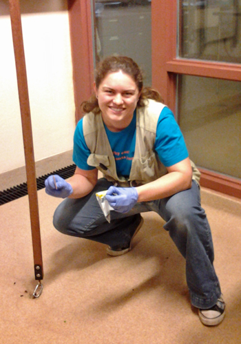
Erin McKenney scooping lemur poop for SCIENCE!
“Mammals don’t secrete the enzymes that are necessary, so no mammal can digest fiber on its own,” McKenney said. “These microbes are performing an incredibly important life process for us.”
At the Duke Lemur Center, McKenney collected fecal samples from three different species of lemur that evolved to eat different foods—a strict leaf-eater, and two omnivores. Using DNA sequencing, she determined the communities of bacteria that are living in their guts at different life stages from birth to adulthood.
Watching microbiomes through time may enable her to answer the question of how the microbiome of each species becomes teeming with 20 trillion bacteria, and if the patterns differ based on diet.
Vegetarian lemurs can eat a surprising variety of stuff we’d find nasty, like pokeweed and even poison ivy. (Duke Lemur Center, David Haring)
The results suggest that all species of baby lemurs, when they are born and nursing from their mothers have similar microbiome profiles that are much less complex than adult profiles. But leaf-eaters that eat the most fiber show adult microbiome profiles as soon as solid foods are introduced, which is in contrast to the other two species that take longer to reach adult microbiome profiles. Additionally, leaf-eaters have more complex microbial communities, which allows them to digest fiber-rich foods.
“So when you start to think about the really big picture, beyond everything the gut microbes do for the hosts they live inside of, we find the microbes have done an incredible service to mammalian speciation. The only way that we have leaf-eaters is because of these gut microbes,” McKenney said.
Fresh off a visiting teaching gig at Duke-Kunshan University and a sabbatical in Australia, canine and primate cognition researcher Brian Hare is about to land in your living room.
Hare, an associate professor of Evolutionary Anthropology and founder of Duke’s canine cognition lab and the Triangle startup Dognition.com, is now a television host too.
He’ll be hosting a three-part series on Nat Geo WILD at 10 p.m. ET this Friday, Saturday and Sunday nights called “Is Your Dog a Genius?”
Hare will introduce viewers to some of the latest knowledge about what our dogs think and understand, as well as sharing some at-home games you can use to reveal your dog’s personality. He’ll also visit with some ordinary and extraordinary dogs to see their problem-solving in action.
Friday’s episode is titled ” Doggy See Doggy Do.” Saturday is “Who’s Your Doggy.” And Sunday is “Talk Doggy to Me.”
Guest post by Gregg Gunnell, Division of Fossil Primates
(A version of this column originally appeared in the Duke Lemur Center newsletter)
Lagerstätten – that word sends a shiver of excitement up and down the spine of every paleontologist.
In German the word means ‘storage place’ or ‘deposits,’ but in paleontology it has come to mean a very rich fossil deposit that contains complete or nearly complete specimens that sample a wide variety of the creatures living at a certain time.
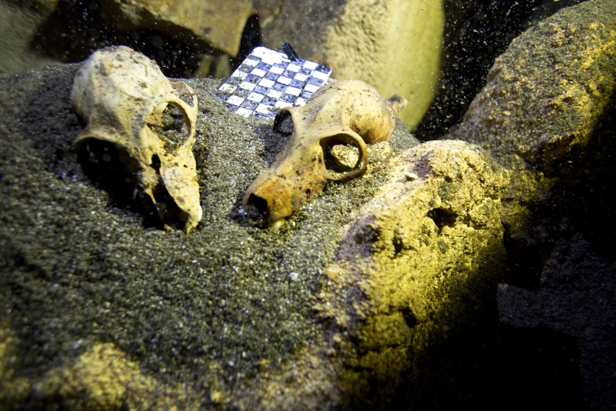
A cave diver and subfossil specimen in Aven Cave, Madagascar. The plastic triangle is a scale for photographs of the specimen in situ. (Image by Phillip Lehman and Pietro Donaggio-Bitner)
As you might imagine, Lagerstätten are quite rare. Some of the more famous examples are the Burgess Shale in Canada which preserves soft body outlines of ancient (530 million years ago) Cambrian animals; the Jurassic (150 Ma) Solenhofen limestones in Germany where the famous Archaeopteryx is found; and the middle Eocene (45 Ma) Messel Oil Shale in Germany which preserves whole skeletons of many birds, mammals, reptiles, amphibians, and insects.
I have had the good fortune to be in on the discovery of two Lagerstätten in addition to studying specimens from two others. The first one our team discovered was in 1998 in Pakistan, a place we named Gandhera Quarry. It preserves a remarkable wealth of early Eocene (52 Ma) mammals from Balochistan Province – an assemblage that has yet to completely studied.
But the latest and most exciting to me as Director of the Division of Fossil Primates in the Duke Lemur Center happened late last year in Southwest Madagascar.
The discovery of subfossils at a place called Aven Cave was known to local people, but not reported to the scientific community until an Australian cave diver named Ryan Dart saw it. The cave and its specimens are underwater. The specimens are called subfossils, because they aren’t old enough to have completed (or in some cases even started) the fossilization process.
A joint team from the University of Antananarivo, Duke University, University of Massachusetts, Brooklyn College and Midwestern University led an expedition to this cave site in October 2014. Cave divers Phillip Lehman and the Dominican Republic Speleological Society dive team helped us find a treasure trove of subfossils.
Lemur skulls, as they were found in the cave, with a scale marker. (Photo courtesy of Phillip Lehman and Pietro Donaggio-Bitner)
Only a preliminary survey has been made of Aven Cave to date, but it is clear already that it is one of the richest subfossil sites ever discovered in Madagascar. The initial list of animal specimens found in the cave includes three genera of extinct lemurs (Pachylemur, Mesopropithecus, and Megaladapis) as well as one species of a living form, Lemur catta, the familiar ring-tailed lemur. In addition to the primates there are abundant specimens of bats (Hipposideros), carnivores (the extinct fossa Cryptoprocta spelea as well as a smaller, still living species, C. ferox), two species of rodents, an extinct pygmy hippopotamus, crocodiles, turtles, and two bird species including the extinct elephant bird Mullerornis.
Not only is there a diverse assembly of species coming from Aven Cave, the sample is also abundant, with many species represented by multiple specimens. Many specimens appear to be complete or nearly complete skeletons.
The expedition was aided by Mr. Lovasoa Dresy, the director of Tsimanampetsotsa National Park, and was generously supported by the National Science Foundation and the National Geographic Society.
We anticipate many more and surprising discoveries in the future. Stay tuned for updates from Aven Cave!
What does famous lemur researcher, Dame Alison Richard, do when she has a burning question she can’t answer?
She visits Duke and appeals to a room full of lemur enthusiasts to help out.
Richard’s question concerns the curious case of the mouse lemurs at Beza Mahafaly in southwestern Madagascar, where she has been involved in a wildlife-monitoring program since the mid-1990s.
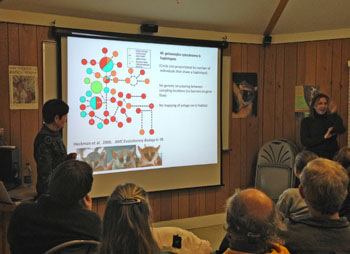
Alison Richard (left) and Lemur Center Director Anne Yoder (right) lead a discussion in the ‘Beach House’ at DLC.
“What do I know about mouse lemurs?” she questioned a group that gathered at the Duke Lemur Center on March 3 as the first of three talks she held at Duke this week as part of the Von der Heyden Fellows Program. “Probably less than you do. But I am incredibly interested in what is going on with them at Beza Mahafaly.”
Everywhere else in Madagascar, mouse lemurs that look indistinguishable are classified as different species due to big variations at the genetic level. But at Beza Mahafaly, Richard is finding that mouse lemurs with major deviations in appearance are genetically the same.
Dame Alison Richard (Photo: HHMI)
For a long time, the general view was that there were two species of mouse lemur in the forests of Beza Mahafaly : the gray-brown mouse lemur and the gray mouse lemur (both being exceptionally adorable).
A few studies in the mid-1990s and early 2000s compared the shapes of certain features such as jawbone shape and leg length, and confirmed this view. Then, researchers started noticing a few trapped animals that had very noticeable differences in coat coloration. These animals were redder than the other two known species. Was this a possible third species?
In 2006, Duke Lemur Center Director, Anne Yoder, and her former Ph.D. student Kellie Heckman examined this same population of mouse lemurs from a genetic standpoint. Comparing sequences of DNA they expected to find major genetic differences between the two known species, and possibly confirm the existence of a third species.
“The genetic data was a disaster for the mouse lemurs,” Richard said.
All the samples collected from animals at Beza Mahafaly, regardless of the animal’s outward appearance, sorted together and seemed to be one species.
Dame Alison and the bedeviled mouse lemur of Beza Mahafaly
“There’s a part of me that’s very distressed about this, but there’s a part of me that thinks this is great,” Richard said. “At Beza Mahafaly we swim upstream. We’re contrarians,” she said laughing. “But we still don’t know how to best explain the diversity that we do see.”
She offered up some suggestions: A glimpse of an ongoing process of change? A replacement by one species over another? The beginning of a new species?
Flashing a picture of a mouse lemur displaying ominous eye shine from a headlamp, she said: “The mouse lemurs are waiting with an evil gleam in their eye to be told the truth about themselves. The question is how should we take this forward?”
Guest Post By Sheena Faherty, Ph.D. Candidate in Biology
Dame Alison Richard is the epitome of someone who puts her money where her mouth is. And her dedication is directed precisely where it’s needed most.
Richard, a protector of lemurs, artisanal salt entrepreneur and endless optimist, is not just doing something about Madagascar’s conservation crisis. She’s doing everything about it.
Alison Richard (Photo: HHMI)
She’ll visit Duke March 3-5 to give three-part lecture series discussing her role in over forty years of community-based conservation efforts in Madagascar.
Members of the Duke community know all too well that our beloved lemurs— many of which can only be found at the Duke Lemur Center or in Madagascar—are in dire straights.
Their plight has been a life’s work for Richard, who is best known for her research on sifakas in the spiny forests of Madagascar. But she also lays claim to having been the first female vice-chancellor at Cambridge. She has now returned to Yale, where she spent most of her career, as a senior research scientist and professor emerita.
“Sometimes I think that because I’m covering so many bases, I end up doing nothing very well,” Richard said. “But it’s what I do and I can’t imagine not doing any of them—so it’s too bad,” she said laughing.
Richard is a conservationist who understands that without considering the local people’s well-being, all attempts to save wildlife habitats will fail.
“There are a variety of ways in which we are trying to facilitate socio-economic enhancements to people’s lives,” Richard said. “[On a recent trip to Madagascar] I met with the association of women salt producers, who are producing artisanal salt by techniques that have been in place for hundreds of years.”
In collaboration with a start-up company that is highly focused on sustainability, she recently shipped the first 500 kilos of the Madagascan salt to the U.S.
Verreaux’s Sifaka, a favorite of Richard’s in Southwestern Madagascar. (Credit: Flickr user nomis-simon, CC)
Taking time away from protecting the lemurs and enhancing the lives of the Malagasy people, Richard said her Duke lectures will have broad appeal for anyone interested in conservation, or for those who just enjoy seeing adorable pictures of lemurs.
She hopes to focus on writing a book, the topic of which will draw from her public lecture on March 5 at 6:00 pm at the Great Hall of the Mary Duke Biddle Trent Semans Center for Health Education. This lecture is set to explore how an array of different sciences has changed our understanding of Madagascar’s history.
And the conservationist who said she does everything has some advice for conserving her own mental sanity.
“One thing I need to do going forward is to find things to stop doing,” she admits. “And I’m not good at that because they are all too interesting and seemingly too important,” she said.
So, what’s next for Alison Richard?
“More of doing everything!” she said.
Richard’s installation as vice chancellor of Cambridge in November 2009 was occasioned by a visit from her majesty Queen Elizabeth II, who’s husband, Prince Philip, is the chancellor.
The National Evolutionary Synthesis Center (NESCent), is tucked away behind the supermarkets and youth-infested restaurants on Ninth Street in Durham. It’s a National Science Foundation brainchild with the purpose of consolidating data collected on small scales to help evolutionary biologists answer larger scale questions.
Allen Rodrigo directs NESCent and is a professor of biology at Duke
NESCent pursues a variety of missions, from answering these big ideas to connecting evolutionary science to linguistics and religious and cultural studies. Behind NESCent’s day-to-day function is evolutionary biologist Allen Rodrigo.
As a response to the question “so, what exactly is it that you do?” Rodrigo laughs. Here at NESCent, he oversees all of the programs, managing NSF grant money and keeping each part of the center on track with its mission. But NESCent is coming to the end of its funded run, and Dr. Rodrigo himself does far more than direct this innovative program.
Rodrigo is also a professor at Duke University and a computational evolutionary biologist. As a student, he was interested in three areas of study: mathematics, computer science, and biology. He continued pursuing all three tracks throughout his higher education, and allowed coincidence to launch him into his field today. The timing of his post-doc perfectly coincided with a late-1980s boom in technologic and scientific advances. With the invention of PCR and the subsequent increase in ability to study genetics, there came a demand for people with the skill and ability to conduct studies computationally, thus propelling Dr. Rodrigo into this growing field.
“There are many benefits to using computational methods,” Rodrigo said. “Suppose you want to compare two potential hypotheses on how a system might look, what patterns you might see. A computational biologist can help you with that.” He advocates for his area of study with a digestible list of its merits: “It helps experimentalists, it helps make inferences, and it helps make predictions about patterns.”
Today Rodrigo teaches classes at Duke, including courses on statistics for biologists and courses on computational science. He applies his passions for computational biology to his own research.
He is currently using computational study to track the evolution of traits of cancer related to their malignancy. “We start with a small set of cells and develop simulations that tell us how these cells change, grow, and divide,” Rodrigo said. “We can simulate how mutations accumulate, and can simulate, for a given collection of cells, what patterns of evolution you’d obtain.”
Chichi Zhu
Working with oncologists from Duke, his job is to use these computational and mathematical methods to search for patterns that oncologists can then use to collect laboratory data. “To do this all in a lab would take quite a long time,” he said. “To apply computational biology is much more efficient.”
A handful of bird species survived the K-T extinction. Chicken genomes have changed the least since that terrible day.
By Karl Leif Bates
In the beginning was the Chicken. Or something quite like it.
At that moment 66 million years ago when an asteroid impact caused the devastating Cretaceous–Tertiary (K-T) extinction, a handful of bird-like dinosaur species somehow managed to survive.
The cataclysm and its ensuing climate change wiped out much of Earth’s life and brought the dinosaurs’ 160-million-year reign to an end.
But this week, the genomes of modern birds are telling us that a few resourceful survivors somehow scratched out a living, reproduced (of course), and put forth heirs that evolved to adapt into all the ecological niches left vacant by the mass extinction. From that close call, birds blossomed into the more than 10,000 spectacularly diverse species we know today.
Erich Jarvis
Not all birds are descended from chickens, but it’s true that chicken genes have diverged the least from the dinosaur ancestors, says Erich Jarvis, an associate professor of neurobiology in the Duke Medical School and Howard Hughes Medical Institute Investigator.
He’s one of the leaders on a gigantic release of scientific data and papers that tells the story of the plucky K-T survivors and their descendants, redrawing many parts of the bird family tree.
From giant, flightless ostriches to tiny, miraculous hummingbirds, the descendants of those proto-birds now rule over the skies, the forests, the cliff-faces and prairies and even under water. While other birds were learning to reproduce songs and sounds, hover to drink nectar, dive on prey at 230 miles per hour, run across the land at 40 miles per hour, migrate from pole to pole or spend months at sea out of sight of land, the chickens just abided, apparently.
Sweet little chickadees had a very big, very scary cousin. “Parrots and songbirds and hawks and eagles had a common ancestor that was an apex predator,” Erich Jarvis says. “We think it was related to these giant ‘terror birds’ that lived in the South American continent millions of years ago.”
The new analyses released this week are based on complete-genome sequencing done mostly at BGI (formerly Beijing Genomics Institute). and DNA samples prepared mostly at Duke. The budgerigar, a parrot, was sequenced at Duke. There are 29 papers in this first release, but many more will tumble out for years to come.
“In the past, people have been using 1, 2, up to 20 genes, to try to infer species relationships over the last 100 million years or so,” Jarvis says. But whole-genome analysis drew a somewhat different tree and yielded important new insights.
More than 200 researchers at 80 institutions dove into this sea of big data like so many cormorants and pelicans, coming up with new insights about how flight developed and was lost several times, how penguins learned to be cold and wet and fly underwater and how color vision and bright plumage co-evolved.
The birds’ genomes were found to be pared down to eliminate repetitive sequences of DNA, but yet to still hold microchromosomal structures that link them to the dinosaurs and crocodiles.
The sequencing of still more birds continues apace as the Jarvis lab at Duke does most of the sample preparation to turn specimens of bird flesh into purified DNA for whole-genome sequencing at BGI. (More after the movie!)
As one of three ring-leaders for this massive effort, Erich Jarvis is on 20 papers in this first set of findings. Eight of those concern the development of song learning and speech, but the other ones are pretty cool too:
Evidence for a single loss of mineralized teeth in the common avian ancestor. Robert W. Meredith, et al.Science. Instead of teeth, modern birds have a horny beak to grab and a muscular gizzard to “chew” their food. This analysis shows birds lost the use of five genes related to making enamel and dentin just once about 116 million years ago.
Complex evolutionary trajectories of sex chromosomes across bird taxa. Qi Zhou, et al. Science. The chromosomes that determine sex in birds, the W and the Z, have a very complex history and more active genes than had been expected. They may hold the secret to why some birds have wildly different male and female forms.
Three crocodilians genomes reveal ancestral patterns of evolution among archosaurs. Richard E Green, et al Science. Three members of the crocodile lineage were sequenced revealing ‘an exceptionally slow rate of genome evolution’ and a reconstruction of a partial genome of the common ancestor to all crocs, birds, and dinosaurs.
Evidence for GC-biased gene conversion as a driver of between-lineage differences in avian base composition. Claudia C. Weber, et al, Genome Biology. The substitution of the DNA basepairs G and C was found to be higher in the genomes of birds with large populations and shorter generations, confirming a theoretical prediction that GC content is affected by life history of a species.
Low frequency of paleoviral infiltration across the avian phylogeny.Jie Cui, et al. Genome Biology. Genomes maintain a partial record of the viruses an organism’s family has encountered through history. The researchers found that birds don’t hold as much of these leftover bits of viral genes as other animals. They conclude birds are either less susceptible to viral infection, or better at purging viral genes.
Genomic signatures of near-extinction and rebirth of the Crested Ibis and other endangered bird species. Shengbin Li, et al Genome Biology. Genomes of several bird species that are recovering from near-extinction show clues to the susceptibilities to climate change, agrochemicals and overhunting that put them in peril. This effort has created better information for improving conservation and breeding these species back to health.
Dynamic evolution of the alpha (α) and beta (β) keratins has accompanied integument diversification and the adaptation of birds into novel lifestyles. Matthew J. Greenwold, et al. BMC Evolutionary Biology. Keratin, the structural protein that makes hair, fingernails and skin in mammals, also makes feathers. Beta-keratin genes have undergone widespread evolution to account for the many forms of feathers and claws among birds.
Comparative genomics reveals molecular features unique to the songbird lineage. Morgan Wirthlin, et al. BMC Genomics. Analysis of 48 complete bird genomes and 4 non-bird genomes identified 10 genes unique to the songbirds, which account for almost half of bird species today. Two of the genes are more active in the vocal learning centers of the songbird brain.
Reconstruction of gross avian genome structure, organization and evolution suggests that the chicken lineage most closely resembles the dinosaur avian ancestor. Michael N. Romanov, et al. BMC Biology. Of 21 bird species analyzed, the chicken lineage appears to have undergone the fewest changes compared to the dinosaur ancestor.
Two Antarctic penguin genomes reveal insights into their evolutionary history and molecular changes related to the cold aquatic environment. Cai Li, et al. Gigascience. The first penguins appeared 60 million years ago during a period of global warming. Comparisons of two Antarctic species show differences and commonalities in gene adaptations for extreme cold and underwater swimming.
Evolutionary genomics and adaptive evolution of the hedgehog gene family (SHH, IHH, and DHH) in vertebrates. Joana Pereira, et al. PLoS ONE. Whole-genome sequences for 45 bird species and 3 non-bird species allow a more detailed tracing of the evolutionary history of three genes important to embryo development.
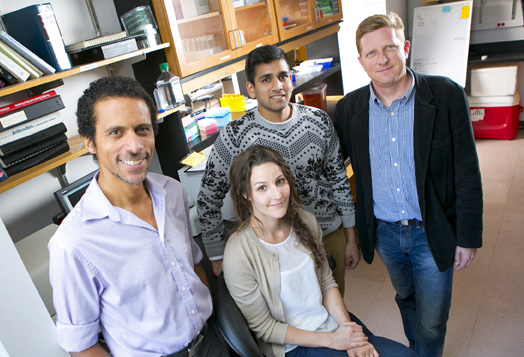
A Duke lab led the effort to isolate bird DNA for sequencing at BGI: (L-R) Erich Jarvis, associate professor of neurobiology and Howard Hughes Medical Institute investigator, lab research analyst Carole Parent, undergraduate research assistant Nisarg Dabhi, and research scientist Jason Howard. (Duke Photo, Les Todd)
So now you are at Duke — one of the world’s best research universities — but now what? You might be taking cool classes, but how can you take advantage of the world-class research happening here? Roughly 50 percent of Duke undergrads do so at some point. Getting involved in research as a freshman might sound intimidating (I know it did to me!), but a little luck and perseverance can get you off to a strong start.
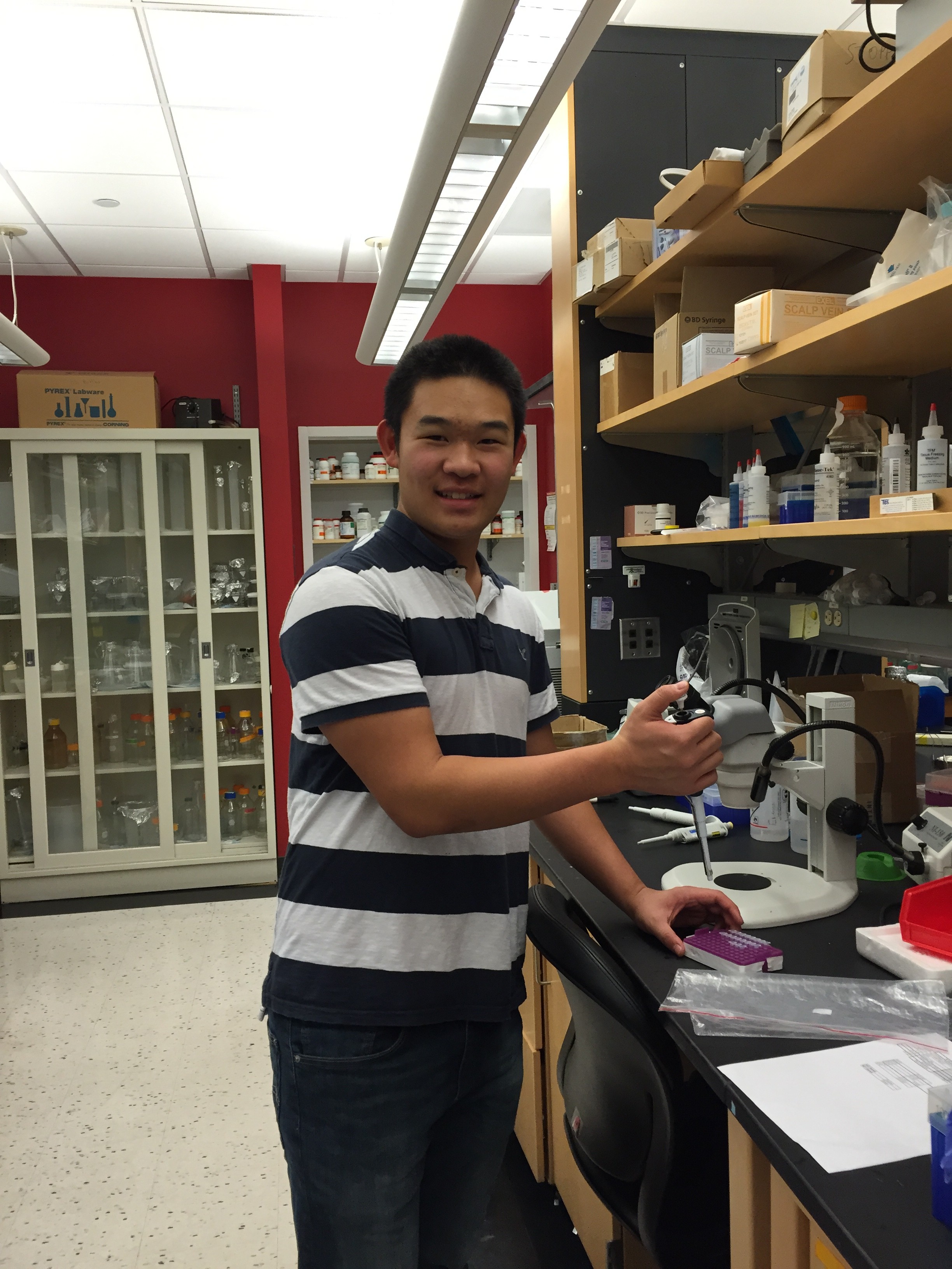
Alan working in Dr. Eroglu’s laboratory.
I had the opportunity to talk with Duke freshman Alan Kong about his experiences trying to get into research labs, and how he successfully ended up finding one to join. Alan is considering majoring in biology whilst on the premed track.
He initially started to look into labs within a month of starting classes at Duke. He spent about two months sending out emails to professors who were working on interesting projects.
“It was a very frustrating search, and initially difficult. I emailed five professors, and emailed each many times,” Alan said. “But perseverance ultimately paid off.”
Alan now works in the lab of Dr. Eroglu who is an assistant professor of cell biology, associated with the Duke School of Medicine. According to the Duke Institute of Brain Science website description, Eroglu’s laboratory “is interested in understanding how central nervous system (CNS) synapses are formed.”
Alan was also accepted into two others labs, but ultimately felt Dr. Eroglu was the best fit. “I picked Eroglu because her research was very interesting, and relevant to my interests,” Alan says. “I felt I could learn more interesting techniques in research, such as working with live animals.”
Now, Alan has been working in Dr. Eroglu’s lab for a month. When I asked him how it was going, with a smile he exclaimed, “It’s great!”
“Right now I am learning techniques such as genotyping, western blot. I even took out the retina of a rat!” Alan said. “I am learning the ropes of the lab, and my mentor said that down the road, if I learn properly, I can eventually work on my own independent project!”
When asked for any advice for other students thinking of getting into research, Alan said “Persistence is key — don’t give up! It’s a difficult process; don’t let small things get in the way. Keep trying until you find one.”
Imagine learning research methods while also getting charged by a rhino, observing a pride of lions hunting warthogs, and glimpsing a cheetah and her cub.
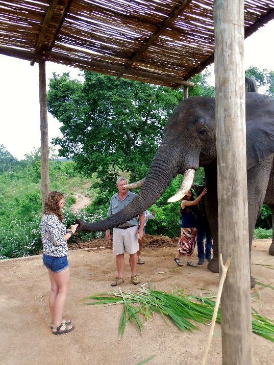
Lizzie feeding an elephant in Kruger National Park in South Africa. Her research attempted to map woody tree coverage in a section of the park since rising elephant populations has resulted in the destruction of woody trees.
Elizabeth “Lizzie” Hoerauf experienced this last summer with five other students from Yale, Vanderbilt, Duke, and Reed Universities. The Organization for Tropical Studies (OTS) brought them to Kruger National Park in South Africa, charging them with researching various plants and animals in the park — with a primary focus on the effects of the park’s growing elephant population.
Culling of elephants – i.e., reducing their populations by selective slaughter — has been banned since 1994, resulting in a significant increase in the elephant population. This seems admirable, yet as the increasingly abundant massive animals roam the savanna they knock down more trees. Fewer trees across the park means lower habitat diversity for the park’s other plants and animals.
Lizzie’s project laid the groundwork for other students’ surveying efforts. To map woody cover, Lizzie took overhead images from previous park data of one of the four supersites that make up the park and overlaid them using Geographic Information Systems (GIS) software.
Her project did not come without its struggles. One moment she would emerge from her tent with a look of triumph telling her peers, “Guys look how amazing this is!” The next, disgruntled and lamenting, “Everything crashed….” However, her ultimately successful data consolidation demonstrated that less than ten percent of the area she studied was now populated with tall trees — a significant decrease from years past.
With some time to reflect Lizzie says that she learned more from this project than in the classroom. When I asked her if she had interests in returning to project in the future she noted a preference to pursue field research projects of her own. Lizzie did let me know that OTS is a wonderful program that Duke offers — it felt rewarding to be a small part of a massive and constantly evolving field research project.
Using Geographic Information Systems (GIS) software, Lizzie was able to map out and analyze woody tree cover for one of the supersites of the park. Her analysis found approximately 6 percent of the supersite to be covered by tall trees, much lower than previous years (12 percent in 1940 or 9 percent in 1974).
Any movie that begins with an extreme close-up of the back side of a fruit fly — the same kind found feeding on over-ripe tomatoes and bananas in your kitchen — may seem like an unlikely candidate for action blockbuster of the year. But this is no typical movie.
https://www.youtube.com/watch?v=fwzIUnKNw0s
Duke biologists Dan Kiehart and Serdar Tulu recorded this 3D close-up of a developing fly embryo using new super-resolution microscope technology developed by Eric Betzig, one of the winners of the 2014 Nobel Prize in Chemistry.
Cutting-edge microscopes available on many campuses today allow researchers to take one or two images a second, but with a new technique called lattice light-sheet microscopy — developed by Betzig and colleagues and reported in the Oct. 24, 2014, issue of Science — researchers can take more than 50 images a second, and in the specimen’s natural state, without smooshing it under a cover slip.
Kiehart and Tulu traveled to the Howard Hughes Medical Institute’s Janelia Farm research campus in Ashburn, Virginia, where the new microscope is housed, to capture the early stages of a fruit fly’s development from egg to adult in 3D.
Fruit fly embryos are smaller than a grain of rice. By zooming in on an area of the fly embryo’s back that is about 80 microns long and 80 microns wide — a mere fraction of the size of the period at the end of this sentence — the researchers were able to watch a line of muscle-like cells draw together like a purse string to close a gap in the fly embryo’s back.
The process is a crucial step in the embryo’s development into a larva. It could help researchers better understand wound healing and spina bifida in humans.
Their movie was assembled from more than 250,000 2D images taken over 100 minutes. The hundreds of thousands of 2D snapshots were then transferred to a computer, which used image-processing software to assemble them into a 3D movie.
“This microscope gives us the highest combination of spatial and temporal resolution that we can get,” Kiehart said.
Betzig won this year’s Nobel Prize for his work on techniques that allow researchers to peer inside living cells and resolve structures smaller than 200 nanometers, or half the wavelength of light — a scale once thought impossible using traditional light microscopes.
Even finer atomic-scale resolution has long been possible with microscopes that use beams of electrons rather than light, but only by killing and slicing the specimen first, so living cells and the tiny structures in motion inside them couldn’t be observed.
Betzig and collaborators Wesley Legant, Kai Wang, Lin Shao and Bi-Chang Chen of Janelia Farm Research Campus all played a role in developing this newest microscope, which creates an image using a thin sheet of patterned light.
The fly movie is part of a collection of videos recorded with the new technology and published in the Oct. 24 Science paper.
One video in the paper shows specialized tubes inside cells called microtubules — roughly 2,000 times narrower than a human hair — growing and shrinking as they help one cell split into two.
Other videos reveal the motions of individual cilia in a single-celled freshwater creature called Tetrahymena, or cells of a soil-dwelling slime mold banding together to form multicellular slugs.
Kiehart and Tulu will be going back to Janelia Farm in January to use the microscope again.
“For this visit we’re going to zoom in to a smaller area to look at individual cells,” Tulu said.
“Waking up the morning of October 8 and hearing on the radio that our paper includes a Nobel Prize winner was pretty special,” Kiehart said.
CITATION: “Lattice light-sheet microscopy: Imaging molecules to embryos at high spatiotemporal resolution,” Chen, B.-C., et al. Science, October 2014. http://www.sciencemag.org/content/346/6208/1257998