Meet Claire Engstrom, a Senior from Pasadena California. Claire is a Biology major who works in the Gersbach Lab at Duke.
Claire first got involved with on-campus research through her pre-orientation program, PSearch that introduces incoming first-years to undergraduate research. Following her experience in PSearch, Claire got her first work-study research position in the Tung Lab where she worked closely with Jenny Tung, an Associate Professor in the Departments of Evolutionary Anthropology and Biology at Duke and a Faculty Associate of the Duke University Population Research Institute.
In the Tung Lab, Claire’s research focused on how DNA methylation is passed through generations. Essentially looking at the inheritance of DNA whose methylation was impacted by environmental factors and how that affects future generations.
Duke has research opportunities available in all disciplines as well as across departments. Approximately 53% of undergraduates graduate with research experience. Not only can students participate in groundbreaking research, but they can receive funding from the university as well to support the work they are doing.
Within the Biology department, there is a fellowship called B-SURF, the Biological Sciences Undergraduate Research Fellowship, an 8-week summer research program for rising sophomores. Claire applied for and was accepted to the fellowship and placed in one of Duke’s biomedical science laboratories. She also received a $4,000 stipend for her summer research.
Claire was placed in Charles Gersbach’s Lab focused on researching Genome Editing for Gene and Cell Therapy. Dr, Gersbach is a Rooney Family Associate Professor of Biomedical Engineering and has conducted groundbreaking work in genome editing.
Members of the Gersbach Lab in Fall 2019
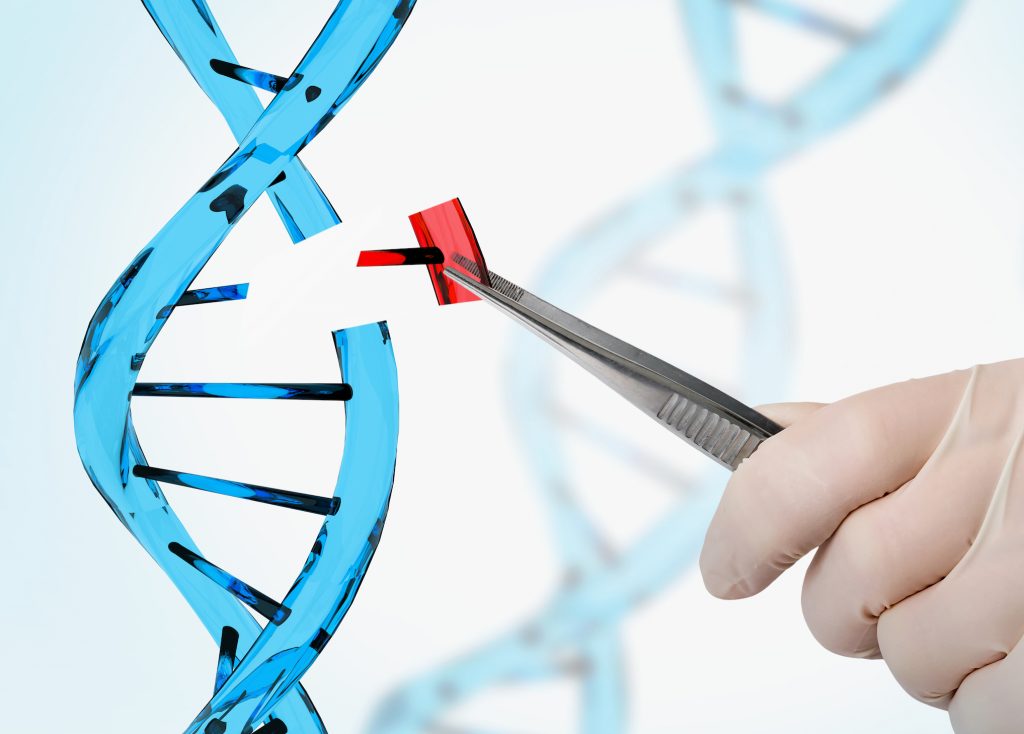
Gersbach is doing research in several different domains of biomedical engineering. Claire’s project focuses on using CRISPR-Cas9, a technology that allows scientists to change an organism’s DNA using clustered regularly interspaced short palindromic repeats and CRISPR-associated protein 9. faster, cheaper, more accurate, and more efficient than other existing genome editing methods.
Prior to joining his lab, Claire had already heard a lot about Gersbach in her course Biology 201 as well as through reading his papers. The project she would spend the next two and a half years working on focused on using and optimizing CRISPR-Cas9 to treat Duchenne’s Muscular Dystrophy and lessen the severity of the symptoms.
Duchenne’s Muscular Dystrophy is a muscle wasting disease that affects one in every five thousand male births.
“People are diagnosed when they are around five and then they lose the ability to walk and their heart can’t pump blood because of the lack of muscles.” Claire explained.
“CRISPR-based genome editing restores dystrophin expression in mouse models of Duchenne muscular dystrophy. Cross-sections of muscle tissue where the dystrophin protein has been labeled green, including normal, healthy tissue (left), tissue from a mouse model of Duchenne muscular dystrophy (middle), and tissue from the same mouse model that has been treated with the CRISPR gene editing system (right). Nelson et al., Science (2016)”
Thus, those affected often die in early adulthood despite current advances in cardiovascular and respiratory treatments. Duchenne’s Muscular Dystrophy generally occurs as a result of a frameshift mutation of the dystrophin gene. As a result, one’s muscles can no longer connect to anything making it nearly impossible to contract and function properly. In the Gersbach lab they are trying to treat the mutation by using CRISPR-Cas9 to remove an exon or coding region of the gene in order to shift the reading frame back into its normal place.
This shift produces a less severe phenotype that lessens the effects of Duchenne’s Muscular Dystrophy. The result will significantly improve the quality of life and life spans for affected patients.
Claire will be continuing her work in the Gersbach lab full time in Spring 2021 as she graduated early, with distinction in the Fall. Her thesis on the work she did in the Gersbach lab was recently approved and her results will be published in a larger paper in the future. After this year she plans to take a gap year an then return to California to hopefully attend grad school and pursue a Ph.D. in Biology.
The COVID-19 epidemic has impacted the Duke research enterprise in profound ways. Nearly all laboratory-based research has been temporarily halted, except for research directly connected to the fight against COVID-19. It will take much time to return to normal, and that process of renewal will be gradual and will be implemented carefully.
Trying to put this situation into a broader perspective, I thought of the 1939 essay by Abraham Flexner published in Harper’s magazine, entitled “The Usefulness of Useless Knowledge.” Flexner was the founding Director of the Institute for Advanced Study at Princeton, and in that essay, he ruminated on much of the type of knowledge acquired at research universities — knowledge motivated by no objective other than the basic human desire to understand. As Flexner said, the pursuit of this type of knowledge sometimes leads to surprises that transform the way we see that which was previously taken for granted, or for which we had previously given up hope. Such knowledge is sometimes very useful, in highly unintended ways.
Gregory Gray, MD MPH
The 1918 influenza pandemic led to 500 million confirmed cases, and 50 million deaths. In the Century since, consider how far we have come in our understanding of epidemics, and how that knowledge has impacted our ability to respond. People like Greg Gray, a professor of medicine and member of the Duke Global Health Institute (DGHI), have been quietly studying viruses for many years, including how viruses at domestic animal farms and food markets can leap from animals to humans. Many believe the COVID-19 virus started from a bat and was transferred to a human. Dr. Gray has been a global leader in studying this mechanism of a potential viral pandemic, doing much of his work in Asia, and that experience makes him uniquely positioned to provide understanding of our current predicament.
From the health-policy perspective, Mark McClellan, Director of the Duke Margolis Center for Health Policy, has been a leading voice in understanding viruses and the best policy responses to an epidemic. As a former FDA director, he has experience bringing policy to life, and his voice carries weight in the halls of Washington. Drawing on faculty from across Duke and its extensive applied policy research capacity, the Margolis Center has been at the forefront in guiding policymakers in responding to COVID-19.
Through knowledge accrued by academic leaders like Drs. Gray and McClellan, one notes with awe the difference in how the world has responded to a viral threat today, relative to 100 years ago. While there has been significant turmoil in many people’s lives today, as well as significant hardship, the number of global deaths caused by COVID-19 has been reduced substantially relative to 1918.
One of the seemingly unusual aspects of COVID-19 is that a substantial fraction of the population infected by the virus has no symptoms. However, those asymptomatic individuals shed the virus and infect others. While most people have no or mild symptoms, other people have very adverse effects to COVID-19, some dying quickly.
This heterogeneous response to COVID-19 is a characteristic of viruses studied by Chris Woods, a professor medicine in infectious diseases. Dr. Woods, and his colleagues in the Schools of Medicine and Engineering, have investigated this phenomenon for years, long before the current crisis, focusing their studies on the genomic response of the human host to a virus. This knowledge of viruses has made Dr. Woods and his colleagues leading voices in understanding COVID-19, and guiding the clinical response.
A team led by Greg Sempowski, a professor of pathology in the Human Vaccine Institute is working to isolate protective antibodies from SARS-CoV-2-infected individuals to see if they may be used as drugs to prevent or treat COVID-19. They’re seeking antibodies that can neutralize or kill the virus, which are called neutralizing antibodies.
Barton Haynes, MD
Many believe that only a vaccine for COVID-19 can truly return life to normal. Human Vaccine Institute Director Barton Haynes, and his colleagues are at the forefront of developing that vaccine to provide human resistance to COVID-19. Dr. Haynes has been focusing on vaccine research for numerous years, and now that work is at the forefront in the fight against COVID-19.
Engineering and materials science have also advanced significantly since 1918. Ken Gall, a professor of mechanical engineering and materials science has led Duke’s novel application of 3D printing to develop methods for creatively designing personal protective equipment (PPE). These PPE are being used in the Duke hospital, and throughout the world to protect healthcare providers in the fight against COVID-19.
Much of the work discussed above, in addition to being motivated by the desire to understand and adapt to viruses, is motivated from the perspective that viruses must be fought to extend human life.
In contrast, several years ago Jennifer Doudna and Emmanuelle Charpentier, academics at Berkeley and the Max Planck Institute, respectively, asked a seemingly useless question. They wanted to understand how bacteria defended themselves against a virus. What may have made this work seem even more useless is that the specific class of viruses (called phage) that infect bacteria do not cause human disease. Useless stuff! The kind of work that can only take place at a university. That basic research led to the discovery of clustered regularly interspaced short palindromic repeats (CRISPR), a bacterial defense system against viruses, as a tool for manipulating genome sequences. Unexpectedly, CRISPR manifested an almost unbelievable ability to edit the genome, with the potential to cure previously incurable genetic diseases.
Charles Gersbach, a professor of Biomedical Engineering, and his colleagues at Duke are at the forefront of CRISPR research for gene and cell therapy. In fact, he is working with Duke surgery professor and gene therapy expert Aravind Asokan to engineer another class of viruses, recently approved by the FDA for other gene therapies, to deliver CRISPR to diseased tissues. Far from a killer, the modified virus is essential to getting CRISPR to the right tissues to perform gene editing in a manner that was previously thought impossible. There is hope that CRISPR technology can lead to cures for sickle cell and other genetic blood disorders. It is also being used to fight cancer and muscular dystrophy, among many other diseases and it is being used at Duke by Dr. Gersbach in the fight against COVID-19.
David Ashley, Ph.D.
In another seemingly bizarre use of a virus, a modified form of the polio virus is being used at Duke to fight glioblastoma, a brain tumor. That work is being pursued within the Preston Robert Tisch Brain Tumor Center, for which David Ashley is the Director. The use of modified polio virus excites the innate human immune system to fight glioblastoma, and extends life in ways that were previously unimaginable. But there are still many basic-science questions that must be overcome. The remarkable extension of life with polio-based immunotherapy occurs for only 20% of glioblastoma patients. Why? Recall from the work of Dr. Woods discussed above, and from our own observation of COVID-19, not all people respond to viruses in the same way. Could this explain the mixed effectiveness of immunotherapy for glioblastoma? It is not known at this time, although Dr. Ashley feels it is likely to be a key factor. Much research is required, to better understand the diversity in the host response to viruses, and to further improve immunotherapy.
The COVID-19 pandemic is a challenge that is disrupting all aspects of life. Through fundamental research being done at Duke, our understanding of such a pandemic has advanced markedly, speeding and improving our capacity to respond. By innovative partnerships between Duke engineers and clinicians, novel methods are being developed to protect frontline medical professionals. Further, via innovative technologies like CRISPR and immunotherapy — that could only seem like science fiction in 1918 (and as recently as 2010!) — viruses are being used to save lives for previously intractable diseases.
Viruses can be killers, but they are also scientific marvels. This is the promise of fundamental research; this is the impact of Duke research.
“We shall not cease from exploration And the end of all our exploring Will be to arrive where we started And know the place for the first time.”
T.S. Eliot, Four Quartets
Post by Lawrence Carin, Vice President for Research
“I don’t believe that [going to] Mars pushes us hard enough.” This was just one of the bold, thought-provoking statements made by Dr. Mae Jemison, who came to speak at Duke on Monday, February 24 as part of the 15th annual Jean Fox O’Barr Distinguished Speaker Series, presented by Baldwin Scholars.
Dr. Jemison is at the pinnacle of interdisciplinary engagement—though she is most famous for serving as a NASA astronaut and being the first African American woman to go into space, she is also trained as an engineer, social scientist and dancer. Dr. Jemison always knew that she was going to space—even though there were no women or people or color participating in space exploration as she was growing up.
Dr. Jemison says that simply “looking up” brought her here. As a child, she would look up at the sky, see the stars and wonder if other children in other places in the world were looking at the same view that she had. Growing up in the 1960’s instilled into Dr. Jemison at an early age that our potential is limitless, and the political culture of civil rights, changing art and music and decolonization were all about “people declaring that they had a right to participate.”
Photo courtesy of Elizabeth Roy
One of the biggest pieces of advice that Dr. Jemison wanted to impart on her audience was the value of confidence, and how to build confidence in situations where people are tempted to feel incapable or forget the strengths they already possess. “They told me if I wanted to lead projects I needed an M.D.,” Dr. Jemison explained. “I went to medical school because I know myself and I knew I would want to be in charge one day.”
At 26 years old, Dr. Jemison was on call 24 hours a day, 7 days a week, 365 days a year as the Area Peace Corps Medical Officer for Sierra Leone and Liberia. She described a case where a man came back with a diagnosis of malaria from Senegal. When Dr. Jemison first took a look, the diagnosis seemed more likely to be meningitis. After making an “antibiotic cocktail,” from what she had on site, she realized this man might lose his life if they didn’t get him to a better hospital. At this point, Dr. Jemison wanted to call a military medical evacuation, and she had the authority to do it. However, another man working with her suggested calling a doctor in Ivory Coast, or a doctor at the hospital in Germany to see what he thought before making the evacuation. Dr. Jemison knew what the patient needed in this situation was to be flown to Germany regardless of the cost of the evacuation. In reflecting on this experience, she says that she could have given someone else her authority, but letting her confidence in herself and what she knew was the right thing to do would have negatively impacted her patient.
So, how do you maintain confidence? According to Dr. Jemison, you come prepared. She knew her job was to save people’s lives, not to listen to someone else. Dr. Jemison also admonished the audience to “value, corral and protect your energy.” She couldn’t afford to always make herself available for non-emergency situations, because she needed her energy for when a patient’s life would depend on it.
Photo courtesy of Elizabeth Roy
Dr. Jemison’s current project, 100 Year Starship, is about trying to ensure we have the capabilities to travel to interstellar space. “The extreme nature of interstellar hurdles requires we re-evaluate what we think we know,” Dr. Jemison explained. Alpha Centauri, the next closest star, is more than 25 trillion miles away. Even if we go 10% the speed of light, it will still take us 50 years to get there. We need to be able to travel faster, the vehicle has to be self-replenishing, and we have to think about space-time changes. What Dr. Jemison calls the “long pole in the tent” is human behavior. We need to know how humans will act and interact in a small spaceship setting for possibly decades of space travel. Dr. Jemison is thinking deeply about how we can apply the knowledge we already possess to fix world problems, and how we can start preparing now for problems we may face in the future. For example, how would health infrastructure in deep space look different? How would we act on a starship that contains 5,000 people when we can’t figure out how to interact with each other on the “starship” we’re on now?
Returning to the childhood love for stargazing that brought her here, Dr. Jemison discussed towards the end of her talk that a stumbling block for the majority of people is insufficient appreciation of our connection across time and space. She has worked with a team to develop Skyfie, an app that allows you to upload photos and videos of your sky to the Sky Tapestry and explore images other people in different parts of the world are posting of their sky. Dr. Jemison’s hope is this app will help people realize that we are interconnected with the rest of the universe, and we won’t be able to figure out how to survive as a species on this planet alone.
Imagine robots that can move, sense and respond to stimuli, but that are smaller than a hair’s width. This is the project that Cornell professor and biophysicist Itai Cohen, who gave a talk on Wednesday, January 29 as a part of Duke’s Physics Colloquium, has been working on with and his team. His project is inspired by the microscopic robots in Paul McEuen’s book Spiral. Building robots at such a small scale involves a lot more innovation than simply shrinking all of the parts of a normal robot. At low Reynolds number, fluids are viscous instead of inertial, Van der Waals forces come into play, as well as other factors that affect how the robot can move and function.
Cohen’s team designs robots that fold similar to origami creatures. Image from Origami.me
To resolve this issue, Cohen and his team decided to build and pattern their micro robots in 2D. Then, inspired by origami, a computer would print the 2D pattern of a robot that can fold itself into a 3D structure. Because paper origami is scale invariant, mechanisms built at one scale will work at another, so the idea is to build robot patterns than can be printed and then walk off of the page or out of a petri dish. However, as Cohen said in his talk last Wednesday, “an origami artist is only as good as their origami paper.” And to build robots at a microscopic scale, one would need some pretty thin paper. Cohen’s team uses graphene, a single sheet of which is only one atom thick. Atomic layer deposition films also behave very similarly to paper, and can be cut up, stretch locally and adopt a 3D shape. Some key steps to making sure the robot self-folds include making elements that bend, and putting additional stiff pads that localize bends in the pattern of the robot. This is what allows them to produce what they call “graphene bimorphs.”
Cilia on the surface of a cell. Image from MedicalXpress.
Cohen and his team are looking to use microscopic robots in making artificial cilia, which are small leg-like protrusions in cells. Cilia can be sensory or used for locomotion. In the brain, there are cavities where neurotransmitters are redirected based on cilial beatings, so if one can control the individual beating of cilia, they can control where neurotransmitters are directed. This could potentially have biomedical implications for detecting and resolving neurological disorders.
Right now, Cohen and his lab have microscopic robots made of graphene, which have photovoltaics attached to their legs. When a light shines on the photovoltaic receptor, it activates the robot’s arm movement, and it can wave hello. The advantage of using photovoltaics is that to control the robot, scientists can shine light instead of supplying voltage through a probe—the robot doesn’t need any tethers. During his presentation, Cohen showed the audience a video of his “Brobot,” a robot that flexes its arms when a light shines on it. His team has also successfully made microscopic robots with front and back legs that can walk off a petri dish. Their dimensions are 70 microns long, 40 microns wide and two microns thick.
Cohen wants to think critically about what problems are important to use technology to solve; he wants make projects that can predict the behavior of people in crowds, predict the direction people will go in response to political issues, and help resolve water crises. Cohen’s research has the potential to find solutions for a wide variety of current issues. Using science fiction and origami as the inspiration for his projects reminds us that the ideas we dream of can become tangible realities.
In the first week of fall semester, four first-year engineering students, Sean Burrell, Teya Evans, Adam Kramer, and Eloise Sinwell, had a brainstorming session to determine how to create a set of physical therapy stairs designed for children with disabilities. Their goal was to construct something that provided motivation through reward, had variable step height, and could physically support the students.
Evans explained, “The one they were using before did not have handrails and the kids were feeling really unstable.”
, Teya Evans is pictured stepping on the staircase her team designed and built. With each step, the lightbox displays different colors.
The team was extremely successful and the staircase they designed met all of the goals set out by their client, physical therapists. It provided motivation through the multi-colored lightbox, included an additional smaller step that could be pulled out to adjust step height, had a handrail to physically support the students and could even be taken apart for easy transportation.
This is a part of the Engineering 101 course all Pratt students are required to take. Teams are paired with a real client and work together throughout the semester to design and create a deliverable solution to the problem they are presented with. At the end of the semester, they present their products at a poster presentation that I attended. It was pretty incredible to see what first-year undergraduates were able to create in just a few months.
The next poster I visited focused on designing a device to stabilize hand tremors. The team’s client, Kate, has Ataxia, a neurological disorder that causes her to have uncontrollable tremors in her arms and hands. She wanted a device that would enable her to use her iPad independently, because she currently needs a caregiver to stabilize her arm to use it. This team, Mohanapriya Cumaran, Richard Sheng, Jolie Mason, and Tess Foote, needed to design something that would allow Kate to access the entire screen while stabilizing tremors, being comfortable, easy to set up and durable.
The team was able to accomplish its task by developing a device that allowed Kate to stabilize her tremors by gripping a 3D printed handlebar. The handlebar was then attached to two rods that rested on springs allowing for vertical motion and a drawer slide allowing for horizontal motion.
“We had her [Kate] touch apps in all areas of the iPad and she could do it.” Foote said. “Future plans are to make it comfier.”
The team plans to improve the product by adding a foam grip to the handlebar, attaching a ball and socket joint for index finger support, and adding a waterproof layer to the wooden pieces in their design.
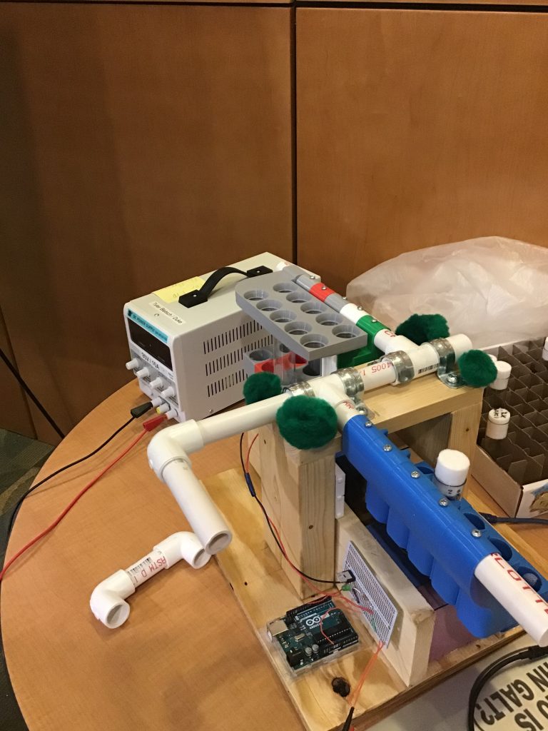
The last project I visited created a “Fly Flipping Device.” The team, C. Fischer, E. Song, L. Tarman, and S. Gorbaly, were paired with the Mohamed Noor Lab in the Duke Biology Department as their client.
Tarman explained, “We were asked to design a device that would expedite the process of transferring fruit flies from one vial to another.”
The Noor lab frequently uses fruit flies to study genetics and currently fly flipping has to be done by hand, which can take a lot of time. The goal was to increase the efficiency of lab experiments by creating a device that would last for more than a year, avoid damaging the vials or flies, was portable and fit within a desk space.
The team came up with over 50 ideas on how to accomplish this task that they narrowed down to one that they would build. The product they created comprised of two arms made of PVC pipe resting on a wooden base. Attached to the arms were “sleeves” 3D printed to hold the vials containing flies. In order to efficiently flip the flies, one of the arms moves about the axis allowing for multiple vials to be flipped that the time it would normally take to flip one vial. The team was very successful and their creation will contribute to important genetic research.
The Fly Flipping Device
It was mind-blowing to see what first-year students were able to create in their first few months at Duke and I think it is a great concept to begin student education in engineering through a hands-on design process that allows them to develop a solution to a problem and take it from idea to implementation. I am excited about what else other EGR 101 students will design in the future.
This is the sixth and final 2019 post written by students at the North Carolina School of Science and Math as part of an elective about science communication with Dean Amy Sheck.
Dr. Patrick Codd is the Director of the Duke Brain Tool Laboratory and an Assistant Professor of Neurosurgery at Duke. Working as a neurosurgeon and helping with the research and development of various neurosurgical devices is “a delicate balance,” he said.
Patrick Codd
Codd currently runs a
minimally invasive neurosurgery group. However, at Massachusetts General
Hospital, he used to run the trauma section. When asked about which role was
more stressful, he stated “they were both pretty stressful” but for different
reasons. At Mass General, he was on call for most hours of the day and had to
pull long shifts in the operating room. At Duke, he has to juggle surgery,
teaching, and research and the development of new technology.
“I didn’t know I was
going to be a neurosurgeon until I was in college,” Codd said. Despite all of
the interesting specialties he learned about in medical school, he said “it was
always neurosurgery that brought me back.”
Currently, he is
exclusively conducting cranial surgery.
Neurosurgeon U.S. Air Force Maj Jonathan Forbes,looks through loupes as he performs brain surgery at the Bagram Air Field in Afghanistan, Oct. 10, 2014.
Though Dr. Codd has earned
many leadership positions in his career, he said he was never focused on advancement.
He simply enjoys working on topics which he loves, such as improving minimally
invasive surgical techniques. But being in leadership lets him unite other
people who are interested in working towards a common goal in research and
development. He has been able to skillfully bring people together from various
specialties and help guide them. However, it is difficult to meet everyone’s
needs all of the time. What is important for him is to be a leader when he
needs to be.
Dr. Codd said there are typically five to eight research papers necessary in to lay the groundwork for every device that is developed. However, some technologies are based on the development of a single paper. He has worked on devices that make surgery more efficient and less minimally invasive and those that help the surgical team work together better. When developing technologies, he tries to keep the original purpose of the devices the same. However, many revisions are made to the initial design plans as requirements from the FDA and other institutions must be met. Ironically, Dr. Codd can’t use the devices he develops in his own operating room because it would be a conflict of interest. Typically other neurosurgeons from across the country will use them instead.
Imagine a live, health-focused version Shark Tank open to the public: presentations
from real health professionals,
presenting real innovations they developed
to address real health care issues.
And yes, there are real money awards
at stake.
At ten minutes ‘til show time, people gather in small groups
clothed in suits, business attire, and white coats. They chat in low voices. The
hum of comfortable conversation buzzes through the room. The sixth floor of the
Trent
Semans Center is quite the setting. Three sides of the room are
encapsulated in glass and you can easily see an expansive view of both Duke’s
West and Medical campuses, as well as luscious green trees comprising parts of
Duke’s Forest. Naturally, there is a glorious view of the Chapel, basked in
sunlight.
This light finds its way into the room to shine on various
research posters at the back displayed on a few rows of mobile walls. Though a
few strays meander through the stationary arrangements – stopping to look more
closely at particular findings – most people make their way into the room and
find a seat as the minutes dwindle away. The hum grows and there is a bit of anticipatory
energy among those readying themselves to present.
At three minutes after 10, the program director of the Duke
Institute for Health Innovation, Suresh Balu,
takes position at the front of the room, standing before the small stage at
center that is surrounded by lots of TV monitors. No seat in the room is a bad
one. Balu indicates that it is time to begin and the hum immediately
dissipates. He explains the general format of the event: six pitches total,
five minutes to present, eight minutes to answer questions from investors, a
show-of-hand interest from investors, and transition to the next pitch,
followed by deliberation and presentation of awards.
After a round of thanks, introduction of the emcee – Duke’s
Chief of Cardiology, Dr. Manesh Patel – the curtains opened – figuratively – on
Duke’s fifth annual Innovation
Jam.
Groups presented on the problems they were addressing, their
proposed innovations, and how the innovations worked. There was also
information about getting products into the market, varying economic analysis,
next steps or detailed goals for the projection of the projects, and analysis
of the investment they are currently seeking and for what purposes.
The first group pitched an idea about patient-centric blood
draw and suggest a device to plug into existing peripheral draws to reduce the frequent
poking and prodding that hospital patients often experience during their hospital
stay when blood is needed for lab tests. Next up was a group who designed an
intelligent microscope for automated pathology that has a programmable system and
uses machine learning to automate pathological blood analysis that is currently
highly time consuming. Third at bat was a group that made a UV light bag to
clean surgical drain bags that frequently become colonized with bacteria and
are quite frankly “nasty” – according to the presenter.
Batting cleanup was PILVAS – Peripherally Inserted Left
Ventricular Vent Anticoagulation System – which is a device that would be
accessory to VA
ECMO support to reduce thromboembolism and
stroke that are risks of ECMO. Fifth was the ReadyView and ReadyLift, a laparoscopic
tool set that is much cheaper than current laparoscopic tools and methods, and
because of its ability to be used with any USB compatible laptop, it would
increase access to laparoscopic surgery in countries that have a high need for
it. Last, but not least, was an innovation that is the first synthetic
biometric osteochondral
graft for knee cartilage repair that hopes to improve knee osteoarthritis surgical
care as the first hydrogel
with the same mechanical properties of cartilage.
Following a quick ten-minute break for investors to huddle
around and discuss who should win the awards – $15,000 for Best Innovation and
$15,000 for Best Presentation – the winners were announced. Drumroll, please.
ReadyView won Best Presentation and the synthetic
osteochondral graft won Best Innovation. A pair of representatives from
Microsoft were also in attendance – a first for the Innovation Jam – and
awarded SalineAI, the group who designed the intelligent microscope with an
independent award package.
Patel, the emcee, says we are in the midst of a fourth
industrial revolution.
“What is the biggest cinema in the world?” Patel asked.
“Netflix,” he says. Industries are reimagining themselves and healthcare is no
exception.
What is the best healthcare system of the future going to
look like? Of course, we really don’t know, but there are certainly people who
are already doing more than just think about it.
Emerging technology has created a new doping technique for athletic performance that is, as of now, perfectly legal.
Coined “neuro-doping,” this method sends electric current through one’s brain to facilitate quicker learning, enhanced muscular strength, and improved coordination. Use of this electronic stimulus has taken off in the sports world as a replacement for other doping methods banned by the World Anti-Doping Agency (WADA). Because it’s relatively new, WADA has yet to establish rules around neuro-doping. Plus, it’s virtually undetectable. Naturally, a lot of athletes are taking advantage of it.
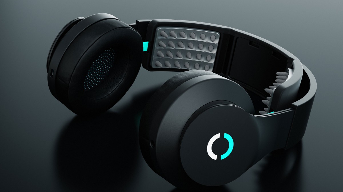
One specific method of neuro-doping is known as Transcranial Direct-Current Stimulation (tDCS). It works by sending a non-invasive and painless electrical current through the brain for around three to 20 minutes, in order to excite the brain’s cortex, ultimately increasing neuroplasticity (Park). This can be done commercially via a headset like device for $200.
The Halo Sport
Weight lifters, sprinters, pitchers, and skiers are just some of many types of athletes who can benefit from tCDS. By practicing with these headphones on, new neural pathways are constructed to help their bodies achieve peak performance. Dr. Greg Appelbaum, director of Opti Lab and the Brain Stimulation Research Center, says it’s especially useful for athletes where technique and motor skills triumph — such as a sprinter getting out of the blocks or an Olympic ski jumper hanging in the air. Top-tier athletes are pushing that fine limit of what the human body can accomplish, but neuro-doping allows them to take it one step further.
Neuro-doping has other applications, too. Imagine insanely skilled Air Force pilots, surgeons with exceptionally nimble hands, or soldiers with perfect aim. tCDS is being used to make progress in things like Alzheimer’s and memory function because of its impact on cognitive functioning in the forms of increased attention span and memory. You could even learn the guitar faster.
In this sort of context, it’s a no brainer that neuro-doping
should be taken advantage of. But how ethical is it in sports?
The
precedent for WADA to ban a substance or technique has been based on meeting
two of the following three criteria: (1) drugs or tools that likely enhance performance to secure a
winning edge; (2) drugs or tools that place athletes’ health at risk; (3) any
substances or techniques that ruin the “spirit-of-sport” (Park). Lots of
research has shown tCDS is pretty legit. As for health risks, tCDS is still in
the experimental stage, so not much can be said about its side effects. Ethically,
it causes a lot of controversy.
Many issues come into play when thinking about allowing athletes to neuro-dope. Given its similarities with other popular drugs, tCDS could introduce unfair advantages. Furthermore, not everyone may have access to the technology, and not everyone may want to use it. However, it’s important to note that sports already have unfair advantages. Access to things like proper coaching and nutrition may not be a reality for everyone. Sports are just inherently competitive.
Back when baseball players doped, it was awesome to watch them crush balls out of the park. Reintroducing performance enhancement through tCDS could mean we start seeing mountain bikers launching insane air and world records being smattered. The human body could achieve newfound heights.
Are the benefits worth it? Does neuro-doping ruin the “spirit of the sport?” Regardless of these important questions, tCDS is a fascinating scientific discovery that could make a difference in this world. So, what do you think?
Post by Will Sheehan
Park, Cogent Social Sciences (2017), 3: 1360462 https://doi.org/10.1080/23311886.2017.1360462
Every seat full. Students perched on the aisle stairs and lining the back walls.
What topic could possibly pull so many away from their final exams? Not “How to Stop Procrastinating” nor “How to Pass Life After Failing Your Exams” but rather “Gene-Editing Human Embryos: Unpacking the Current Controversy” on the Duke campus.
On December 6, the University Program in Genetics and Genomics and the Molecular Genetics and Microbiology department co-hosted a panel responding to He’s claims. Charles A. Gersbach from the Biomedical Engineering department lead the discussion of what exactly happened and then joined the panel which also contained Misha Angrist, a senior fellow in the Science & Society initiative; Heidi Cope, a genetic counselor; Giny Fouda, an assistant professor in pediatrics; and Vandana Shashi, a genetic counselor.
Dr. He Jiankui announced he had used CRISPR to edit genes in twin embryos that were then born at full term.
But what exactly has He potentially done to these twin girls? Can they fly? Breathe underwater? Photosynthesize? Not exactly. He said he deleted a gene called CCR5 to increase their HIV resistance. Two percent of Northern Europeans naturally have a mutation that removes the CCR5 gene from their DNA and as a result do not display any traits other than increased HIV resistance.
Many researchers have explored blocking CCR5 activity as a potential HIV treatment. Using CRISPR-Cas9, a genetic engineering technology that can cut and paste specific sequences in the DNA, He targeted CCR5 during in vitro fertilization. According to his tests, he successfully removed both copies of the CCR5 gene in one of the girls. However, in the other girl, the CCR5 remained normal on one chromosome and on the other, CRISPR had deleted more than intended. The effects of that additional deletion are unknown. Both the girls are mosaics, meaning the genetic change occurred in some of their cells and not in others, leading to still more uncertainties.
Researchers have conducted genetic engineering experiments on both somatic cells and human embryo cells that were never brought to term. (Somatic cells constitute all parts of the body other than the eggs and sperm.) But because He altered the twin girls as embryos and then they grew to full term, their children could inherit these changes. This alters their family line, not just a single individual, increasing the ethical implications.
According to Shashi, He’s experiment becomes difficult to justify. Additionally, embryos have not consented to these changes in their genetics, unlike a patient undergoing genetic therapy.
Many doctors, scientists, and journalists have also questioned He’s lack of transparency because he hid this work until his grand announcement, which caused China to arrest him. In addition, as Cope explained, “it is not typically the PI who does the informed consent process” as He did with these parents.
While He defends his work by saying that the girls’ father carries HIV and wished to increase the girls’ safety, the twins were not actually at great risk for HIV. Their father’s medical history does not increase their chances of contracting the virus, and the overall risk for HIV in China is low. As Fouda emphasized in the panel, “there was no justification for this experiment.” While He discussed the potential for genetic engineering to help society, for these two individuals, no medical need existed, and that increases the ethical dilemma.
A final concern of researchers is the current inability to ensure technical competency and accuracy. As seen by the additional deletion in one of the girls, CRISPR-Cas9 still makes errors. Thus using it to alter not only a human being but all of that individual’s progeny would demand a much higher standard, something close to a life-or-death scenario.
But, the panelists also noted, if it hadn’t been He, it would have been somebody else. Perhaps somebody else may have done it more ethically with more transparency and a more traditional consent process, Angrist said.
While He’s claims have yet to be proven, the fact that they could reasonably be true has many concerned. The World Health Organization has announced that they will begin greater oversight of genetic engineering of the human germline.
On campus over the last weeks, I’ve heard mixed reviews on He’s work with some joking about future superhero babies while others have reacted with fear. The technology does live among us; however, the world is working on writing the guidebook and unrolling the yellow tape.
The brain is the body’s most complex organ, and consequently the least understood. In fact, researchers like Michael Tadross, MD, PhD, wonder if the current research methods employed by neuroscientists are telling us as much as we think.
Michael Tadross is using novel approaches to tease out the causes of neuropsychiatric diseases at a cellular level.
Current methods such as gene editing and pharmacology can reveal how certain genes and drugs affect the cells in a given area of the brain, but they’re limited in that they don’t account for differences among different cell types. With his research, Tadross has tried to target specific cell types to better understand mechanisms that cause neuropsychiatric disorders.
To do this, Tadross developed a method to ensure a drug injected into a region of the brain will only affect specific cell types. Tadross genetically engineered the cell type of interest so that a special receptor protein, called HaloTag, is expressed at the cell membrane. Additionally, the drug of interest is altered so that it is tethered to the molecule that binds with the HaloTag receptor. By connecting the drug to the Halo-Tag ligand, and engineering only the cell type of interest to express the specific Halo-Tag receptor, Tadross effectively limited the cells affected by the drug to just one type. He calls this method “Drugs Acutely Restricted by Tethering,” or DART.
Tadross has been using the DART method to better understand the mechanisms underlying Parkinson’s disease. Parkinson’s is a neurological disease that affects a region of the brain called the striatum, causing tremors, slow movement, and rigid muscles, among other motor deficits.
Only cells expressing the HaloTag receptor can bind to the AMPA-repressing drug, ensuring virtually perfect cell-type specificity.
Patients with Parkinson’s show decreased levels of the neurotransmitter dopamine in the striatum. Consequently, treatments that involve restoring dopamine levels improve symptoms. For these reasons, Parkinson’s has long been regarded as a disease caused by a deficit in dopamine.
With his technique, Tadross is challenging this assumption. In addition to death of dopaminergic neurons, Parkinson’s is associated with an increase of the strength of synapses, or connections, between neurons that express AMPA receptors, which are the most common excitatory receptors in the brain.
In order to simulate the effects of Parkinson’s, Tadross and his team induced the death of dopaminergic neurons in the striatum of mice. As expected, the mice displayed significant motor impairments consistent with Parkinson’s. However, in addition to inducing the death of these neurons, Tadross engineered the AMPA-expressing cells to produce the Halo-Tag protein.
Tadross then treated the mice striatum with a common AMPA receptor blocker tethered to the Halo-Tag ligand. Amazingly, blocking the activity of these AMPA-expressing neurons, even in the absence of the dopaminergic neurons, reversed the effects of Parkinson’s so that the previously affected mice moved normally.
Tadross’s findings with the Parkinson’s mice exemplifies how little we know about cause and effect in the brain. The key to designing effective treatments for neuropsychiatric diseases, and possibly other diseases outside the nervous system, may be in teasing out the relationship of specific types of cells to symptoms and targeting the disease that way.
The ingenious work of researchers like Tadross will undoubtedly help bring us closer to understanding how the brain truly works.


 By Anna Gotskind


/cdn.vox-cdn.com/uploads/chorus_image/image/62603270/AP_18332234595077.0.jpg)